
Remember me
The High-Performance Liquid Chromatography (HPLC) method employed in this study successfully separated and quantified Vitamin D3. The experimental conditions for the chromatographic analysis were adopted from existing literature [20]. The same mobile phase experimental conditions were applied for the analysis of both Vitamin D3 and Azulene. The retention time for Vitamin D3 was determined to be 4.021 min under the specified chromatographic conditions (Fig. 2a), while Azulene was determined to be 1.979 (Fig. 2b). The two figures show sharp chromatogram peaks that did not overlap significantly with the main drug peaks.
Fig. 2
Representative HPLC chromatograms and calibration curve (a) Chromatogram of Vitamin D3 with a retention time of 4.02 min. (b) Chromatogram of Azulene with a retention time of 1.9 min. Both compounds were effectively separated using a validated method with a shared mobile phase. (c) Calibration curve of Vitamin D3 demonstrating a linear relationship between concentration (0.25–10 µg/mL) and peak area, with a correlation coefficient (R2) of 0.998
To establish the linearity in a calibration curve, a series of Vitamin D3 working standard solutions, ranging from 0.25 µg/mL to 10 µg/mL, was prepared through serial dilution. The calibration curve was established by plotting the peak area against the concentration of each standard solution. The linearity of the calibration curve was confirmed, demonstrating a reliable relationship between the concentration of Vitamin D3 and its corresponding peak area (R2 = 0.998) (Fig. 2c).
Vitamin D3 saturated solubility in liquid lipidsVitamin D3 demonstrated negligible solubility in water (1.03 × 10⁻⁶ mg/mL at room temperature), classifying it as practically insoluble [64]. This poor aqueous solubility supports the rationale for encapsulating Vitamin D3 within lipid-based carriers to enhance its solubility and bioavailability. To identify suitable lipid excipients, the saturated solubility of Vitamin D3 was assessed in various liquid lipids, including PEG-35 Castor Oil (CO), Flaxseed Oil (FSO), Propylene Glycol Monolaurate (Lauroglycol FCC) (PML), and Medium Chain Triglycerides Caprylic/Capric Triglyceride (CCT). The testing was conducted at room temperature to avoid the need for lipid melting, considering the instability of Vitamin D3.
The experimental approach involved the addition of an excess amount of Vitamin D3 (3 mg) to 1 mL of each liquid lipid (CO and FSO), and 7 mg for each (CCT and PML) until apparent total dissolution was achieved. CO and FSO were not injected into the HPLC due to their high viscosity, which could potentially block the HPLC column. Therefore, their quantification via HPLC was not feasible in this study. CCT and PML demonstrated the ability to solubilize Vitamin D3 without affecting its retention time. The HPLC analysis revealed that Vitamin D3 was retained at 3.9 min for both CCT and PML. While CO and FSO were excluded from HPLC analysis due to concerns about device viscosity, CCT and PML were found to effectively solubilize Vitamin D3 without impacting its retention time. Utilizing the quantification equation derived from the Vitamin D3 calibration curve and considering the area under the curve for each liquid lipid and Vitamin D3, the concentration of Vitamin D3 was determined. The calculated values are expressed as Y in the equation Y = 162965X + 24,453. The Vitamin D3 areas under the curves are Y = 484,187 for PML and Y = 817,110 for CCT.
For PML: 484,187 = 162965X + 24,453, resulting in X≈2.82 µg/mL.
For CCT: 817,110 = 162965X + 24,453, resulting in X≈4.86 µg/mL.
After considering the dilution factor (200x), the solubility of Vitamin D3 in 1 mL of CCT is 972 µg/mL, whereas in 1 mL of PML, it is 564 µg/mL. These findings indicate a higher solubilization of Vitamin D3 in CCT compared to PML. Additionally, peaks on the chromatogram correspond to the concentration of Vitamin D3, and the area under the peaks can be used for quantitative comparisons.
Compatibility test of solid and liquid lipidsThe compatibility study between solid lipids Glyceryl Monostearate (Kolliwax® GMS II) and Tefose® 1500 (PRG-6 Stearate/PEG-32 Stearate) (GMS and PEG), respectively, and liquid lipid Medium Chain Triglycerides Caprylic/Capric Triglyceride (CCT) was conducted using a Laser Confocal Microscope Olympus LEXT OLS5100. The intended formulation ratio of 1:1 was employed for the lipid mixture (Fig. 3). Upon slight heating above the solid lipid’s melting point, the observations revealed that both GMS and PEG exhibited compatibility with CCT (Fig. 4).
Fig. 3
Optical microscopy images showing the compatibility study between Glyceryl Monostearate (GMS) and Tefose® 1500 (PEG). (a and b) Solid lipids Glyceryl Monostearate (Kolliwax® GMS II) (GMS) and (c and d) Tefose® 1500 (PRG-6 Stearate/PEG-32 Stearate) (PEG) images under the optical microscopy
Fig. 4
Optical microscopy images of lipid mixtures. (a and b) Glyceryl Monostearate and Caprylic/Capric Triglyceride (GMS-CCT) mixture, which shows a clear, homogeneous dispersion with minimal crystallization, indicating strong compatibility. In contrast, (c and d) Tefose® 1500 and Caprylic/Capric Triglyceride (PEG-CCT) mixture exhibits visible oil droplets and delayed solidification, as marked by arrows, suggesting lower compatibility at this ratio. The images illustrate the miscibility and physical compatibility of solid and liquid lipids at a 1:1 ratio, The images were taken shortly after cooling from just above the solid lipid’s melting point
The microscopic analysis, conducted under both transmitted and polarized light, provided valuable insights into the compatibility of the lipid mixtures. The clear, homogenous appearance in both the GMS-CCT and PEG-CCT lipid mixture, as well as softer consistency and reduced crystallization, were observed, indicating good compatibility at a 1:1 ratio. However, a slight distinction emerged between the two solid lipids when changing the ratio. Specifically, GMS and CCT demonstrated a higher level of compatibility compared to PEG and CCT, as evidenced by the presence of visible oil drops in the PEG-CCT lipid mixture. In addition, PEG did not solidify at room temperature when mixed with CCT immediately; however, it took approximately 15 min to solidify (Fig. 5). This highlighted the potential influence of the lipid ratio on the physical characteristics of the mixture. These findings underscore the importance of lipid compatibility in pharmaceutical and cosmetic formulations, with GMS emerging as a more suitable solid lipid partner when combined with liquid lipid (CCT). Further optimization of lipid ratios may offer opportunities for modifying the properties of lipid-based formulations to meet specific application requirements.
Fig. 5
Optical microscopy images showing different ratios of lipid mixtures. (a) CCT-GMS at a 3:7 ratio, (b) CCT-GMS at a 7:3 ratio, (c) CCT-PEG at a 3:7 ratio, and (d) CCT-PEG at a 7:3 ratio. The images were captured to evaluate the effect of varying solid-to-liquid lipid ratios on miscibility and physical homogeneity. The GMS-based mixtures (a, b) display more uniform structures and better dispersion, while the PEG-based mixtures (c, d) exhibit visible oil droplets and phase separation, particularly at higher CCT content. Arrows indicate regions of phase separation or oil droplet formation in PEG-containing mixtures. Among the ratios tested, the 1:1 GMS-CCT mixture (see Fig. 4(a, b)) exhibited the highest degree of compatibility, suggesting it as the most suitable composition for subsequent formulation development
Formulating a lipid nanoparticle dispersion: size and zeta potential evaluationThe development of the placebo lipid nanoparticle dispersion utilizing the Hot Homogenization and Ultrasonication method yielded a monomodal distribution, as evidenced by a single peak in the particle size distribution analysis (Fig. 6). The initial formulation, composed of 5% Ethoxylated Oleyl Alcohol 20 OE (EOA – Chemonic OE20), 2% Medium Chain Triglycerides (Caprylic/Capric Triglyceride), 2% Glyceryl Monostearate (GMS – Kolliwax GMS II), and 50% Polyvinyl Pyrrolidone k30 aqueous dispersion (PVPk30 – Kollidon 30), demonstrated promising results with favorable collaborative assessments. The emulsification process, involving controlled heating and gradual addition of the aqueous phase into the oil phase, was critical in achieving a stable dispersion.
Fig. 6
The illustration demonstrates the size and PDI of the placebo LNP formulation
To impart a charge to the formulation for enhanced stability, Sodium Lauryl Sulfate (SLS), an anionic surfactant, was introduced in a ratio of (1:10) from the non-ionic surfactant. The next phase of optimization will focus on refining both the process parameters and the formulation to further enhance the nanoparticle dispersions’characteristics and performance for potential applications in drug delivery systems.
Process development for lipid nanoparticlesExperimental Design and Analysis: The DoE data were analyzed using Minitab software with a Box-Behnken Design, resulting in 15 experiments, each performed in triplicate. Stability was observed to decrease after 7 days, so only data from days 0 and 1 were considered. Observations at Day 0 are that the particle size was excessively small, which may be due to an excess of surfactant leading to the formation of micelles rather than lipid nanoparticles [43, 65].
Analysis of 1-Day Data: Response Surface Regression (RSR) was performed on the 1-day data. The Analysis of Variance indicated that the model as a whole was statistically significant (p < 0.001) for all parameters: z-average, PDI, and Zeta Potential. Factors such as stirring, ultrasound time, and ultrasound amplitude showed significant linear, quadratic, and interaction effects. Therefore, the RSR was used to optimize the parameters for 1-day data. Targets were set for Zeta Potential (−40 mV), PDI (0.3), and Z-average (200 nm). The software identified a solution with desirability of 97.3%, recommending stirring at 752 rpm, an ultrasound time of 15 min, and an ultrasound amplitude of 44%.
Formulation development for lipid nanoparticlesThe goal was to target specific ranges for PDI and particle size at 0 days. The response optimization parameters were set with a PDI target range between 0.2037 and 0.474 and a particle size range between 45.37 nm and 157.533 nm, each with a weight and importance of 1. It is important to note that this range reflects the broader design space used during optimization; however, formulations with PDI values above 0.3 were not considered optimal. For the surfactant EOA, the optimal solution was found at a surfactant concentration of 2.71%w/w and an oil phase concentration of 5.64%w/w, yielding a PDI of 0.300 and a particle size of 130.0 nm with a composite desirability of 1.000. Another significant solution for the surfactant TW, with a surfactant concentration of 4.97%w/w and an oil phase concentration of 1.63%w/w, achieved a PDI of 0.2793 and a particle size of 91.9 nm with a composite desirability of 0.6571. For the surfactant ECO, the optimal parameters were a surfactant concentration of 6.35%w/w and an oil phase concentration of 5.45%w/w, resulting in a PDI of 0.2841 and a particle size of 129.934 nm with a composite desirability of 0.9136. These formulations were recommended for further testing, noting that these values are valid only at t = 0 days, as most formulations exhibited an increase in particle size over time, indicating potential destabilization. However, this trend was observed during the optimization phase; once the final optimized formulation was identified, it demonstrated stable particle size and PDI over 30 days. The multiple response predictions for these optimal settings provided 95% confidence intervals for PDI and particle size, ensuring robust evaluation and proof for development (Fig. 7).
Fig. 7
The illustration demonstrates the size and PDI of optimized LNP formulations
Size and polydispersity index (PDI) of optimized lipid nanoparticle (LNP) formulations prepared with different surfactants: EOA, TW, and ECO. The figure illustrates the particle size and PDI at day 0 for each optimized formulation, demonstrating compliance with the target design space. These results supported the selection of stable and effective formulations for further development and long-term evaluation.
Imran et al., Nasiri et al., and Stefanov & Andonova have demonstrated the critical role of systematic optimization in improving nanoparticle performance. For instance, the work on α-tocopherol-loaded solid lipid nanoparticles emphasized the importance of formulation variables, such as lipid concentration and surfactant composition, in achieving desirable physicochemical characteristics [22, 66, 67]. Similarly, our study employed a design of experiments (DoE) approach to optimize surfactant and oil phase concentrations, leading to enhanced particle stability, reduced PDI, and improved encapsulation efficiency.
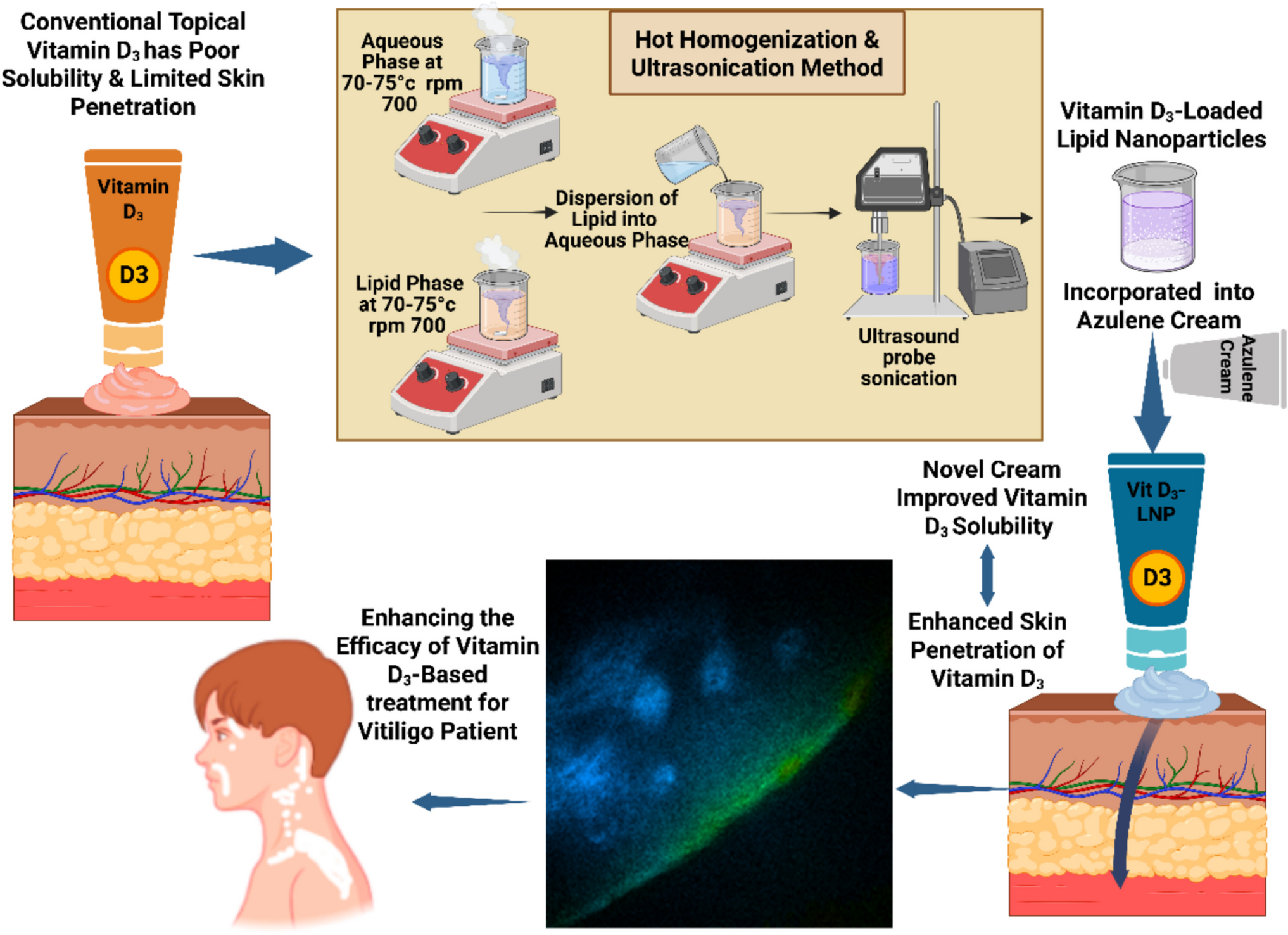
Cream formulation vehicle for the lipid nanoparticlesThe formulation demonstrated stability for more than one year, making it suitable to serve as a vehicle for the dispersion of Vitamin-D3 nanoparticles and azulene [68,69,70,71,72]. To this formulation, Vitamin D3 (cholecalciferol) was incorporated at a concentration of 0.01% w/w (1 mg per 100 g of cream), based on evidence demonstrating its topical efficacy in stimulating melanocyte activity, which is crucial for repigmentation in the treatment of vitiligo. A study using 100 µg topical Vitamin D3 showed increased melanocyte DOPA-positivity, supporting its biological activity at low doses [73]. Additionally, clinical findings with Topical-vitamin D gel delivering 125 µg/g Vitamin D3 confirmed the safety and effectiveness of the transdermal route for systemic and local delivery [74]. Azulene was incorporated at 0.03% w/w (0.3 mg/g cream), based on its demonstrated anti-inflammatory properties and its ability to reduce UVB-induced erythema comparably to a 0.5% hydrocortisone cream [2].
Characterization of LNP loaded with Vitamin D3Physical characterizationThe DLS analysis provided key information on the LNPs’ hydrodynamic diameter and size distribution, with PDI values indicating the uniformity of the formulation. A PDI value below 0.3 is generally considered ideal for lipid nanoparticles, as it reflects a narrow size distribution and correlates with enhanced physical stability [19]. Zeta potential values greater than + 30 mV or less than −30 mV suggest sufficient electrostatic repulsion to prevent aggregation. Furthermore, the inclusion of non-ionic surfactants with high ethylene oxide content contributes to steric stabilization. These mechanisms collectively help to reduce flocculation, coalescence, and Ostwald ripening, thereby supporting the long-term stability of the dispersion [75, 76]. Based on the optimization results and subsequent stability testing, the formulation with surfactant ECO at a concentration of 6.35% and an oil phase concentration of 5.46% was recommended for further development. This formulation not only meets the initial target criteria for PDI and particle size but also demonstrates enhanced stability over time, making it a promising candidate for long-term applications [30]. When Vitamin D3 was incorporated into the ECO surfactant formulation, the particle size was observed to be 153.9 nm, with a PDI of 0.216 (Fig. 8a), which TEM confirmed (Fig. 8c,d, and e), and the zeta potential was measured as −54.3 mV (Fig. 8b).
Fig. 8
Characterization of Lipid Nanoparticles (LNP) loaded with Vitamin D3. (a) Dynamic Light Scattering (DLS) analysis showing particle size distribution and polydispersity index (PDI) of the Vit D3-LNP formulation, (b) Zeta potential distribution measured by DLS, (c) Transmission Electron Microscopy (TEM) image of LNP morphology at 12,000 × magnification, (d) TEM image at 30,000 × magnification, and (e) TEM image at 80,000 × magnification, highlighting nanoparticle structure and distribution
Our formulation achieved a particle size below 200 nm with high encapsulation efficiency, consistent with trends reported in the literature. For example, Ramezanli et al. study on polymeric nanospheres for Vitamin D3 topical delivery emphasized the need for nanoscale carriers to improve skin penetration and loading efficiency [77]. Furthermore, Hussain, Xu et al. research on nano-based strategies for Vitamin D delivery supports the notion that reducing particle size enhances bioavailability and cellular uptake [78], which is in agreement with the design and performance outcomes of our optimized lipid nanoparticle system.
Thermogravimetric characterisation of Vitamin D3 in LNPDSC analysis of dried samples (5–10 mg) was performed under nitrogen, the ramp rate ranged from 30 °C to 600 °C at 10°C/min to assess the thermal behaviour of pure Vitamin D3 (Vit-D3), Vitamin D3-loaded lipid nanoparticles (Vit D3-LNP), and lipid nanoparticles (LNP). The DSC thermogram (Fig. 9a) showed a sharp endothermic peak for pure Vitamin D3 around 82–86°C, indicating its crystalline nature and characteristic melting behavior. However, in Vit D3-LNP, this peak was shifted, broadened, or diminished, suggesting encapsulation and interactions with formulation components such as PVP, GMS, and CCT. For LNP, the characteristic GMS melting peak (55–65°C) was observed. Still, it appeared broadened and slightly shifted, likely due to interactions with liquid CCT, which altered the crystalline structure and increased lipid matrix fluidity. The absence of a distinct Vitamin D3 peak in LNP confirmed successful encapsulation, indicating that Vitamin D3 was well-integrated into the lipid matrix rather than existing as a separate crystalline phase.
Fig. 9
(a) Differential Scanning Calorimetry for Vitamin D3, LNP, and for Vit-D3-LNP, (b) Thermogravimetric (TGA) of Vitamin D3, LNP, and Vit-D3-LNP. (a) Differential Scanning Calorimetry (DSC) thermograms of pure Vitamin D3, lipid nanoparticles (LNP), and Vitamin D3-loaded lipid nanoparticles (Vit-D3-LNP). The DSC analysis reveal a sharp endothermic peak for pure Vitamin D3 at 82–86 °C (corresponding to the Vit-D3 melting point), indicating its crystalline nature, while the absence or broadening of this peak in Vit-D3-LNP confirm successful encapsulation and interaction with formulation components, including Caprylic/Capric Triglyceride (CCT), Glyceryl Monostearate (GMS – Kolliwax® GMS II), and Polyvinylpyrrolidone K30 (PVP K30 – Kollidon® 30). (b) Thermogravimetric Analysis (TGA) of pure Vitamin D3, LNP, and Vit-D3-LNP. TGA results demonstrate enhanced thermal stability of the formulations, with LNP and Vit-D3-LNP exhibiting a more gradual degradation profile compared to pure Vitamin D3. These findings confirm the successful integration of Vitamin D3 into the lipid-PVP matrix, improving its stability and encapsulation efficiency for potential topical delivery
The Thermogravimetric Analysis (TGA) (Fig. 9b) further supported these findings by demonstrating the thermal behaviour of the formulations. Pure Vitamin D3 exhibited significant weight loss at lower temperatures, while both LNP and Vit D3-LNP showed a more gradual degradation pattern, indicating improved thermal characteristics. The presence of PVP and lipids (GMS, CCT) in the LNP system likely contributed to this effect by promoting an intimate interaction between the components, which may have restricted molecular mobility and slowed down the degradation process. While amorphization can sometimes lead to decreased stability, in this case, the positive interactions between the PVP, lipids, and Vitamin D3 appeared to enhance the formulation’s thermal behavior [79]. These results confirmed that Vitamin D3 was effectively incorporated into the lipid-polymer matrix, supporting its potential for topical delivery at physiological temperatures (32°C–37°C).
Determination of encapsulation efficiencyThe drug-loaded lipid nanoparticles (DLPL) encapsulation efficiency assessment was conducted to determine the amount of Vitamin D3 successfully encapsulated within the LNP. Using high-performance liquid chromatography (HPLC) to quantify the encapsulated Vitamin D3, the total amount of Vitamin D3 in the LNP (n = 3) was 5.345 µg/mL. In contrast, free Vitamin D3 (n = 3) was 0.161 µg/mL.
The encapsulation efficiency was 96.98%, indicating a high level of Vitamin D3 encapsulation within the lipid nanoparticles.
To validate the method, a mass balance analysis was also performed by summing the encapsulated and free drug quantities and comparing them with the initial amount added. The mass balance recovery was 103.08%, confirming the accuracy and reliability of the encapsulation efficiency determination.
Chemical stability of Vitamin D3 loaded in LNPThe HPLC analysis revealed the percentage reductions in Vitamin D3 concentration (starting at 10 µg/mL) in lipid nanoparticles (LNP) under different storage conditions after 30 days (Fig. 10). At room temperature, samples stored in the dark storage showed a 6% reduction in Vitamin D3 content, confirming the protective effect against light exposure. In contrast, room temperature storage under continuous light exposure resulted in a 9% reduction, highlighting Vitamin D3’s photosensitivity and the need for storage in dark environments to prevent light-induced degradation and inactive isomer formation [80, 81].
Fig. 10
HPLC Analysis Results for Vitamin D3 Stability Under Different Storage Conditions after 30 Days. (a) Vitamin D3 reference concentration (initially 10 µg/mL). (b) Vitamin D3 degradation in LNPs stored in the dark: minimal loss (6%), confirming the protective effect against light exposure. (c) Under continuous light exposure, degradation increased to 9%, demonstrating Vitamin D3’s photosensitivity and supporting the need for dark storage to avoid light-induced isomer formation. (d) Refrigerated storage at 4 °C resulted in a 13% reduction, indicating moderate improvement in stability. (e) Freezing at –20 °C resulted in a 15% reduction, indicating only a slight enhancement over refrigeration. (f) Elevated temperature storage at 40 °C caused the most significant degradation (40%), highlighting the temperature sensitivity of Vitamin D3 and the importance of avoiding heat during storage
The effect of temperature was even more pronounced. Storage at 40 °C led to the highest degradation, with a 40% reduction, indicating that high temperatures drastically accelerate Vitamin D3 breakdown. In cool storage conditions, degradation was 13% at 4 °C and 15% at −20 °C, suggesting that while refrigeration slows degradation, cooling offers only a slight improvement in stabilization. These findings emphasize that proper storage in dark, room-temperature conditions is crucial to minimizing Vitamin D3 degradation and maintaining stability [80, 82, 83]. Consistent with our results, previous studies have shown that Vitamin D3 predominantly undergoes photochemical degradation into biologically inactive isomers, including tachysterol, lumisterol, and isotachysterol, under standard storage conditions. Notably, no pharmacologically active degradation products have been identified as a result of non-enzymatic degradation pathways of Vitamin D3 [82, 83]. This supports the chemical stability profile observed in our formulation, in which no additional peaks corresponding to active degradation products were detected by HPLC analysis.
Characterization of cream loaded with LNPThe rheological analysis of both the base cream and the LNP-loaded cream (LNP-Cream) confirmed non-Newtonian, shear-thinning (pseudoplastic) behavior. As shear rate increased from very low values (~ 0.00008 s⁻1 for cream and ~ 0.00068 s⁻1 for LNP-Cream) to high shear rates (~ 5350 s⁻1 for LNP-Cream and ~ 474 s⁻1 for cream), the viscosity of both formulations decreased markedly, spanning several orders of magnitude (Fig. 11). This shear-thinning profile is characteristic of semisolid topical systems, indicating that both formulations exhibit reduced resistance to flow under mechanical stress, such as rubbing or spreading during application. This behavior supports the creams’ suitability for topical use, as they provide high viscosity at rest for stability, while allowing ease of application under shear. The yield stress of the LNP-cream was also lower than that of the base, indicating a reduced force requirement for initial flow. This property can enhance the user experience during the application. These trends are consistent with previous findings where nanoparticle inclusion modified the internal cream structure without compromising its physical stability [34, 38, 84].
Fig. 11
Rheological profiles of the base cream and LNP-loaded cream (LNP-Cream)at 25 °C. These profiles show shear-thinning (pseudoplastic) behavior (a) for the cream and (b) for the LNP-cream. Viscosity decreases with increasing shear rate, demonstrating reduced flow resistance under mechanical stress
Texture profile analysis further supported this observation (Fig. 12a and b). The LNP-loaded cream exhibited a reduction in hardness (12.97 g) compared to the control cream (17.97 g) (P < 0.05), indicating a softer texture (Fig. 12c). Adhesiveness and cohesiveness also declined slightly in the LNP formulation, which may improve tactile perception by reducing stickiness while maintaining structural integrity. Springiness showed only a minimal decrease, indicating that the formulation retained adequate elasticity after deformation. These outcomes align with those of Dabbaghi et al., who emphasized the importance of maintaining viscoelastic properties to ensure consistent product performance and user acceptability [33].
Fig. 12
Texture profile analysis of the base cream and LNP-loaded cream (LNP-Cream). (a) and (b) show the force-distance curves obtained from double compression cycle tests. (c) Quantitative comparison of texture parameters including hardness, adhesiveness, cohesiveness, and springiness. The LNP-Cream exhibited significantly lower hardness (P < 0.05) compared to the base cream, indicating a softer texture. Adhesiveness, cohesiveness, and springiness showed slight, non-significant (NS) reductions, suggesting preserved structural integrity and elasticity
Together, these results confirm that the incorporation of lipid nanoparticles into the topical cream vehicle did not compromise its stability or performance. On the contrary, the observed reduction in viscosity and softening of texture in the LNP-loaded cream imparted functional advantages, potentially enhancing spreadability, user comfort, and overall application performance.
In vitro skin penetration testThe cumulative permeation results across all time points (0, 2, 4, 6, and 24 h) revealed a significant difference between the free Vitamin D3 and the cream formulation, P < 0.05 (Table 4) and (Fig. 13a and b). Free Vitamin D3 exhibited negligible permeation across the epidermis, with only a minimal cumulative amount being detected at 24 h (0.001–0.002 µg/cm2). In contrast, markedly higher permeation was recorded for the cream formulation, with cumulative amounts reaching 0.006–0.009 µg/cm2 at 24 h. These findings were interpreted to highlight the ability of the cream formulation to enhance skin permeation and delivery compared to free Vitamin D3, likely due to the lipid nanoparticle (LNP) carrier system and the cream base’s capacity to facilitate Vitamin D3’s diffusion through the stratum corneum.
Table 4 In vitro permeation test using three different donors: Mean Cumulative amount (µg/cm2)—All time points. Mean cumulative amount of Vitamin D3 permeated (µg/cm2) at each time point (0, 2, 4, 6, and 24 h) following in vitro skin permeation test (IVPT) using three different donors. Values are presented as mean and standard deviation (SD)Fig. 13
In vitro permeation test using three different donors: (a) and (b) Mean Cumulative amount (µg/cm2—All time points. (a) and (b) Mean cumulative drug amount (µg/cm2) at all time points (0, 2, 4, 6, and 24 h) following the in vitro penetration test (IVPT). The results show a significant difference (P < 0.05) between free Vitamin D3 and the cream formulation. Free Vitamin D3 exhibited minimal permeation, with only 0.001–0.002 µg/cm2 detected at 24 h, whereas the cream formulation demonstrated significantly higher permeation, reaching 0.006–0.009 µg/cm2. This suggests that the lipid nanoparticle (LNP) cream formulation enhances Vitamin D3 delivery by facilitating diffusion through the stratum corneum
Similarly, differences in drug retention in the donor phase, stratum corneum (SC), and deeper skin layers were indicated by the results (Table 5). Free Vitamin D3, high donor phase drug amounts (1.1–1.38 µg/cm2), and very low SC and skin content (0.006–0.085 µg/cm2) confirmed limited diffusion and retention. Conversely, the cream formulation exhibited lower residual drug in the donor phase (0.18–0.67 µg/cm2) (P < 0.05), significantly higher SC content (0.76–1.35 µg/cm2) (P < 0.05), and superior drug retention in deeper skin layers (0.25–0.47 µg/cm2) (P < 0.05) (Fig. 14) suggesting its enhanced delivery potential.
Table 5 In vitro permeation test using three different donors. Mean Donor Drug Amount (µg/cm2). Vitamin D3 distribution across the donor phase, stratum corneum (SC), and deeper skin layers after 24 h of in vitro permeation using three different donors. Results are expressed as mean drug amount (µg/cm2) and standard deviation (SD)
Comments (0)