Screen the active ingredients and targets of dendrobium huoshanense
We obtained the active components of Dendrobium huoshanense and their targets of action from the database via cancer HSP [12] (https://old.tcmsp-e.com/CancerHSP.php) and Symmap [13] (https://www.symmap.org). Their targets for the components were further predicted by SwissTargetPrediction [14] (http://swisstargetprediction.ch) based on their chemical structures. The active ingredients were screened based on drug-like properties (DL) ≥ 0.18 and oral bioavailability (OB) ≥ 30%, and then we translated the target names of these active ingredients into gene names via the Uniprot database (https://www.uniprot.org), and finally we used Cytoscape 3.9.1 software to construct a Dendrobium huoshanense active ingredients and the relationship network between the target genes.
The analysis of PPI network, KEGG, GO, and Hallmark
We analyzed the interactions between the potential therapeutic targets of Dendrobium huoshanense through the STRING database (https://cn.string-db.org) and used Cytoscape software to construct a protein–protein interaction (PPI) network. To further investigate the biological functions of the targets of Dendrobium huoshanense and to explore the possible molecular mechanisms of Dendrobium huoshanens, we used R4.0.4 software and Metascape (https://metascape.org) for Gene Ontology (GO), Kyoto Encyclopedia of Genes and Genomes (KEGG), and Hallmark enrichment analysis.
Study cohort and expression analysis
A cohort of LUAD patients from The Cancer Genome Atlas (TCGA), which contains RNA sequencing data and phenotypic information from 576 LUAD patients, was included in this study. We used GEPIA2 (http://gepia2.cancer-pku.cn) for differential expression analysis and survival analysis of four genes, Fragile X-related protein-1 (FXR1), actin-related protein 3 (ACTR3/ARP3), altered tubulin alpha 8 (TUBA8) and nuclear factor kappa-B subunit 2 (NF-κB2).
GSEA
We performed GSEA (GSEA V4.2.3) enrichment analysis on RNA-seq data from 576 LUAD patients from TCGA. The samples were classified as high-expression (> 50%) or low-expression (< 50%) based on the level of IL-35 expression. The c2.cp.kegg.v7.4.symbols.gmt gene set was selected for GSEA analysis of the above genes separately. Based on gene expression profiles and phenotypic groupings, a minimum gene set of 5 and a maximum gene set of 5000 were set with 1000 resampling, and a P value of < 0.05 and an FDR of < 0.25 were considered statistically significant.
Single-cell RNA-seq data and analysis
We obtained single-cell transcriptome sequencing data for two sets of LUAD, GSE139555 and GSE131907, from the GEO database and analyzed them using the scTIME Portal (http://sctime.sklehabc.com/unicellular/home).
Chemicals
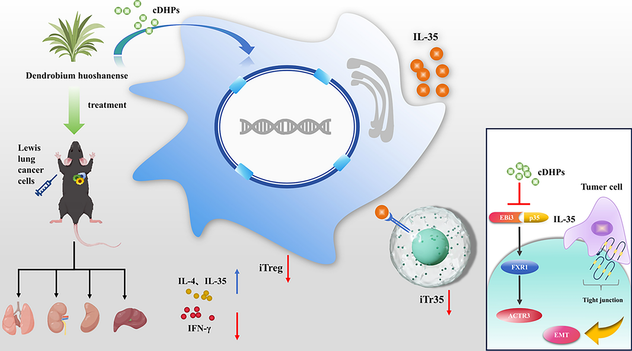
cDHPs was purchased from the Sichuan Weikeqi Biological Technology Co., Ltd. (WKQ-0027085, Chengdu, China) with a purity > 90% tested by high-performance liquid chromatograph.
Cell line, tumor models and treatment
Lewis lung carcinoma (LLC) was purchased from ATCC and cultured in Dulbecco's modified Eagle medium (DMEM) containing 10% fetal bovine serum (FBS), 100 units/mL of penicillin and 100 µg/mL of streptomycin at 37 °C in a humidified incubator with 5% CO2. Routine testing of LLC confirmed the absence of mycoplasma contamination, and limited generation cultures were performed.
Thirty C57BL/6 female mice, 3 weeks old, 15–17 g, were purchased from Changzhou Cavens Laboratory Animal Co., LTD. (Changzhou, China). The animals were housed and maintained under optimal conditions of light, temperature, and humidity, with free access to food and water. To create an LLC model, 1 × 10⁶ LLC was resuspended in 100 µL PBS and inoculated under the skin in the right axilla of mice after 1 week of adaptive feeding. Thirty mice were randomly divided into 3 groups (n = 10): normal, model, and cDHPs groups. Three days after inoculation, 200 mg/kg cDHPs were given to the cDHPs group and equal amounts of distilled water were given to the normal and model groups by daily gavage. All experimental procedures were authorized by the Wannan Medical College’s Animal Experimentation Ethics Committee and followed the “Principles of Laboratory Animal Care”. After 21 days of treatment, the mice were euthanized. Single-cell suspensions of primary cell isolates were prepared from spleens using a mechanical grinding method. Spleen and blood specimens were taken, ground, and then centrifuged and suspended in 1640 medium using red cell lysate. Flow cytometry was used to detect iTr35, CD4+ Foxp3-Tconv, and lung tissue and remaining tumor tissue were stored in 4% paraformaldehyde or ˗ 80 °C in a refrigerator to measure other biochemical parameters.
Flow cytometry assay
Antibodies used for flow cytometry analysis of CD4+Foxp3- Tconv, iTr35, were sorted using a BD flow cytometer as follows: PerCP-Cy 5.5 Rat Anti-Mouse Foxp3 (563,902, BD Bioscience, USA), FITC Rat Anti-Mouse CD4 (553046, BD Bioscience, USA), Human/Mouse IL-12/IL-35 p35 PE-conjugated Antibody (IC2191P, R&D System, USA), Mouse IL-27/IL-35 EBI3 Subunit APC-conjugated Antibody (IC18341A, R&D System, USA), CD16/CD32 Monoclonal Antibody(93) (14-0161-81, Invitrogen, USA), Cell Stimulation Cocktail (500 ×) (00-4970, Invitrogen, USA), Protein Transport Inhibitor Cocktail (500 ×) (00–4980, Invitrogen, USA), Transcription Factor Buffer Set (51-9008100, BD Biosciences, USA).
Quantitative real-time PCR (RT-PCR)
Total RNA was extracted from spleens using TRIzol (15596026, Invitrogen, USA), reverse transcribed by HyperScript III RT SuperMix for qPCR with gDNA Remover (R202, NovaBio, China) and PCR amplified using NovoStart SYBR qPCR SuperMix Plus (E096, novoprotein, China). PCR products were amplified using the following thermal cycling: 95 °C for 1 min, 95 °C for 20 s, 60 °C for 1 min, 40 cycles, using 2% agarose containing ethidium bromide The PCR amplification products were observed on gels, and parameters were used for amplification data analysis using the ΔΔCt method. Primers were designed using Primer Premier 5 software. The primers used are listed in Table 1.
Table 1 Primers used for RT-PCR amplificationsWestern blotting
Spleen tissue (50 mg) was ground and lysed with 500 μL of RIPA lysate (P0013B, Beyotime, China). The resulting cell lysates were centrifuged at 12,000 rpm for 15 min at 4 °C. The BCA kit (P0012S, Beyotime, China) was used to detect protein expression in the lysate, and equal amounts of protein were separated by SDS-PAGE and transferred to PVDF membranes (IPVH00010, Millipore, USA), and the membranes were closed with 5% skimmed milk powder before the addition of primary antibody (1:1000). The membrane was incubated overnight at 4 °C and then washed with 0.1% Tween-20 in tris-buffered saline. Secondary antibodies were mixed with HRP (1:2000) and incubated at 37 °C for 1 h. Protein expression was determined using the ECL kit and chemiluminescent image detection system and quantified using ImageJ software. In the western blot analysis, the following antibodies were used: IL-12A rabbit mAb (A20383, ABclonal, China), EBI3 rabbit mAb (A19613, ABclonal, China), and β-actin antibody (#4970, CST, USA).
Enzyme-linked immunosorbent assay (ELISA)
Following serum collection, ELISA kits were used to detect the concentrations of IFN-γ (CSB-E04578m, CUSABIO, China), IL-35 (CSB-E13145m, CUSABIO, China), and IL-4 (CSB-E04634m, CUSABIO, China).
Natural killer (NK) cell activity assay
The spleen tissue was ground, and NK cells were extracted after lysing the red blood cells. The target cells of NK cells were co-cultured with NK cells at 100:1, 50:1, 25:1, and the killing ability of NK cells was detected by adding Cell Counting Kit-8 (BS350B, Biosharp, China) after 20 h.
Statistical analysis
Statistical analysis was performed using Student's t-test for comparison of two groups or one-way analysis of variance for comparison of more than two groups followed by Tukey's multiple comparison test. For multiple testing, a Bonferroni post hoc test of p values was done. Statistical calculations were performed using GraphPad Prism (GraphPad, San Diego, USA). The data were expressed as the means standard deviations of at least three independent experiments. p value < 0.05 was considered statistically significant.


Comments (0)