Materials
Doxil® was manufactured by Baxter Healthcare (Deerfield, IL, USA) and Doxorubicin Hydrochloride Liposome Injection formulations (2 mg/mL) manufactured by Sun Pharmaceuticals (Goregaon, Mumbai, India), Dr. Reddy’s Laboratories (Hyderabad, Telangana, India) and Zydus LifeSciences Global FZE (Ahmedabad, Gujarat, India) were purchased from the University of Michigan Hospital Pharmacy. Daunorubicin hydrochloride was purchased from Sigma Aldrich (St. Louis, MO, USA). Doxorubicinone and daunorubicinone were purchased from Cayman Chemical (Ann Arbor, MI, USA). Hydrogenated soy phosphatidylcholide (HSPC) and 1,2-distearoyl-sn-glycero-3-phosphoethanolamine-N-[amino (polyethylene glycol)−2000] (ammonium salt) (DSPE-PEG 2000) were purchased from Lipoid GmbH (Ludwigshafen, Germany). Lyso-PC (16:0) and Lyso-PC (18:0) were purchased from Avanti Polar Lipids (Birmingham, AL, USA). Cholesterol and ammonium acetate were purchased from Sigma Aldrich (St. Louis, MO, USA). Poly-Prep chromatography columns (Cat No. 731–1550 and 731–1553) were purchased from Bio-Rad Laboratories (Hercules, CA, USA). TOYOPEARL HW-55F resin in ethanol was purchased from Tosoh Bioscience (Grove City, OH, USA). Holey carbon grids (R 1.2/1.3, 300 Mesh, Copper) were obtained from Quantifoil (Großlöbichau, Germany). Round cryo grid boxes with lids were obtained from MiTeGen (Ithaca, NY, USA). C-clips and c-clip rings were purchased from Thermo Fisher (Waltham, MA, USA). Liquid nitrogen and dewars were provided by the University of Michigan Life Sciences Institute Cryo-EM Core Facility (Ann Arbor, MI, USA). Axygen 96-well full-skirt clear PCR microplate and nonsterile, 96 Round Well Sealing Mat for PCR microplates were purchased from Corning (Corning, NY, USA). All other solvents and reagents were of analytical grade and were purchased from Fisher (Waltham, MA, USA).
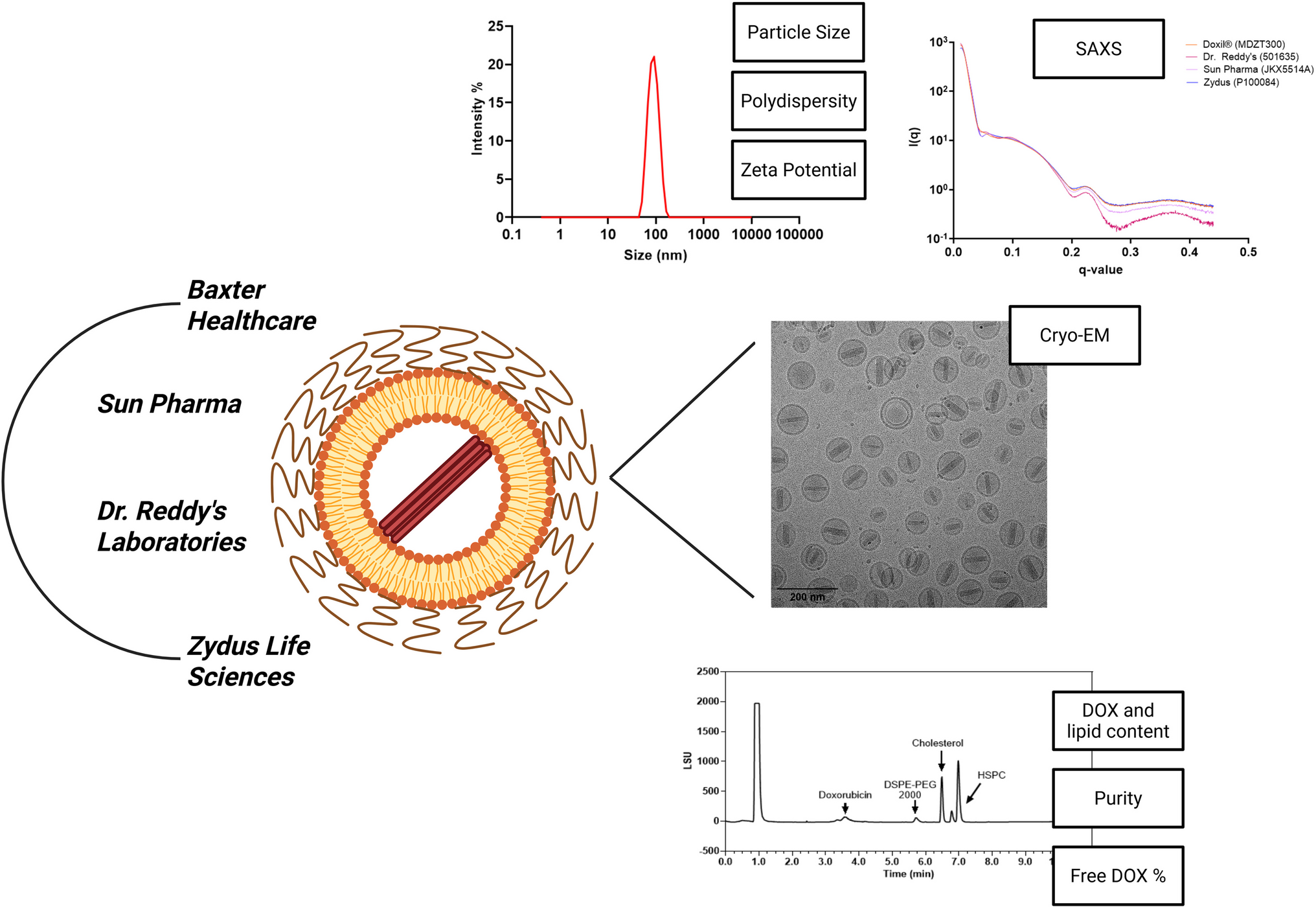
MethodsParticle Size
Particle size was measured using the Malvern ZetaSizer Nano ZSP (Worcestershire, England, UK). Liposomal doxorubicin samples were diluted tenfold in deionized water and transferred to Plastibrand® semi-micro disposable cuvettes (BRAND GMBH, Wertheim, Germany) for analysis. Water and liposome were selected as the dispersant and material, respectively, and the measurement temperature was 25°C. The refractive index for water and liposome were set to 1.33 and 1.45. The viscosity was 0.8872 cp for water at 25°C and sample equilibrium time was set to 120 s. All samples were measured in triplicates, and the particle size data obtained was zeta average, an intensity-based harmonic mean, plotted as a function of intensity (%) versus particle size (nm). Particle size data was directly obtained from the built-in software.
Zeta Potential
Zeta potential measurements were performed using the samples prepared for particle size analysis. A 1 mL syringe was used to handle and transfer the sample solution from semi-micro disposable cuvettes to DTS1070 folded capillary cells (Malvern Panalytical Ltd, England). Sufficient volume of solution covered the electrodes on both sides of the capillary cell. Zeta potential was measured using the Malvern ZetaSizer Nano ZSP under the same system parameters for the particle size method and the dielectric constant was set to 78.5. Data was reported in millivolts.
Doxorubicin Content
DOX content was quantified using ultra performance liquid chromatography (UPLC). An Acquity UPLC-TUV detector system with Empower 3 software was used with a BEH C18 column (100 × 2.1 mm, 1.7 μm) (Waters Corporation, Milford, MA, USA) under isocratic conditions consisting of deionized water with 0.1% formic acid as aqueous solvent (A) and methanol with 0.1% formic acid as organic solvent (B). Liposomal DOX samples were diluted 100-fold in methanol. A 45% to 55% ratio of mobile phase for solvent A to B was employed. Flow rate was set to 0.3 mL/min, injection volume was 10 µL, detection wavelength was 254 nm, and the run time lasted 15 min. A method description for standard curve generation is provided in Supplementary Materials.
Lipid Content
Lipid concentration was quantified using an Acquity UPLC with an evaporative light scattering detector (ELSD) system with Empower 3 software (Waters Corporation, Milford, MA, USA). A gradient mobile phase was used to separate HSPC, DSPE-PEG (Mw 2000) and cholesterol, and consisted of two solvent systems: A) a mixture of 40% v/v deionized water and 60% v/v methanol with 10 mM ammonium acetate buffer pH 6.4, and B) a mixture of 90% v/v isopropanol and 10% v/v methanol with 10 mM ammonium acetate buffer pH 6.4. The 10 mM ammonium acetate buffer was prepared by adding 77 mg of ammonium acetate to 100 mL of deionized water, and the pH of the solution was adjusted to 6.4.
Liposomal DOX samples were diluted 40-fold in methanol and vortexed to mix. An Xbridge C18 column (4.6 × 150 mm, 3.5 μm) (Waters Corporation, Milford, MA, USA) was used to separate the lipids. The flow rate and column temperature were set to 1 mL/min and 45°C. Injection volume was 10 µL and the run time was 15 min. ELSD gas pressure was set to 40 psi, data rate was set to 10 points per second and the temperature of the drift tube was set to 55°C. Gain was set to 150 and the nebulizer was set to heating at 45%. Compressed gas was used for ELSD operation. A method description for the gradient and standard curve generation is provided in Supplementary Materials.
Doxorubicin Purity
Another UPLC-TUV method was developed to identify DOX impurities. The mobile phase consisted of two solvents: A) deionized water with 0.1% formic acid, and B) a mixture of 80% v/v acetonitrile and 20% v/v methanol with 0.1% formic acid. Daunorubicin hydrochloride, doxorubicinone, and daunorubicinone are designated as doxorubicin process and product-related impurities by the United States Pharmacopeia (USP). Liposomal DOX samples were prepared by diluting the samples tenfold in methanol. A UPLC BEH C18 column (100 × 2.1 mm, 1.7 μm) was used to separate impurities from doxorubicin. Column temperature and flow rate were set to 35°C and 0.5 mL/min. An injection volume of 10 µL, run time of 10 min, and dual detection wavelengths (220 and 254 nm) were used to identify doxorubicin and impurity peaks. The relative retention time (RRT) for each impurity standard in reference to the doxorubicin standard was calculated and impurities were identified by assigning corresponding peaks in each sample based on standard RRT ± 5%. Doxorubicin and impurity concentration were calculated using the linear regression equation from the standard curve (Supplemental Materials).
Lipid Purity
Lipid purity was determined using a UPLC assay with an evaporative light scattering detector (ELSD) under the same conditions detailed in the "Lipid Content" section. Lysophosphatidylcholine (Lyso-PC) 16:0 and 18:0 are two common lipid degradants from hydrolysis that often occurs in dissolution. Impurity stock solutions (1 mg/mL) were prepared in methanol and diluted to 0.05 mg/mL. Liposomal DOX samples were diluted 40-fold in methanol. The UPLC-ELSD method for lipid purity was performed and a doxorubicin standard was spiked with Lyso-PC (16:0) and Lyso-PC (18:0) to identify the retention time of each impurity peaks in our sample. The amount of cumulative lipid impurity was quantified by calculating the percentage ratio of cumulative peak area for lipid impurity to the cumulative lipid peak area.
Free, Non-Encapsulated Doxorubicin in Liposomal Formulations
Separation of free, non-encapsulated DOX from liposomal DOX was achieved using size exclusion chromatography. Poly-Prep columns were washed with methanol and water. 600 µL of TOYOPEARL HW-55F resin (in ethanol) was added to the column and installed on a vacuum manifold processing station. Excess water was drained from the columns before loading the sample and the resin surface was kept flat. Liposomal DOX (10 µL) was carefully added to the resin surface. A free DOX solution (20 µL) prepared at a concentration of 1 mg/ml in methanol was loaded onto an equivalent resin bed volume (600 ul) for equivalent measurement of free doxorubicin recovery. Deionized water (400 µL) was slowly added to the column and the wash solution was collected under vacuum into a microtube. The water wash was repeated three times and 1.2 mL of water wash solution was collected. Next, 400 µL of a 50:50 acetonitrile/methanol solution was slowly added to the column. The same wash procedure was performed using the organic solution and 1.2 mL of organic wash solution was collected. The wash solution tubes were transferred to a nitrogen evaporator set to 50°C to dry the samples. Dry sample residues were reconstituted with 250 µL of methanol. A reference sample was prepared by adding 240 µL of methanol to 10 µL of liposomal DOX. Samples were transferred to UPLC vials and free, non-encapsulated doxorubicin content was quantified using the previously developed UPLC-TUV method described in the "Doxorubicin Purity" section. Free doxorubicin content was calculated using the following equation:
$$\% Free\;Doxorubicin=\frac\times 100$$
(1)
Cryogenic Transmission Electron Microscopy (Cryo-TEM)
Doxorubicin liposome morphology was determined utilizing the cryo-TEM technique in collaboration with the University of Michigan (U-M) Life Sciences Institute (LSI) CryoEM Core Facility. Liposomal DOX formulations at a doxorubicin concentration of 2 mg/mL were plunged frozen in liquid ethane cooled to freezing point in liquid nitrogen using a Vitrobot plunge freezer (ThermoFisher Scientific, Waltham, MA, USA). 3 µL of liposomal DOX samples were deposited onto glow-discharged (PelcoEasiGlow discharger) Cu 300 R 1.2/1.3 holey carbon grids (Quantifoil, Großlöbichau, Germany) and samples were frozen on the grids using the following Vitrobot parameters (10 s wait time, 5 s blot time, 100% humidity, 4°C). Samples were visualized under cryogenic conditions on a Talos Arctica (ThermoFisher Scientific, Waltham, MA, USA) operated at 200 kV. Images were recorded using a Gatan K2 Summit direct electron detector (Gatan, Pleasanton, CA, 94,588) in counting mode at 17.5 kx magnification, corresponding to a pixel size of 2.48 Å/pixel. SerialEM v4.1 was used to control microscope and camera functionality (10, 11). The exposure time was set to deliver 20 electrons/Å2 cumulative dose to the area of interest. Representative images of each sample were recorded from the center of the holes. FIJI/ImageJ was used to manually measure particle dimensions of length, width, and membrane thickness using a minimum sample size of 500 liposomes (12).
Small Angle X-Ray Scattering (SAXS)
The SAXS technique was used to characterize the microstructure similarity of DOX liposomes present in liposomal DOX formulations developed by different manufacturers. Microstructure was analyzed using the high-throughput SAXS technique (HT-SAXS) and similarity between batches was compared using the SAXS Similarity tool developed and conducted by the SIBYLS Beamline (12.3.1) of the Advanced Light Source at the Lawrence Berkeley National Laboratory (Berkeley, CA, USA), supported by the US Department of Energy and National Institutes of Health (13). Liposomal DOX samples were added to an AXYGEN 96-well full-skirt clear PCR microplate with a consistent volume of 30 µL of buffer and sample at three concentrations between 1–10 mg/mL. Labeled concentration for liposomal doxorubicin hydrochloride formulations is 2 mg/mL. Therefore, the following concentrations were prepared for SAXS analysis: 1 mg/mL (low concentration), 1.5 mg/mL (medium concentration) and 2 mg/mL (high concentration). Buffer preparation is critical for SAXS analysis as it can impact the scattering profile of the sample. The prepared formulation buffer should be as closely resemblant of the buffer present in the original sample. Therefore, the prepared formulation buffer was comprised of a 9.4% sucrose solution with 1.55 mg/mL histidine at pH 6.5 similar to the buffer used in the manufacturing of the commercial formulation. 100 mL of buffer was prepared by adding 9.4 g of sucrose and 155 mg of L-histidine to deionized water. The pH of the solution was adjusted to 6.5 with 1 N of sodium hydroxide and/or hydrochloric acid. Prepared buffer and sample solutions were transferred to two 96-well PCR microplates, sealed, and kept under cold conditions prior to analysis by the SIBYLS Beamline at the Lawrence Berkeley Laboratory.
In Vitro Dissolution
In vitro dissolution testing provides critical information to characterize the rate and extent of drug release from a finished dosage form, which can be correlative of in vivo drug performance. From a regulatory perspective, dissolution testing is used in formulation evaluation during product development for batch-to-batch quality control and assurance (14). A dissolution method with discriminative capability, capable of characterizing release profiles of formulations of the same composition but from different manufacturers when compared to the reference product, is crucial to assess a product’s performance and efficacy. Yuan et al. developed a conventional release method using dialysis membranes and the referenced method was employed in the present work to evaluate dissolution profiles for one batch per manufacturer (15).
Briefly, 0.8 mL of each liposomal DOX formulation (C DOX = 2 mg/mL) was added to 300 kDa Float-a-lyzer dialysis tubes which were inserted into 50-mL centrifuge tubes containing 39.2 mL release media containing 100 mM ammonium bicarbonate, 75 mM MES buffer, 5% w/v hydroxypropyl-β-cyclodextrin, 5% w/v sucrose, 0.02% sodium azide at pH 6. The centrifuge tubes were placed on an orbital shaker (ThermoFisher MaxQ 6000) operating at 320 rpm speed and incubated at 45°C. A free DOX solution was prepared with doxorubicin hydrochloride in the same release media at an equivalent concentration (C DOX = 2 mg/mL) as the liposomal formulation. 0.8 mL of the free DOX solution was added directly to a 50-mL centrifuge tube containing 39.2 mL of release media and was used as a positive control. At 0, 1, 2, 4, 8, 12, and 24 h, 150 μL of release media was sampled for UV detection at 480 nm on a plate reader (Synergy NEO HTS Multi-Mode Microplate Reader (Bio-Tek) and replaced with an equal amount of fresh release media. Samples were prepared and analyzed in triplicate. The cumulative percent DOX release was calculated using the following equation:
$$Cumulative\;DOX\;release\;\left(\%\right)=\frac\;of\;liposome\;formulations}\;of\;free\;DOX\;control}\times100\%$$

Comments (0)