
Remember me
Studies 1 (EudraCT number: 2018-003947-48) and 2 (EudraCT number: 2018-000274-29) were conducted at a clinical pharmacology unit in Munich, Germany, while Study 3 (Regulatory Agency Identifier Number: 140537) was conducted at two study centers in the USA. Study 1 was a single-center study with two parts. Part 1 of Study 1 (electronic supplementary material [ESM], Section 1) was an open-label, non-randomized, single ascending dose study, with two subjects per dose, to select the oral and intravenous dose to be applied in Part 2. Part 2 of Study 1 was a randomized, open-label, four-way crossover study in different subjects from Part 1. Oral and intravenous dosing in Part 2 was based on Part 1 using the following criteria: 1) a PK profile that enabled a reliable calculation for the elimination half-life (t½); and 2) a maximum plasma concentration (Cmax) not exceeding the well-tolerated plasma concentration measured at the top doses of 3000−4000 μg in the first-in-humans study of mosliciguat (Bayer AG, data on file). The inhaled dose of 1000 µg was based on data from Study 2 showing that this dose provided a measurable PK profile. The IV and oral doses were estimated from consideration of pre-clinical PK data obtained with IV and oral formulations in various species. Four mosliciguat treatments were evaluated in Study 1 Part 2, designated as: treatment A (1000 μg inhaled dry powder formulation with charcoal block); treatment B (1000 μg inhaled dry powder formulation without charcoal block); treatment C (100 μg IV; 2-h infusion); and treatment D (1000 μg oral solution). Each treatment was given once to each subject under fasting conditions, separated by a washout of ≥ 7 days. The charcoal block was administered relative to the study drug in the following sequence: 10 g of charcoal 15 min before inhalation of study drug; 10 g of charcoal immediately after inhalation of study drug (after rinsing mouth with 200 mL of water) and 2 h after inhalation of study drug; and 5 g of charcoal 4 and 8 h after inhalation of study drug. Charcoal block is used to stop any gastrointestinal absorption of inhaled drug that may be swallowed during administration, allowing investigators to establish that systemic absorption reflects only the proportion of drug absorbed from the respiratory tract [20, 21]. The European Medicines Agency requires regulatory submissions to include two PK studies: one with and one without validated charcoal block; the use of charcoal block is not mandatory in PK studies for US Food and Drug Administration submissions [22].
Study 2 was a single-blind, randomized, placebo-controlled, parallel-group, multiple-dose escalation study. A single inhaled dose of mosliciguat 480 μg or placebo was administered on day 1 and each of days 3–8. This dose was based on human scaling from pre-clinical studies in animal models of PH, including a PH minipig model, unilateral ventilation in minipigs, a hypoxic dog model, and a bronchoconstriction model in rats [19]. After thorough evaluation of the safety, tolerability, and PK results from the previous dose step, new subjects then received inhaled mosliciguat 1000 μg or 2000 μg or placebo on day 1 and days 3–8 of their treatment. Subjects were observed on days 9 and 10 and discharged on day 11. Inhalations took place 1.5–2 h after start of intake of a standardized continental breakfast. The time to the next meal was approximately 5.5 h after dosing.
Study 3 was a randomized, placebo-controlled, single-blind, group comparison study. Inhaled mosliciguat 1000 μg once daily or placebo was administered for 14 days. On the screening visits and the follow-up visit, the subjects came to the study site in a fasting state. During their stay at the study site (day − 1 to day 18) they were served breakfast approximately 1.5–2 h before dosing, followed by a standardized lunch, snack, and dinner 4, 8, and 12 h after dosing, respectively. The meals were standardized on the pre-dose day and the PK profile days only. On the other days, non-standardized meals were served.
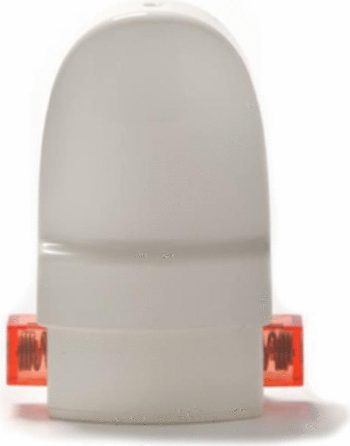
2.2 Formulations Used in the StudiesFor pulmonary application of mosliciguat, a dry powder formulation was developed by blending micronized mosliciguat as the active pharmaceutical ingredient (API) with a specifically engineered carrier made of pharmaceutical inhalation-grade lactose acting as the carrier particle for the API. There were no other ingredients. The API is poorly soluble in any aqueous medium over a wide pH range, but it is soluble in organic solvents. For these studies, the dry powder formulation was contained in inhalation-grade hard capsules of hydroxypropylmethylcellulose, administered with a commercially available, manually operated, breath-activated, low-resistance inhaler (Plastiape RS01 Model 7, Plastiape S.p.A., Osnago Lecco, Italy) (Fig. 1) [23].
Fig. 1
The inhaler used in the current studies
The inhaler is operated by:
Tilting the head slightly backward and exhaling
Taking the mouthpiece between the lips
Inhaling deeply and comfortably
Holding the breath for approximately 3 s
Releasing the mouthpiece from the lips and exhaling
Based on human-dose estimations, different dose strengths for the studies were developed (480 µg and 1000 µg nominal doses), optimizing the fine particle dose (FPD) < 4.5 µm to specified target values (i.e., the delivered mass of drug particles sized < 4.5 µm under standardized conditions, characterized as the delivered drug amount in the deep airways). The dry powder formulation was designed to produce a fine particle fraction < 4.5 µm of ≥ 20% related to the nominal dose and ≥ 30% related to the delivered dose (DD). The DD (i.e., the total mass of drug particles delivered from the device mouthpiece) was characterized as the total dose delivered to the patient orally. The FPD < 4.5 µm is the subfraction of the DD that is expected to reach the site of action in the deep lung. Subfractions with larger particle size are expected to impact in the upper airways and the oropharyngeal region and subsequently enter the gastrointestinal system by swallowing, and are assumed not to be available for local deposition at the site of action in the deeper lung. The in vitro aerosol performance of mosliciguat dry powder inhaler (DPI) capsules was characterized across all three studies based on compendial methods described in the European Pharmacopoeia (European Pharmacopoeia Online [edqm.eu]) and the United States Pharmacopoeia (usp.org). Nominal dose content was determined by reversed-phase high-performance liquid chromatography (RP-HPLC) with ultraviolet detection. In vitro DD measurement of the capsule formulations was determined according to the European Pharmacopoeia monograph “Preparations for Inhalation – Powders for inhalation” using the specified sample collection tube. The aerodynamic particle size distribution (APSD) to measure the fine particle dose < 4.5 µm (and fraction), as well as residual drug contents in the empty capsule and the used device, was determined with the RS01 DPI device and inhalation capsules according to European Pharmacopoeia 2.9.18 “Aerodynamic assessment of fine particles” using a next-generation impactor. For both DD and APSD analyses, drug contents in the dose unit sampling adapter and on the filter stages of the next-generation impactor were determined by RP-HPLC with ultraviolet detection.
The effectiveness of the charcoal block for absorbing mosliciguat in the gastrointestinal tract in Study 2 was confirmed in an in vitro study in which 15 mg of a lactose–mosliciguat mixture was dissolved in 100 mL of water/ethanol (90/10, pH 8.5) to reach a concentration of 300 μg/100 mL. After addition of 10 g of medicinal charcoal, the mixture was stirred. Samples were taken after 10 and 30 min, centrifuged, and the concentration of mosliciguat was determined in the supernatant.
2.3 Randomization and BlindingIn Study 1 Part 2, subjects were randomized open-label to four treatment sequences: A−D−C−B, B−C−D−A, C−A−B−D, and D−B−A−C. Studies 2 and 3 were performed in a single-blind design, with the investigator aware of treatment assignments. Randomization was performed by the investigator using a computer-generated randomization list generated by the study sponsor. To achieve blinding, capsules containing mosliciguat or placebo were identical in size, shape, and color.
2.4 SubjectsSubjects in all three studies were healthy men (as determined by medical evaluation including medical history, physical examination, laboratory tests, and cardiac monitoring) aged 18–45 years with body mass index 18–29.9 kg/m2. Women were excluded because data from reproductive toxicity studies were not yet available and to avoid gender-related effects on the results. Studies 1 and 2 were restricted to subjects of white race. Subjects of reproductive potential had to agree to use two reliable and acceptable methods of contraception simultaneously when sexually active (ESM, Section 2). Exclusion criteria are presented in the ESM (Section 3). All subjects provided written informed consent before any study-specific tests or procedures were carried out.
2.5 Endpoints and AssessmentsIn Study 1, the primary endpoints were the frequency of adverse events (AEs) in Part 1 (secondary endpoint in Part 2), and dose-adjusted area under the concentration–time curve (AUC/D) and dose-adjusted maximum plasma concentration (Cmax/D) in Part 2. For all studies reported here, AUC refers to the area under the concentration–time curve from zero to infinity after a single dose (AUC0−inf). In both study parts, blood samples for analysis of mosliciguat PK in plasma were taken pre-dose, then at 0.25, 0.5, 0.75, 1, 1.5, 2, 2.25 (IV only), 2.5, 2.75 (IV only), 3, 4, 6, 8, 12, and 15 h post-dose; at 24, 28, 32, 36, and 48 h post-dose; and at the follow-up visit within 7–10 days after the last administration of study drug. Urine samples for PK analysis were collected up to 48 h post-dose in Part 2. After inhalation, the device and the used capsules were collected and analyzed to assess any residual mosliciguat contained in the individual inhalers and emptied capsules. A follow-up visit was conducted in both Part 1 and Part 2 of Study 1 at 7–10 days after the last administration of study drug.
Key PK parameters in Studies 2 and 3 included AUC, AUC/D, Cmax, and Cmax/D on day 1, and AUC from time 0–24 h for the planned dose interval after first dose of multiple dosing (AUC[0–24]τ,md), dose-adjusted AUC(0–24)τ,md (AUC[0–24]τ,md/D), maximum observed drug concentration after multiple dose administration (Cmax,md), and dose-adjusted Cmax,md (Cmax,md/D) on day 8 (Study 2) or day 14 (Study 3). In Studies 2 and 3, accumulation ratios were calculated from AUC, Cmax, and drug concentration at the end of the dose interval (Ctrough). In both studies, a follow-up visit was conducted 7–10 days after discharge.
In Study 2, blood samples for PK analysis were taken pre-dose, then at 0.25, 0.5, 0.75, 1, 1.5, 2, 2.5, 3, 4, 6, 8, 12, 15, 24, 28, 36, and 48 h post-dose, then Ctrough on days 4, 5, 6, and 7. On day 8, blood samples were taken pre-dose and then at 0.25, 0.5, 0.75, 1, 1.5, 2, 2.5, 3, 4, 6, 8, 12, 15, 24, 28, 32, 36, 48, 60, and 72 h post-dose. On day 1 of Study 3, blood samples for PK analysis were taken pre-dose, then at 0.25, 0.5, 0.75, 1, 1.5, 2, 2.5, 3, 3.5, 4, 6, 8, 12, 15, 24, and 48 h post-dose, then once daily on days 4–13. On day 14, blood samples were taken pre-dose and at 0.25, 0.5, 0.75, 1, 1.5, 2, 2.5, 3, 3.5, 4, 6, 8, 12, 15, 24, and 48 h post-dose, as well as on days 17, 18, and 21. Details of formulae for calculation of PK parameters in all three studies are included in the ESM, Section 4. Quantitative analysis of mosliciguat in plasma, as well as analysis of residual mosliciguat in capsules and devices, was performed using a fully validated high-performance liquid chromatography and tandem mass spectrometric detection (LC-MS/MS).
In Study 1, mouth-rinse samples from subjects who received inhaled mosliciguat with charcoal block containing 0.2% Tween®20 were diluted with plasma, followed by protein precipitation with acetonitrile. Mosliciguat concentrations were then assessed using LC-MS/MS. The amount of mosliciguat in the mouth rinse after inhalation was derived from the measured volume and concentration in the mouth rinse as:
$$}\left( }} \right) = }\left( }} \right)/1000 \times }\left( }/}} \right)$$
If the measured concentration was below the lower limit of quantification (LLOQ) (50.0 ng/mL), the amount was set to zero. For further information on the bioanalytic methods of all studies, see ESM, Section 5.
Unless stated otherwise, the doses described are the nominal doses, which are the doses filled into the capsules delivered by the inhaler. The administered dose is the nominal dose minus the sum of the amounts remaining in the capsule and the device. The specifications of the formulation are shown in Table 1, and the in vitro data in ESM Table S1.
Table 1 Specifications (ranges) of dry powder inhaler formulations used in Studies 1–32.6 Statistical MethodsNo formal statistical sample size estimation was performed for the three studies. Two subjects in each dose cohort of Study 1, Part 1 were used to confirm the non-clinical PK assumptions and safety of the respective doses in a small group of subjects. Sixteen subjects were randomized in Study 1, Part 2, and 15 had data valid for PK analysis; this was considered sufficient to fulfill the objectives of the study. In Studies 2 and 3, respectively, 12 randomized subjects per dose step (active treatment, n = 9; placebo, n = 3) and 20 randomized subjects per study part (active treatment, n = 16; placebo, n = 4) were considered to be sufficient for evaluation of the study objectives.
Statistical analyses were performed using SAS release 9.2 (Studies 1 and 2) or 9.4 (Study 3) (SAS Institute Inc., Cary, NC, USA). All variables were analyzed by descriptive statistical methods, and all statistical analyses were exploratory.
The definition of the PK analysis set differed between studies. In Study 1, all subjects in Part 1 without validity findings affecting the PK analysis and with a valid PK profile were included. All subjects in Part 2 of Study 1 without validity findings affecting the PK analysis and with valid PK profiles for at least two treatments relevant for the evaluation of absolute bioavailability were included. In Study 2, all subjects who received active study drug and for whom at least one valid PK profile was available were included. In Study 3, all subjects who received active study drug, and for whom valid sets of PK samples on at least one profile day were taken, were included.
In each study, the following summary statistics of mosliciguat plasma concentrations were calculated for each sampling point: geometric mean; geometric standard deviation (SD) (retransformed SD of the logarithms) and coefficient of variation (CV); arithmetic mean, SD, and CV; median (minimum, maximum); and number of measurements. Means at any time were calculated only if at least two-thirds of the individual data were measured and were above LLOQ. For the calculation of the mean value, a data point below LLOQ was substituted by one-half of this limit. In Study 1, the amount of mosliciguat in the mouth rinse and the remaining amount measured in the empty capsules and DPIs after inhalation were described with summary statistics.
In each study, PK parameters were calculated using a model-independent (compartment-free) method. In Studies 1 and 3, Phoenix 8.1 software (Certara, Princeton, NJ, USA) was used in conjunction with the Non-Compartmental Analysis Tool plugin (release 1.0). In Study 2, WinNonlin 5.3 software (Certara) was used in conjunction with the Automation Extension (version WinAE 2.90; Bayer AG, Wuppertal, Germany).
To assess dose proportionality in Study 2, an explorative analysis of variance (ANOVA) (including the factor treatment) was performed on the log-transformed parameters AUC/D, Cmax/D, AUC(0–24)τ,md/D, and Cmax,md/D. Based on these analyses, point estimates (least squares means) and exploratory two-sided 90% confidence intervals for the treatment ratios 1000 μg/480 μg, 2000 μg/480 μg, and 2000 μg/1000 μg were calculated by retransformation of the logarithmic data.
AUC/D and Cmax/D were analyzed assuming log-normally distributed data. The logarithms of these characteristics were analyzed using ANOVA including sequence, participant (sequence), period, and treatment effects. The absolute bioavailability of mosliciguat for oral, inhaled, and inhaled with charcoal block was calculated based on these analyses’ point estimates (least squares means) and exploratory 90% confidence intervals for the following ratios: 1 mg inhaled (with charcoal block)/0.1 mg IV, 1 mg inhaled (without charcoal block)/0.1 mg IV, and 1 mg oral/0.1 mg IV.
The relative bioavailability for inhalation was calculated based on these analyses’ point estimates (least squares means) and exploratory 90% confidence intervals for the ratio 1 mg inhaled (without charcoal block)/1 mg inhaled (with charcoal block), calculated by retransformation of the logarithmic data using the intra-individual SD of the ANOVA.
For any calculations requiring body weight, the subject’s actual weight was measured by a member of the investigator’s team with the subject in underwear or a light hospital gown and without shoes after having emptied his bladder.
Safety was assessed with standard parameters including heart rate, electrocardiographic parameters, blood pressure, and clinical safety and biomarker values derived from blood and urine samples taken at defined intervals before and after dosing. Lung function parameters were measured via body plethysmography in Studies 1 and 2 and by spirometry in Study 3. AEs and serious AEs were summarized using Medical Dictionary for Regulatory Activities terms.
Comments (0)