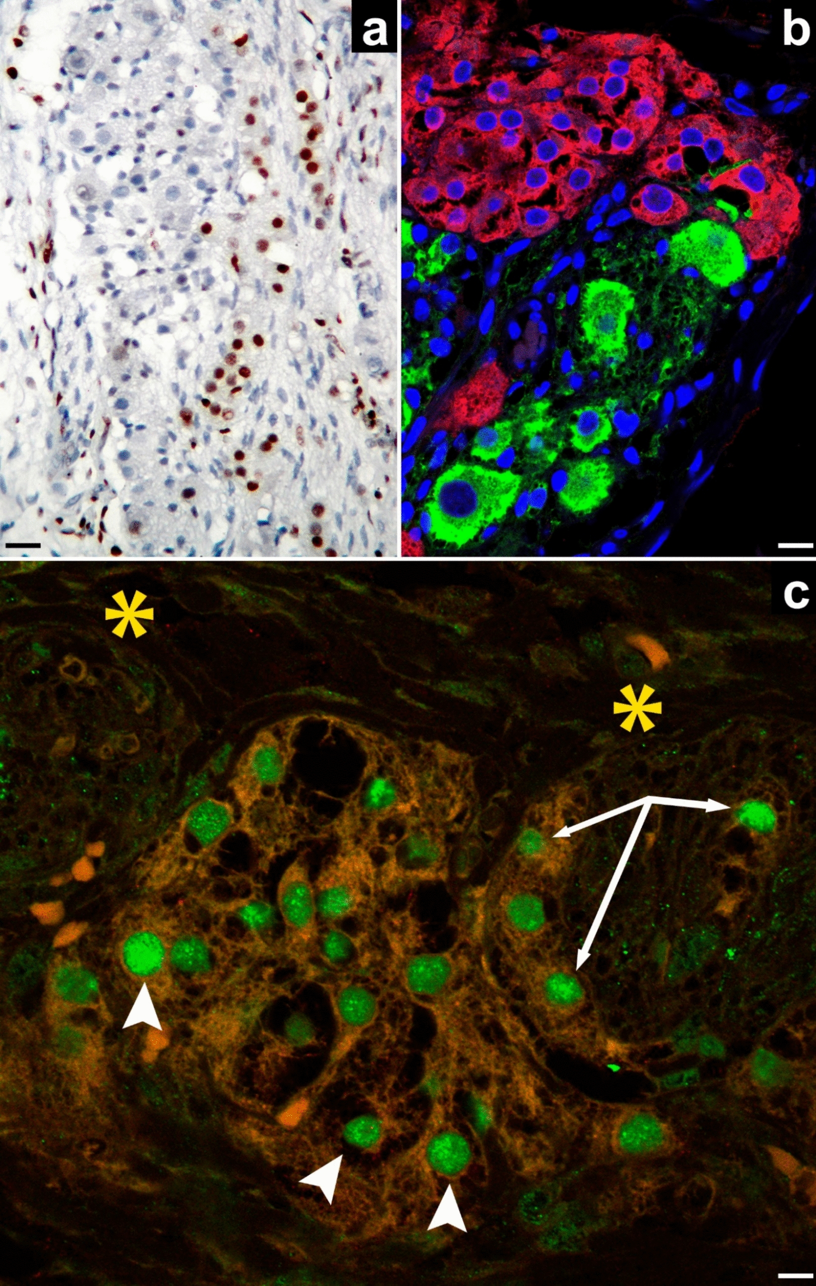
The studied material of mature cystic ovarian teratoma belongs to a 12-year-old Caucasian female (for details, please see Carrasco-Juan, 2024) [4]. The ovaries studied with similar techniques have nodular hyperplasia of OLC. These ovaries belong to a 70-year-old woman who underwent a total hysterectomy with bilateral adnexectomy due to atypical endometrial hyperplasia associated with endometrioid carcinoma infiltrating the endometrium (Type I).
Ethical approval for this study was obtained from the Ethics Committee of La Laguna University (Comité de Ética de la Investigación y de Bienestar Animal, CEIBA 2018 - 0289), including the dissociation of the samples from any information that could identify the patient. The authors, therefore, had no access to identifiable patient information.
Immunohistochemistry
The histological sections obtained, 3 μm thick, were attached to silanized slides. After pretreatment to enhance labeling, sections were blocked with 3% hydrogen peroxide and then incubated with primary antibodies (10–40 min). Primary antibodies from Roche Diagnostics, F. Hoffman-La Roche Ltd., used were: anti-AR, mouse monoclonal anti-human, clone SP107, (dilution 1.7 μg/ml), catalog no. 06523838001; anti-calretinin, mouse monoclonal anti-human, clone SP65 (dilution 6 μg/ml), catalog no. 05992184001; anti-epithelial membrane antigen (EMA), mouse monoclonal anti-human, clone E29 (dilution 0.54 µg/ml), catalog no. 05878900001. The immunoreaction was based on peroxidase activity (PAP) and counterstaining with diaminobenzidine (DAB), subsequently briefly counterstained with hematoxylin, dehydrated in ethanol series, cleared in xylene, and mounted in Eukitt®. Positive and negative controls were used.
Immunofluorescence in confocal microscopy
For double immunofluorescence, 3-μm-thick tissue sections were obtained as described above. For antigen retrieval, sections were deparaffinized and boiled for 20 min in 10 mM sodium citrate buffer (pH 6), rinsed in Tris-buffered saline (TBS, pH 7.6, 0.05 M), and were incubated with a mixture of monoclonal and polyclonal primary antibodies diluted in TBS overnight in a humid chamber and at room temperature. The rabbit polyclonal antibodies used were: anti-tyrosine hydroxylase (TH) (1/500, code no. ab112, Abcam); anti-SF1 (dilution 1/100, code no. AB217317, Abcam); anti-AR (dilution 1/100, code no. SAB5500006, Merck). The mouse monoclonal antibody used was: anti-calretinin (dilution 1/300, code no. 6B3, Swant); anti-SF1 (dilution 1/100, code no. PP-N1665 - 0 C, Perseus Proteomics); anti-class III β-tubulin (βIII-tubulin) (dilution 1/100, code no. T8660, Sigma); anti-chromogranin A (CGA), clone LK2H10, (dilution 1 μg/ml, code no. 0567056001, Roche). The following day, slides were rinsed in TBS and incubated for 1 h at room temperature in the dark with Alexa Fluor 488 goat anti-rabbit IgG (H + L) antibody (1:300, code #A11001, Invitrogen), and Cyanine3 goat anti-mouse IgG (H + L) antibody (1/300, code no. M30010, Invitrogen), for one hour at room temperature in the dark. They were rinsed in TBS and covered with Fluoromount-G with DAPI (REF: 00–4959- 52, Invitrogen from Thermo Fisher Scientific).
Acquisition and processing of images
Images were taken with a Canon EOS 500D digital camera (Canon España S.A., Madrid) coupled to a Leica DMLS microscope (Leica Microsystems, Wetzlar, Germany). Brightness/contrast adjustments, as well as the digital reconstructions (the photo merge), were performed with the Adobe Photoshop CS5 Extended program. Fluorescence immunosignals were obtained using a laser scanning confocal imaging system (Leica TCS SP8, Leica Microsystems, Wetzlar, Germany).


Comments (0)