
Remember me
Sequences for human RECQL5, CSA, CSB, UVSSA, DDB1, CUL4A, RBX1, ELOF1 and UbcH5b were amplified from human cDNA. The sequence for human Pol II CTD was amplified from the codon-optimized RPB1 sequence (GenScript). Sequences for RECQL5, Pol II CTD, CSB and CUL4A were cloned into the 438C vector (Addgene, 55220) with an N-terminal His6-MBP tag; those for CSA and RBX1 were cloned into the 438A vector with no tag (Addgene, 55218); those for DDB1 and UVSSA were cloned into the 438B vector (Addgene, 55219) with an N-terminal His6 tag, and those for ELOF1 and UbcH5b were cloned into the 1C vector (Addgene, 29654) with an N-terminal His6-MBP tag using ligation-independent cloning60. Sequences for CSA and DDB1 as well as CUL4A and RBX1 were further combined into single vectors.
The following primers were used for cloning: RECQL5_Fw, TACTTCCAATCCAATGCAATGAGCAGCCACCATACCACC; RECQL5_Rv, TTATCCACTTCCAATGTTATTATCATCTCTGGGGGCCACAC; CSA_Fw, TACTTCCAATCCAATCGATGCTGGGGTTTTTGTCCGCAC; CSA_Rv, TTATCCACTTCCAATGTTATTATCATCCTTCTTCATCACTGCTGCTCC; CSB_Fw, TACTTCCAATCCAATGCAATGCCAAATGAGGGAATCCCCCA; CSB_Rv, TTATCCACTTCCAATGTTATTATTAGCAGTATTCTGGCTTGAGTTTCCAAATTCC; DDB1_Fw, TACTTCCAATCCAATGCAATGTCGTACAACTACGTGGTAACGG; DDB1_Rv, TTATCCACTTCCAATGTTATTACTAATGGATCCGAGTTAGCTCCTCCACA; UVSSA_Fw, TACTTCCAATCCAATGCAATGGATCAGAAACTTTCGAAGTTGGTAGAAGAG; UVSSA_Rv, TTATCCACTTCCAATGTTATTACTAGTTCAGTGCGTAGTTAAACTGGTTTGAAAAC; ELOF1_Fw, TACTTCCAATCCAATGCAATGGGGCGCAGAAAGTCAAAAC; ELOF1_Rv, TTATCCACTTCCAATGTTATTACTACTGATTGGCCGCCTCG; UbcH5b_Fw, TACTTCCAATCCAATGCAATGGCTCTGAAGAGAATCCACAAGGAAT; UbcH5b_Rv, TTATCCACTTCCAATGTTATTATTACATCGCATACTTCTGAGTCCATTCCC; CUL4A_Fw, TACTTCCAATCCAATGCAATGGCGGACGAGGCCC; CUL4A_Rv, TTATCCACTTCCAATGTTATTATCAGGCCACGTAGTGGTACTGATTC; RBX1_Fw, TACTTCCAATCCAATCGATGGCGGCAGCGATGGAT; RBX1_Rv, TTATCCACTTCCAATGTTATTACTAGTGCCCATACTTTTGGAATTCCCACT; Pol II-CTD_Fw, TACTTCCAATCCAATGCATACTCCCCAACCTCCCCTG; Pol II-CTD_Rv, TTATCCACTTCCAATGTTATTAGTTTTCCTCGTCGGAGTC.
Protein sequences cloned into the 438 vector series were expressed in High Five cells (Gibco, B85502). One liter of High Five cells in Sf-900 II SFM medium (Gibco) were infected with P2 virus generated in SF9 cells (Gibco, 12659017) and grown for 50–72 h. Cells were collected by centrifugation at 1,000g for 18 min, frozen in liquid nitrogen and stored at −80 °C before protein purification. All mutants of RECQL5 were generated using QuikChange61 and were expressed and purified as the wild-type proteins.
Proteins cloned into the 1C vector were expressed in BL21 (DE3)-RIL cells (Agilent, 230245) in LB medium. Expression was induced with 1 mM IPTG when the cell density reached an OD of 0.6–0.8. UbcH5b was expressed overnight at 18 °C, whereas ELOF1 was expressed at 37 °C for 4 h before collection and storage at −80 °C.
Protein purificationFor all RECQL5 constructs, cell pellets were resuspended in lysis buffer (50 mM Tris-HCl, pH 7.5, 500 mM NaCl, 30 mM imidazole, 5% glycerol, 1 mM dithiothreitol (DTT)) supplemented with 1 mM PMSF and EDTA-free protease inhibitor tablets (Roche). The resuspended cells were sonicated, clarified by centrifugation and loaded onto a 5-ml HisTrap HP column (Cytiva). The column was washed with lysis buffer and eluted with lysis buffer supplemented with 300 mM imidazole. The eluate was applied onto a home-packed amylose column (NEB), washed with lysis buffer and eluted with amylose elution buffer (50 mM Tris-HCl, pH 7.5, 300 mM NaCl, 50 mM maltose, 5% glycerol, 1 mM DTT). The His6-MBP tag was cleaved with TEV protease overnight and applied to a HisTrap HP column to remove TEV protease, undigested protein and His6-MBP tag. Flowthrough was collected and further purified with HiLoad 16/600 Superdex 200-pg or HiLoad 16/600 Superdex 75-pg columns (Cytiva) in RECQL5 size exclusion chromatography (SEC) buffer (20 mM HEPES-NaOH, pH 7.5, 200 mM NaCl and 1 mM DTT). The peak fractions were concentrated using Amicon Ultra Centrifugal Filters (Merck). For purification of His6-MBP-tagged wild-type RECQL5, the eluate from the amylose column was diluted four times with buffer A (50 mM Tris-HCl, pH 7.5, 5% glycerol, 1 mM DTT) and applied onto a HiTrap Heparin HP column (Cytiva). The protein was eluted with a gradient of buffer A with 1 M NaCl before being subject to a final size exclusion purification step on the HiLoad 16/600 Superdex 200-pg column (Cytiva) in RECQL5 SEC buffer. Peak fractions were concentrated, frozen in liquid nitrogen and stored at −80 °C.
The His6-MBP-tagged human Pol II CTD was purified the same way as RECQL5 with a HisTrap HP column (Cytiva) followed by an amylose column (NEB). Following amylose elution, protein was diluted to 100 mM NaCl with buffer A, applied to a HiTrap Q HP column and eluted with a gradient of buffer A with 1 M NaCl. Peak fractions were concentrated and treated with lambda phosphatase in the presence of 1 mM MnCl2 to remove insect cell-derived phosphorylation. The lambda phosphatase-treated CTD was applied onto a Superdex 200 increase 10/300 GL column (Cytiva), and peak fractions were concentrated, frozen in liquid nitrogen and stored at −80 °C.
The porcine Pol II was purified from the Sus scrofa domesticus thymus as described35. In brief, the thymus was homogenized in a blender followed by polyethylenimine precipitation and ion exchange chromatography using Macro-Prep High Q resin (Bio-Rad). The eluate was precipitated using ammonium sulfate followed by affinity purification with an 8WG16 antibody column and a further UNO Q1 column (Bio-Rad). The final porcine Pol II sample was stored in Pol II storage buffer (20 mM HEPES, pH 7.5, 150 mM NaCl, 10 μM ZnCl2, 2 mM DTT). Porcine Pol II has a 99.9% sequence identity to human Pol II.
The Twin-Strep-tagged human Pol II was purified from HEK293 cells as described62, using the Twin-Strep-tag at the N terminus of RPB1. Cells were lysed in 0 M buffer (50 mM Tris-HCl, pH 7.9, 5 mM MgCl2, 0.5 mM EDTA, 10% glycerol, 2 mM DTT, 1 mM Na2S2O5, 1 mM PMSF) supplemented with EDTA-free protease inhibitor tablets (Roche) using sonication. Subsequently, an equal volume of 0.6 M ammonium sulfate buffer (0 M buffer with 0.6 M ammonium sulfate) supplemented with EDTA-free protease inhibitor tablets (Roche) and 0.01 mg ml−1 DNase (Sigma-Aldrich) was added, and the cells were sonicated further before centrifugation. Supernatant was precipitated with 50% saturated ammonium sulfate for 1 h at 4 °C, redissolved at 0.5 M ammonium sulfate, applied onto the Strep-Tactin XT 4Flow column (IBA) and eluted with 0.18 M buffer (0 M buffer with 0.18 M ammonium sulfate) supplemented with 50 mM biotin. The eluate was further purified with a UNO Q1 column (Bio-Rad) and eluted with a gradient of 0.18 M to 0.5 M buffer. Fractions containing Pol II were concentrated and buffer exchanged to Pol II storage buffer.
Human P-TEFb, PAF, DSIF, RTF1 and SPT6 were purified as described2. P-TEFb was expressed in High Five cells and purified using GSTrap 4B columns (Cytiva), followed by TEV cleavage and further purification using GSTrap 4B columns to remove the GST tag before the final size exclusion chromatography using a HiLoad 16/600 Superdex 200-pg column (Cytiva) in protein storage buffer (20 mM HEPES-NaOH, pH 7.5, 300 mM NaCl, 10% glycerol, 1 mM DTT). PAF was expressed in High Five cells and purified with a HisTrap HP column (Cytiva) followed by a HiTrap Q HP column (Cytiva). The eluate was treated with lambda phosphatase at 1 mM MnCl2 to remove insect cell-derived phosphorylation. Following TEV cleavage, the protein was reapplied onto the HisTrap HP column and further purified using a HiLoad 16/600 Superdex 200-pg column (Cytiva) in protein storage buffer. RTF1 and SPT6 were expressed in insect cells and purified with a HisTrap HP column followed by a home-packed amylose column (NEB). Following lambda phosphatase treatment and TEV cleavage, the proteins were reapplied onto the HisTrap HP column and further purified using the HiLoad 16/600 Superdex 200-pg column in protein storage buffer. DSIF was expressed in Escherichia coli and purified with a HisTrap HP column before TEV cleavage overnight. The protein was reapplied onto the HisTrap HP column followed by a HiTrap Q HP column and a final purification step with a HiLoad 16/600 Superdex 200-pg column in protein storage buffer.
Human TCR proteins were purified as described38. CSB was expressed in High Five cells and purified using a HisTrap HP column followed by a home-packed amylose column (NEB). The His6-MBP tag was cleaved with TEV protease overnight, and the protein was further purified with HiTrap Heparin HP and HiLoad 16/600 Superdex 200-pg columns in TCR storage buffer (20 mM HEPES-NaOH, pH 7.5, 400 mM NaCl, 5% glycerol, 1 mM DTT). UVSSA was expressed in High Five cells and purified using a HisTrap HP column before TEV cleavage overnight. The cleaved protein was applied onto a HisTrap HP column followed by a HiTrap Heparin HP column and a final purification step on a HiLoad 16/600 Superdex 200-pg column in TCR storage buffer. DDB1–CSA were coexpressed in High Five cells and purified with a HisTrap HP column and a HiTrap Q HP column before TEV cleavage overnight. The cleaved proteins were applied onto a HisTrap HP column and a HiTrap Heparin HP column before the final size exclusion step on the Superdex 200 Increase 10/300 GL column (Cytiva) in TCR storage buffer with 300 mM NaCl. ELOF1 was expressed in E. coli and purified with a HisTrap HP column and a home-packed amylose column before TEV cleavage overnight. The protein was reapplied onto the HisTrap HP column and further purified on a HiLoad 16/600 Superdex 75-pg column in protein storage buffer with 5% glycerol. CUL4A–RBX1 were coexpressed in High Five cells and purified using a HisTrap HP column followed by a home-packed amylose column (NEB). The His6-MBP tag was cleaved with TEV protease overnight, and the protein was further purified with a HisTrap HP column and a HiLoad 16/600 Superdex 200-pg column in TCR storage buffer with 300 mM NaCl. UbcH5b was purified as described for RECQL5, but the pH of the buffers was adjusted to 8.0, and the storage buffer was 20 mM HEPES-NaOH, pH 8.0, 100 mM NaCl, 1 mM DTT.
Typical yields of the purification are listed in the table below:
Protein/complex
Typical yield (mg)
Normalized starting material
Sus scrofa Pol II
2
1 kg thymus
Human Pol II
0.1
1 l HEK293
RECQL5
5
1 l High Five
PAF
1.5
1 l High Five
SPT6
18
1 l High Five
DSIF
1
1 l E. coli
RTF1
9
1 l High Five
P-TEFb
0.4
1 l High Five
CSA–DDB1
0.5
1 l High Five
CSB
1.6
1 l High Five
UVSSA
0.2
1 l High Five
CUL4A–RBX1
13.5
1 l High Five
UbcH5b
0.72
1 l E. coli
ELOF1
2.5
1 l E. coli
His6-MBP-CTD
23
1 l High Five
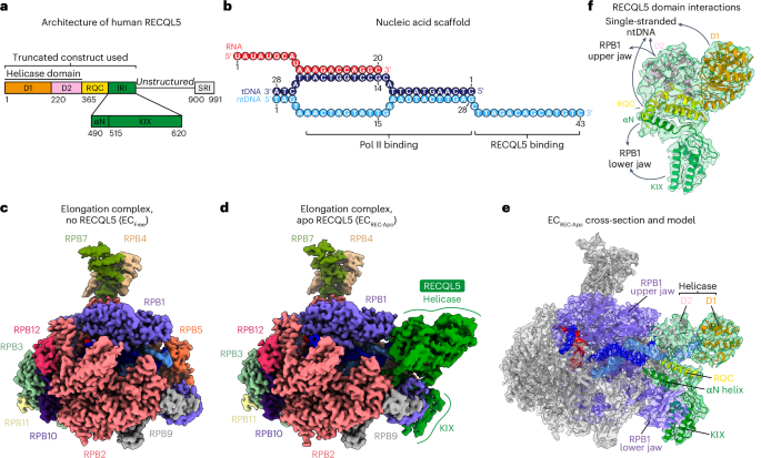
Sample preparation for cryo-EMEC–RECQL5 and EC*–RECQL5 complexes were formed on a DNA–RNA scaffold with a mismatch bubble of 11 nucleotides. The same RNA and template DNA were used for both complexes: RNA, 5′-GAGAGGGAACCCACU-3′; template DNA, 5′-GCTCCCAGCTCCCTGCTGGCTCCGAGTGGGTTCTGCCGCTCTCAATGG-3′. For EC–RECQL5, a non-template DNA of the same length as the template DNA was used: 5′-CCATTGAGAGCGGCCCTTGTGTTCAGGAGCCAGCAGGGAGCTGGGAGC-3′. For EC*–RECQL5, a non-template DNA with a 15-nucleotide 3′ overhang was used: 5′-CCATTGAGAGCGGCCCTTGTGTTCAGGAGCCAGCAGGGAGCTGGGAGCCTTAGACAGCATGTC-3′. All oligonucleotides were synthesized by IDT and resuspended in water.
For the EC–RECQL5 complex, RNA was annealed with an equimolar amount of template DNA by incubating at 60 °C for 5 min, followed by a gradual decrease in temperature at a rate of 1 °C min−1 to a final temperature of 30 °C in 20 mM HEPES, pH 7.5, 100 mM NaCl and 3 mM MgCl2. Porcine Pol II (75 pmol) was incubated with the RNA–DNA hybrid (150 pmol) at 30 °C for 10 min, followed by addition of the non-template DNA (300 pmol) and incubation at 30 °C for another 10 min. The complex was phosphorylated with GSK3B (300 pmol) and ATP (1 mM) in SEC100 buffer (20 mM HEPES, pH 7.5, 100 mM NaCl, 3 mM MgCl2, 1 mM DTT) at 30 °C for 30 min, followed by incubation with RECQL5 (300 pmol) on ice for 30 min. The assembled EC–RECQL5 complex was applied to a Superdex 200 Increase 3.2/300 column (Cytiva) equilibrated in SEC150 buffer (20 mM HEPES, pH 7.5, 150 mM NaCl, 3 mM MgCl2 and 1 mM DTT). The peak fraction of EC–RECQL5 was used for freezing grids.
For the EC*–RECQL5 complex, the Pol II elongation complex was prepared essentially as described for the EC–RECQL5 complex. Final protein amounts used for complex formation were 100 pmol porcine Pol II, 200 pmol RNA–DNA hybrid and 400 pmol non-template DNA. The complex was mixed with elongation factors (SPT6, DSIF and PAF, 200 pmol each) and phosphorylated with P-TEFb (33 pmol) and ATP (1 mM) in SEC100 buffer for 30 min at 30 °C. The assembled EC* complex was applied to a Superose 6 Increase 3.2/300 column (Cytiva) equilibrated in K50 buffer (20 mM HEPES, pH 7.5, 50 mM KCl, 4 mM MgCl2, 1 mM DTT). The peak fraction of EC* was incubated with a 2× molar excess of RECQL5 and 1 mM ADP on ice for 30 min, cross-linked with 0.05% (vol/vol) glutaraldehyde for 45 min on ice and used directly for grid freezing.
The EC–TCR–RECQL5 complex was formed on a DNA–RNA scaffold with a mismatch bubble of 15 nucleotides43: template DNA, 5′-CGCTCTGCTCCTTCTCCCATCCTCTCGATGGCTATGAGATCAACTAG-3′; non-template DNA, 5′-CTAGTTGATCTCATATTTCATTCCTACTCAGGAGAAGGAGCAGAGCG-3′; RNA, 5′-ACAUCAUAACAUUUGAACAAGAAUAUAUAUACAAAAUCGAGAGGA-3′. The DNA and RNA oligonucleotides were synthesized by IDT. RNA was annealed with equimolar template DNA by incubating at 90 °C for 2 min, followed by a gradual decrease in temperature at a rate of 1 °C min−1 to a final temperature of 30 °C in 20 mM HEPES, pH 7.5 and 100 mM NaCl. Porcine Pol II (75 pmol) was incubated with the RNA–DNA hybrid (150 pmol) at 30 °C for 10 min, followed by the addition of the non-template DNA (300 pmol) and incubation at 30 °C for an additional 10 min. The complex was mixed with elongation factors (SPT6 and PAF, 150 pmol each) and phosphorylated with P-TEFb (25 pmol) and ATP (1 mM) in SEC100 buffer for 30 min at 30 °C before being subject to a Superose 6 Increase 3.2/300 column (Cytiva) equilibrated in K50 buffer. Peak fraction was incubated with a 2× molar excess of RECQL5 on ice for 30 min, followed by incubation with a 2× molar excess of TCR components (CSB, CSA–DDB1 and UVSSA), a 4× molar excess of ELOF1 and 1 mM ADP at 30 °C for 10 min. The assembled EC–TCR–RECQL5 complex was cross-linked with 0.05% (vol/vol) glutaraldehyde for 45 min on ice and used directly for grid freezing.
The sample (2.5 μl) was applied to R3.5/1 carbon grids (Quantifoil) with a continuous carbon support (~2.5 nm) for the EC–RECQL5 and EC*–RECQL5 complexes and no support layer for the EC–TCR–RECQL5 complex. The grids were glow discharged for 13–15 s with the Sputter Coater S150B (Edwards Vacuum). The grids were incubated with the sample for 30 s and blotted for 1–1.5 s before plunge-freezing in liquid ethane with a Vitrobot Mark IV (Thermo Fisher) operated at 4 °C and 100% humidity.
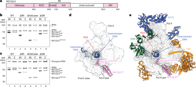
Pulldown assayFor pulldown of non-phosphorylated Pol II with different RECQL5 constructs, Twin-Strep-tagged human Pol II (12 pmol) was incubated with wild-type or mutant RECQL5 (36 pmol) on ice for 30 min in G-SEC150 buffer (20 mM HEPES, pH 7.5, 150 mM NaCl, 3 mM MgCl2, 5% glycerol, 1 mM DTT). Samples were incubated with Strep-Tactin XT 4Flow high-capacity resin (IBA) at 4 °C for 2 h. The resin was washed with G-SEC150 buffer and eluted with G-SEC150 buffer supplemented with 50 mM biotin. The eluate was separated on a 4–12% NuPAGE Bis-Tris gel (Invitrogen) and stained with InstantBlue (Abcam). Pulldowns using phosphorylated Pol II with different RECQL5 constructs were performed in the same way except that Pol II was incubated with P-TEFb (4 pmol) and ATP (1 mM) in SEC100 buffer for 30 min at 30 °C before mixing with RECQL5 constructs in G-SEC150 buffer.
For pulldown of the Pol II CTD with RECQL5SRI, His6-MBP-tagged Pol II CTD (100 pmol) was phosphorylated with P-TEFb (25 pmol) and ATP (1 mM) in SEC100 buffer for 30 min at 30 °C. P-TEFb storage buffer (20 mM HEPES, pH 7.5, 300 mM NaCl, 10% glycerol, 1 mM DTT) was used instead of P-TEFb as a negative control for the non-phosphorylated CTD condition. The CTD was incubated with the SRI domain of RECQL5 (500 pmol) on ice for 30 min, followed by incubation with amylose resin (NEB) equilibrated in G-SEC150 buffer at 4 °C for 1 h. The resin was washed with G-SEC150 buffer and eluted with 20 mM maltose in G-SEC150 buffer. The eluate was loaded on a 4–12% NuPAGE Bis-Tris gel (Invitrogen) and stained with InstantBlue (Abcam).
For pulldown of EC or EC* with RECQL5, the elongation complex was prepared in the same way as described for sample preparation for cryo-EM, except that Twin-Strep-tagged human Pol II was used. Final protein amounts used for complex formation were 12 pmol human Pol II, 24 pmol RNA–DNA hybrid and 48 pmol non-template DNA. The phosphorylation reaction was performed with P-TEFb (4 pmol) and ATP (1 mM) in SEC100 buffer for 30 min at 30 °C for either Pol II alone or with elongation factors (SPT6, DSIF and PAF, 24 pmol each). Samples were incubated with Strep-Tactin XT 4Flow high-capacity resin (IBA) at 4 °C for 2 h. The resin was washed with SEC100 buffer and eluted with 50 mM biotin in SEC100 buffer. The eluate was separated on a 4–12% NuPAGE Bis-Tris gel (Invitrogen) and stained with InstantBlue (Abcam).
For pulldown of RECQL5 with the elongation factors SPT6, DSIF, PAF and RTF1, elongation factors (100 pmol) were phosphorylated with P-TEFb (17 pmol) and ATP (1 mM) in SEC100 buffer for 30 min at 30 °C, followed by addition of His6-MBP-tagged RECQL5 (50 pmol) and incubation on ice for 30 min. Samples were incubated with amylose resin (NEB) equilibrated in K50 buffer at 4 °C for 1 h. The resin was washed with K50 buffer and eluted with K50 buffer supplemented with 20 mM maltose. The proteins were separated on a 4–12% NuPAGE Bis-Tris gel (Invitrogen) and stained with InstantBlue (Abcam).
RNA extension assayAll RNA extension assays were performed with a complementary DNA scaffold as follows: template DNA, 5′-CTGGACTACTGCGCCCTAGACGTGCAGCAAGCTTGGGCTGCAGGTAACCAGTTCTACATGCTAGATACTTACCTGGTCGGAGGCCGACGG-3′; non-template DNA, 5′-CCGTCGGCCTCCGACCAGGTAAGTATCTAGCATGTAGAACTGGTTACCTGCAGCCCAAGCTTGCTGCACGTCTAGGGCGCAGTAGTCCAG-3′; RNA, 5′-/6-FAM/-UUUUUUCCAGGUAAG-3′. All oligonucleotides were synthesized by IDT and resuspended in water.
RNA was annealed with an equimolar amount of template DNA by incubating at 90 °C for 2 min, followed by a gradual decrease in temperature at a rate of 1 °C min−1 to a final temperature of 30 °C in 20 mM HEPES, pH 7.5 and 100 mM NaCl. All concentrations refer to the final concentrations used in the assay. Porcine Pol II (150 nM) was incubated with the RNA–DNA hybrid (100 nM) at 30 °C for 10 min, followed by the addition of the non-template DNA (200 nM) and incubation at 30 °C for an additional 10 min. The complex was mixed with elongation factors (SPT6, DSIF and PAF, 150 nM each) and phosphorylated with P-TEFb (100 nM) and ATP (1 mM) in 20 mM HEPES, pH 7.5, 100 mM NaCl, 3 mM MgCl2, 4% glycerol and 1 mM DTT at 30 °C for 30 min. The reactions were incubated with wild-type or mutant RECQL5 (1.5 µM) on ice for 30 min. Transcription extension was started by adding 100 μM NTP at 20 °C. The reactions were quenched at various time points in equal volume of 2× stop buffer (6.4 M urea, 50 mM EDTA, pH 8.0, 1× TBE). Reactions were treated with 0.13 unit per μl proteinase K (NEB) for 20 min at 37 °C and applied onto 15% denaturing gels (15% acrylamide/Bis-acrylamide 19:1, 7 M urea and 1× TBE, run in 0.5× TBE at 200 V for 100 min). The gels were visualized using the 6-FAM label on the RNA with the Typhoon FLA 9500 Imager (GE Healthcare). Extension assays with additional TCR factors were performed in the same way with the exception that EC* was incubated with 0.75 µM RECQL5, and 0.75 µM TCR factors were added to the reaction after extension by EC*–RECQL5 for 2 min.
Electromobility shift assayElectromobility shift assays for wild-type and D157A mutant RECQL5 were performed with two types of DNA scaffolds, one with a 3′ overhang and one with a splayed duplex. DNA was annealed in 20 mM HEPES, pH 7.5 and 100 mM NaCl by heating at 94 °C for 4 min and slowly cooling down to 30 °C at a rate of 1 °C min−1. Double-stranded DNA at a final concentration of 0.5 μM was mixed with 0–2.5 μM of wild-type or D157A RECQL5 in 20 mM HEPES, pH 7.5, 66 mM NaCl, 5% glycerol, 1 mM DTT and 0.25 mg ml−1 BSA and incubated on ice for 30 min. The samples were then mixed with Novex Hi-Density TBE Sample Buffer (Thermo Fisher) and loaded onto a 6% DNA retardation gel (Invitrogen). Electrophoresis was performed at 4 °C and 100 V in 0.5× TBE buffer for 105 min. Gels were stained with SYBR Gold (Thermo Fisher) and imaged with a Typhoon FLA 9500 Imager (GE Healthcare).
Pol II ubiquitination assayPol II elongation complexes were formed on the same DNA–RNA scaffold as that for the cryo-EM studies of the EC–TCR–RECQL5 complex. RNA was annealed with equimolar template DNA by incubating at 60 °C for 5 min, followed by a gradual decrease in temperature at a rate of 1 °C min−1 to a final temperature of 30 °C in 20 mM HEPES, pH 7.5, 100 mM NaCl and 3 mM MgCl2. All concentrations refer to the final concentrations used in the assay. Human Pol II (150 nM) was incubated with the RNA–DNA hybrid (300 nM) at 30 °C for 10 min, followed by addition of the non-template DNA (600 nM) and incubation at 30 °C for another 10 min. Subsequently, RECQL5 constructs (0 or 450 nM) were added to the elongation complex and incubated on ice for 30 min before adding CSB (300 nM), CSA–DDB1 (300 nM), CUL4A–RBX1 (300 nM), ELOF1 (600 nM) and UVSSA (360 nM) for a further incubation at 30 °C for 15 min. Ubiquitination reactions were initiated upon addition of UBE1 (150 nM, R&D Systems), UbcH5b (1.75 μM), ubiquitin (150 μM, R&D Systems) and ATP (2 mM) in 50 mM Tris, pH 8.0, 50 mM NaCl, 10 mM MgCl2 and 1 mM DTT at 37 °C. The reactions were quenched at various time points by mixing with 4× SDS loading dye (20 mM Tris-HCl, pH 6.6, 8% SDS, 40% glycerol, 0.8% bromophenol blue and 400 mM DTT). Reactions were loaded on a 3–8% Tris-acetate gel (Invitrogen) and transferred onto a 0.2-µm nitrocellulose membrane (Cytiva). The membrane was blocked with 5% (wt/vol) milk in PBS for 30 min at room temperature and incubated with F-12 anti-RPB1 antibody (1:1,000 dilution, Santa Cruz Biotechnology, sc-55492) or anti-CSB antibody (1:1,000 dilution, Santa Cruz Biotechnology, sc-166042) overnight at 4 °C. The membranes were washed with PBST (PBS with 0.2% Tween-20) and incubated with HRP-conjugated anti-mouse secondary antibody (1:10,000 dilution, Proteintech, SA00001-1) for 45 min at room temperature. The membranes were washed with PBST, developed with the Pierce ECL chemiluminescent substrate (Thermo Fisher) and filmed with an OPTIMAX processor (Protec).
Competition assayPol II elongation complex was prepared in the same way as described for the ubiquitination assays. Final protein amounts used for complex formation were 12 pmol human Pol II, 24 pmol RNA–DNA hybrid and 48 pmol non-template DNA. The elongation complex was either assembled using non-phosphorylated Pol II or phosphorylated with 4 pmol P-TEFb and 1 mM ATP in SEC100 buffer for 30 min at 30 °C. The assembled elongation complex was either incubated with wild-type RECQL5 or RECQL5ΔSRI (36 pmol) first on ice for 20 min and then with the TCR complex (36 pmol CSB and CSA–DDB1, 50 pmol UVSSA and ELOF1) on ice for 20 min in SEC100 buffer or vice versa. His6-MBP-tagged wild-type RECQL5 was used to avoid band overlapping with DDB1 in the competition assay using non-phosphorylated Pol II. RECQL5ΔSRI was used in the competition assay with phosphorylated Pol II to eliminate the SRI–CTD interaction and focus on the competition at the Pol II jaw. The elongation complex was incubated with RECQL5 (36 pmol) alone in SEC100 buffer on ice for 20 min as a positive control. The complexes were incubated with Strep-Tactin XT 4Flow high-capacity resin (IBA) at 4 °C for 2 h. The resin was washed with SEC100 buffer with 5% glycerol and eluted with 50 mM biotin in SEC100 buffer with 5% glycerol. The eluate was separated on a 4–12% NuPAGE Bis-Tris gel (Invitrogen) and stained with InstantBlue (Abcam).
Cryo-EM data collection and processingCryo-EM data were collected on the 300-kV Titan Krios with a Falcon 4i direct electron detector (Thermo Fisher). Automated data acquisition was performed with EPU (Thermo Fisher) at a nominal magnification of ×96,000 (0.8156 Å per pixel). Image stacks of 40 frames were collected with a defocus range of −0.5 μm to −2.0 μm in electron-counting mode and at a dose rate of 0.92–1.06 e− Å−2 per frame. A total of 8,602 image stacks were collected for the EC–RECQL5 complex. A total of 60,808 image stacks were collected across two datasets for the EC*–RECQL5 complex. A total of 11,156 image stacks were collected for the EC–TCR–RECQL5 complex.
Motion correction and estimation of the contrast transfer function (CTF) were done in RELION 5.0 (refs. 63,64). Particles in 400 pixels × 400 pixels for EC–RECQL5 and in 480 pixels × 480 pixels for EC*–RECQL5 and EC–TCR–RECQL5 were selected by automatic particle picking in Warp65. For EC–RECQL5 and EC*–RECQL5, further processing steps were performed in RELION 5.0 with final local refinement in cryoSPARC66. Further processing steps for EC–TCR–RECQL5 were performed in cryoSPARC.
For the EC–RECQL5 complex, two-dimensional (2D) classification, followed by three-dimensional (3D) refinement and 3D classification with fine-angle sampling, was performed to remove bad particles from the dataset (Extended Data Fig. 2a,b). Signal subtraction with a soft mask near the Pol II jaw (where the extra density of RECQL5 is) followed by focused 3D classification without alignment was performed to separate EC–RECQL5 particles from EC-only particles (Extended Data Fig. 2c,d). Particles containing densities corresponding to RECQL5 were reverted to original particles and 3D refined, followed by CTF refinement with per-particle defocus estimation and Bayesian polishing to correct beam-induced particle motion (Extended Data Fig. 2e). Polished particles were subjected to an additional round of signal subtraction and focused 3D classification on RECQL5 to further remove the EC-only particles (Extended Data Fig. 2f). Subsequent signal subtraction with a soft mask on the downstream DNA and focused 3D classification without alignment were performed to exclude any apo Pol II–RECQL5 particles (Extended Data Fig. 2g,h). Particles containing densities of downstream DNA were reverted to original particles and imported into cryoSPARC for local refinement, resulting in the final EC–RECQL5 reconstruction with 66,771 particles at an overall resolution of 2.8 Å (Extended Data Fig. 2i).
For the two datasets of the EC*–RECQL5 complex, particles after cleanup in 2D and 3D classification were CTF refined and Bayesian polished separately (Extended Data Fig. 3a–c,e–g). Polished particles in dataset 2 were further cleaned up by an additional round of 3D classification with fine-angle sampling (Extended Data Fig. 3h,i). Signal subtraction with a soft mask on the RECQL5 IRI domain and focused 3D classification without alignment were performed to separate EC*–RECQL5 particles from EC*-only particles for both datasets (Extended Data Fig. 3d,j). Particles containing densities corresponding to RECQL5 from both datasets were combined, reverted to original particles and 3D refined (Extended Data Fig. 3k). Following signal subtraction with a soft mask on PAF and focused 3D classification without alignment (Extended Data Fig. 3l), particles containing PAF densities were selected, reverted to original particles and imported into cryoSPARC for local refinement. This resulted in the final EC*–RECQL5 reconstruction with 314,016 particles at an overall resolution of 2.0 Å (Extended Data Fig. 3m).
To improve the resolution of elongation factors, soft masks were applied individually onto SPT6core, SPT6core–Pol IIstalk, SPT6tSH2, PAF (CTR9–SKI8 region), PAF1–LEO1, SPT4 and SPT5 (KOW2–KOW3 region). Following particle subtraction, 3D classification without alignment was performed for SPT6tSH2, PAF1–LEO1, SPT4 and SPT5 (KOW2–KOW3 region), resulting in medium-resolution maps of each region. These maps allowed docking of existing structures and AlphaFold67-predicted models of the elongation factors into respective regions. For SPT6core, SPT6core–Pol IIstalk and PAF (CTR9–SKI8 region), 3D classification did not find any classes lacking densities corresponding to the elongation factors. Therefore, all subtracted particles were imported into cryoSPARC for local refinement, resulting in a resolution of 2.5 Å for all three maps of SPT6core, SPT6core–Pol IIstalk and PAF (CTR9–SKI8 region) (Extended Data Fig. 3n–p).
For the dataset of the EC–TCR–RECQL5 complex, particles after cleanup in 2D classification were CTF refined (Extended Data Fig. 9a,b), followed by focused 3D classification without alignment using a soft mask on the RECQL5 IRI domain to separate EC–TCR–RECQL5 particles from EC–TCR-only particles (Extended Data Fig. 9c). Particles containing densities of RECQL5 were selected and 3D refined (Extended Data Fig. 9d). Subsequent focused 3D classification without alignment using a soft mask on TCR was performed to exclude EC–RECQL5-only particles (Extended Data Fig. 9e). This resulted in the final EC–TCR–RECQL5 reconstruction with 19,458 particles at an overall resolution of 3.5 Å (Extended Data Fig. 9f).
Local resolution of the maps was estimated using cryoSPARC except for the EC*–RECQL5 and EC–TCR–RECQL5 overall maps, which were estimated in RELION to obtain locally filtered and sharpened maps. All resolution calculations were based on gold-standard FSC using the FSC = 0.143 criterion. A summary of all EM reconstructions obtained in this paper is shown in Table 1.
Model building and refinementInitial models of S. scrofa Pol II (PDB 7B0Y, ref. 35) and EC* (PDB 6GMH, ref. 2) as well as AlphaFold67 predictions of RECQL5 and elongation factors were rigid body fitted into the overall map in Chimera68. The Pol II and RECQL5 models were manually adjusted in Coot69 using the sharpened overall map of EC*–RECQL5 (2.0 Å), and the structure was real-space refined in PHENIX70. The SPT6core model was manually adjusted in Coot using the sharpened local refined SPT6 map (2.5 Å) and real-space refined in PHENIX. The refined SPT6core model was then fitted into the sharpened local refined SPT6core–Pol IIstalk map (2.5 Å), adjusted in Coot and real-space refined in PHENIX. The PAF model was manually adjusted in Coot using the sharpened local refined PAF map (2.5 Å), and the structure was real-space refined in PHENIX. The rest of the elongation factors were rigid body docked into the overall map in Chimera. The resulting complete model of EC*–RECQL5 was then real-space refined in the locally filtered and sharpened overall map using PHENIX and structure restraints of Pol II, SPT6core–Pol IIstalk and PAF.
An additional density was found to bind the tSH2 domain of SPT6, and the tSH2 domain was in a horizontal conformation compared to previous EC* structures2 (Extended Data Fig. 3q). De novo model building using ModelAngelo71 identified this unassigned density to be CDC73. This allowed us to identify a previously uncharacterized interface between the tSH2 domain of SPT6 and CDC73 of PAF (Extended Data Fig. 5e), consistent with biochemical analysis in yeast72.
The EC–RECQL5 model was fitted into the sharpened overall map of EC–RECQL5, manually adjusted in Coot and real-space refined in PHENIX.
The EC–RECQL5 model from EC*–RECQL5 and the TCR model (8B3D)38 were rigid body fitted into the sharpened overall map of EC–TCR–RECQL5 and real-space refined using ADP refinement and the locally filtered and sharpened overall map in PHENIX.
Figures were generated using PyMOL (PyMOL Molecular Graphics System, version 2.0, Schrödinger) and ChimeraX73. Sequence alignment was performed using Jalview74.
Cross-linking-coupled mass spectrometrySamples for cross-linking-coupled mass spectrometry were prepared essentially as described for cryo-EM studies of EC*–RECQL5, except that peak fractions of EC* were combined and incubated with a 1.5× molar excess of RECQL5 on ice for 30 min, cross-linked with 1 mM BS3 on ice for 1 h and quenched with 50 mM Tris-HCl, pH 8.0.
The quenched solution was reduced with 5 mM DTT and alkylated with 20 mM iodoacetamide. The SP3 protocol as described in refs. 75,76 was used to clean up and buffer exchange the reaction. Briefly, the complex was washed with ethanol, resuspended in 100 mM NH4HCO3 and digested with trypsin (Promega) at an enzyme-to-substrate ratio of 1:20 and with 0.1% ProteaseMAX (Promega) overnight at 37 °C. Digested peptides were purified using HyperSep SpinTip P-20 C18 columns (Thermo Scientific), eluted with 60% (vol/vol) acetonitrile (ACN) and dried using a Speed Vac Plus concentrator (Savant). Dried peptides were resuspended in 30% (vol/vol) ACN and separated using a Superdex 30 Increase 3.2/300 column (Cytiva) at a flow rate of 10 μl min−1 in 30% (vol/vol) ACN and 0.1% (vol/vol) trifluoroacetic acid. Fractions containing cross-linked peptides were collected and dried with the Speed Vac Plus concentrator (Savant). Dried peptides were suspended in 3% (vol/vol) ACN and 0.1% (vol/vol) formic acid and analyzed by nanoscale capillary LC–MS/MS using an Ultimate U3000 HPLC system (Thermo Scientific) to deliver a flow of 300 nl min−1. Peptides were trapped on a C18 Acclaim PepMap 100, 5-μm, 0.3-μm × 5-mm cartridge (Thermo Scientific) before separation on an Aurora Ultimate C18, 1.7-μm, 75-μm × 25-cm column (IonOpticks). Peptides were eluted on a 90-min gradient and interfaced via an EASY-Spray ionization source to a Q Exactive Plus mass spectrometer (Thermo Scientific). Data were acquired in data-dependent mode using a Top-15 method, in which high-resolution full-mass scans were carried out (R = 70,000, m/z 400–1,500) followed by a higher-energy collision dissociation of 30 V. The tandem mass spectra were recorded (R = 60,000, automatic gain control target = 5 × 10⁵, maximum injection time = 100 ms, isolation window = 1.2 m/z, dynamic exclusion = 40 s).
Xcalibur raw files were converted to MGF files using ProteoWizard77, and cross-links were analyzed with xiSEARCH78. Search conditions used a maximum of three missed cleavages with a minimum peptide length of 5. Variable modifications used were carbamidomethylation of cysteine (57.02146 Da) and oxidation of methionine (15.99491 Da). The false discovery rate was set to 5%. The sequence database was assembled from all proteins within the complex. Cross-link sites were visualized with xiVIEW79 and the PyXlinkViewer plugin80 in PyMOL version 2.0.
Reporting summaryFurther information on research design is available in the Nature Portfolio Reporting Summary linked to this article.
Comments (0)