Cell culture
The Human umbilical vein endothelial EA.hy926 cell lines were kindly provided by Cell Bank, Chinese Academy of Sciences, supplemented with DMEM/F12 complete medium(Gibco, Thermo Fisher) that containing 10% fetal bovine serum (Newzerum FBS, New Zealand) and 1% Penicillin-streptomycin mixture (keygen BIO, China) and incubated under 37℃, and 5% CO2 standard condition until density of cells reached to 80-90% confluence. Cells divided into four groups: Control group, RSL3 group, RSL3 + low-concentration EMPA intervention group, and RSL3 + high-concentration EMPA intervention group. The stock solution of EMPA (HY-15409, MedChem Express) or RSL3 (HY-100218 A, MedChem Express) was prepared in DMSO and subsequently diluted with culture medium containing 2% FBS.
Cell viability
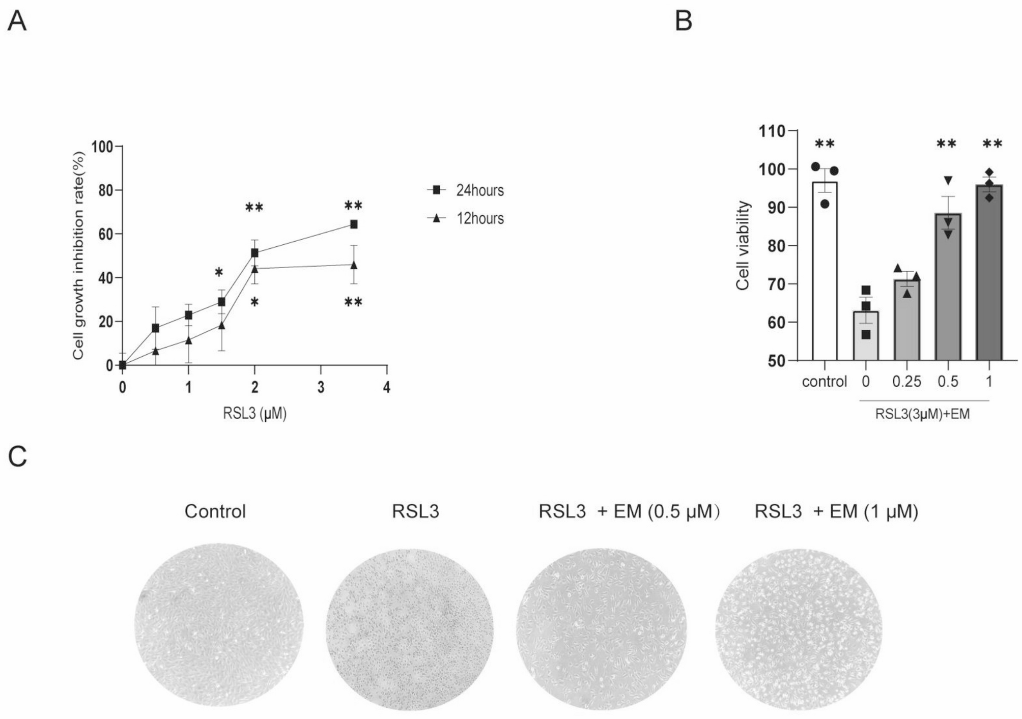
To screen for the optimal concentration of RSL3 and EMPA. Drug-activated cell viability was detected by CCK8 reagent (APE x BIO, USA). The density of cell was adjusted to 1 × 10⁵/mL and then 100 µL of cell suspension was added to each well of 96-well plates. The cells with 80-90% confluence was separately exposed to the diluted concentration of RSL3 solution (0.5, 1, 1.5, 2, and 3.5 µM) for 12–24 h. EMPA (0.25, 0.5 and 1 µM) and both drugs combined for 24 h. Finally, cells were incubated with 110 µL of medium contains 10% CCK-8 for each well and placed in a constant temperature 37℃ for 1–1.5 h, taking care to avoid light throughout. The OD values of 96-well plates were measured at 450 nm using a microplate reader (Thermo Fisher).
Detection of lipid peroxidation
BODIPYTM581/591C11 (D3861, Thermo Fisher) is a lipid peroxidation sensor, which can be detected by flow cytometry (OLYMPUS) and fluorescence microscope (OLYMPUS). The EA.hy926 cells were stained with 10 µM BODIPYTM581/591C11 dilution and incubated in a dark environment. After waiting for 30 min, the cells were then washed with phosphate-buffered saline (PBS) 2–3 times in order to wash away dyestuffs.
Cells were digested with 0.25% trypsin-EDTA and centrifuged at 900 × g for 5 min at 4 °C using a refrigerated centrifuge. After supernatant removal, the pellet was washed twice with PBS through sequential resuspension and centrifugation at 900 × g for 3 min. The fluorescence intensity was quantified with negative controls setting photomultiplier tube voltages. Cellular lipid peroxidation levels were determined by gating intact cell populations and analyzing median fluorescence intensity (MFI) using FlowJo™ software [15].
To assess the inner lipid peroxidation of the EA.hy926 cells through qualitative method, the cells were continually stained with DAPI (P0131, Beyotime Biotechnology) to mark nucleus and observed by fluorescence microscope in a dark enviroment throughout. Finally, analyzed by ImageJ.
Determination of intracellular Fe2+ and MDA
FerroOrange (F314, DOJINDO) was used to detect intracellular Fe2+ sensitively. Firstly, the EA.hy926 cells were washed with HBSS balanced salt solution (C0219, Beyotime Biotechnology) three times, then stained with appropriate amount of DAPI for 3–5 min and aspirated out, and washed with HBSS balanced solution for 2–3 times. Secondly, stained with working solution concentration of 1 µM of FerroOrange for 30 min in 24-well plate. After that, the plate was placed in the incubator at 37℃ for 30 min. Images were captured by fluorescence microscope, and the whole process was avoided from light. The images were taken using a fluorescence microscope, protected from light throughout the process, and quantitatively analyzed by ImageJ software.
Malondialdehyde assay kit (A003-1-2, Jiancheng, Nanjing, China) was used to measure MDA level in cell lysate. The wavelength used for measurement was 532 nm, and then MDA concentration was calculated by creating a standard curve and normalizing it to the protein content.
Small interfering transfection
The sequence of siRNA-NRF2 (GenePharma, China; F:5’-CCAGAACACU-CAGUGGAAUTT-3’ and R:5’-AUUCCACUGAGUGUUCUGGTT-3’) was used t-o knock down the expression of NRF2. The siRNA complexes were prepared by mixing 100 pmol siRNA with 5 µL GP-transfection-mate reagent (GenePharm-a, China) at 1:1 (V/V) ratio in serum-free medium After 15 min incubation at room temperature, the complexes were added to the cells for 4–6 h and subsequently replaced by complete medium. The EA.hy926 cells were divided into four groups: si-NC, si-NRF2, si-NC + RSL3/EMPA, si-NRF2 + RSL3/EMPA.
Quantitative real-time-PCR
The total RNA was isolated from EA.hy926 cells using Trizol reagent (Themor Fisher, USA) and reversely transcribed into cDNA according to kit’s manufacturer’s instruction (RR047A, Takara, Japan). Gene expression level were quantified by StepOnePlus real-time PCR system (Thermor Fish, USA). The primers sequence was as followed: GPX4: (F)5’-CCAGTGAGGCAAGACCGAAGT-3’ (R)5’-TTGGGTTGGATCTTCATCCAC-3’; ACSL4: (F)5’-CCAGTGAGGCAAGACCGAAGT-3’ (R)5’-TTGGGTTGGATCTTCATCCAC-3’; SLC7A11: (F)5’-CCCTGAACTTGCGATCAAGCT-3’ (R)5’-GCTCCAGCTGACACTCATG CT-3’; and housekeeping gene GAPDH: (F)5’-GCCAAGGTCATCCATGACAAC-3’ (R)CAGCGTC AAAGGTGGAGGAGT. The relative amount of mRNA was determined using 2−∆∆Ct method and normalized to the level of GAPDH.
Western blot
The EA.hy926 cells were rinsed twice with ice-cold PBS. After removing PBS, cells in the 6-well plate were lysed with 100 µL/well of ice-cold RIPA lysis buffer (P0013B, Beyotime Biotechonogy) supplemented with 1 mM phenylmethylsulfonyl fluoride (PMSF, ST506, Beyotime Biotechonogy). Then the cell lysate were scraped down with a cell scraper, transferred to 1.5 mL microcentrifuge tubes and homogenized through a 22-gauge needle for 10–15 cycles on ice. Centrifugated at 3000 rpm/min and 4 °C for 15 min, the protein was detected with BCA protein colorimetric assay kit (Elaboscience, Wuhan, China). The Protein of all samples were then electrophoresed in 12.5% SDS-PAGE gel, transferred to PVDF, and blocked in 5% BSA for 1 h at room temperature. The PVDF were incubated with the primary antibody of anti-GPX4 (1:5000, Abcam), anti-ACSL4 (1:3000, Proteintech), anti-SLC7A11 (1:2000, Abcam), anti-NRF2 (1:3000, Proteintech), anti-HO-1 (1:3000, Proteintech), anti-GAPDH (1:50000, Proteintech) or anti-βactin (1:3000, Abcam) at 4℃ overnight. The combined primary antibody were washed with 1 × TBST (Servicebio, Wuhan, China) three times, and incubated with HRP-conjugated goat anti-mouse IgG (1:50000, Proteintech) or HRP-conjugated goat anti-rabbit IgG (1:50000, Proteintech) for 2 h at room temperature, and then were washed three times with 1 × TBST for 10 min each time. After the ECL luminescent solution (Tano, Shanghai, China) was prepared, the PVDF was evenly dropped on protein side to be exposed under Gel Imager (BIO RAD, USA). Using ImageJ software to analyze the grey value of the immunoreactive bands.
Statistical analysis
The values of each group were expressed as mean ± standard deviation. Statistical data were analyzed using GraphPad Prism, and one-way ANOVA was used to compare the differences between the groups, with P-value less than 0.05 being considered a statistically significant difference.


Comments (0)