Structural characteristics of 3D asymmetry in different types of skeletal patterns need to be considered to establish an adequate surgical plan. This study aimed to compare structural asymmetry between patients with mandibular prognathism and retrognathism. Since the CT data from each institute have uniform DICOM file formats [21], the pixel data transmission, transformation and 3D analyses were easily and successfully performed.
The 3D morphology of deviated mandibular prognathism has been frequently reported. Patients with prognathism without asymmetry [4, 5, 22, 23] or normal occlusion [6] served as controls in previous studies. However, differences between the pattern of 3D skeletal asymmetry in prognathism and retrognathism have been seldom reported. According to a previous report [11] on 3D skeletal differences between mandibular retrognathic and prognathic patients with facial asymmetry, both groups showed shorter ramus lengths (condylar head to gonion) on the deviated side (short side) than the contralateral side. Since the result of only six measurements were presented, it was difficult to understand the factors related to asymmetric mandibles in different skeletal patterns.
You et al. [5] and Thiesen et al. [24] investigated the characteristics of facial asymmetry with mandibular prognathism. Both group also reported 3D skeletal features of asymmetry with retrognathism separately, using the same study variables in both consecutive studies [20, 25]. Their studies yielded similar findings to the aforementioned results of other report [11]. However, these studies did not directly compare the different subject groups [5, 20, 25] or had limited number of study variables and subject numbers [24].
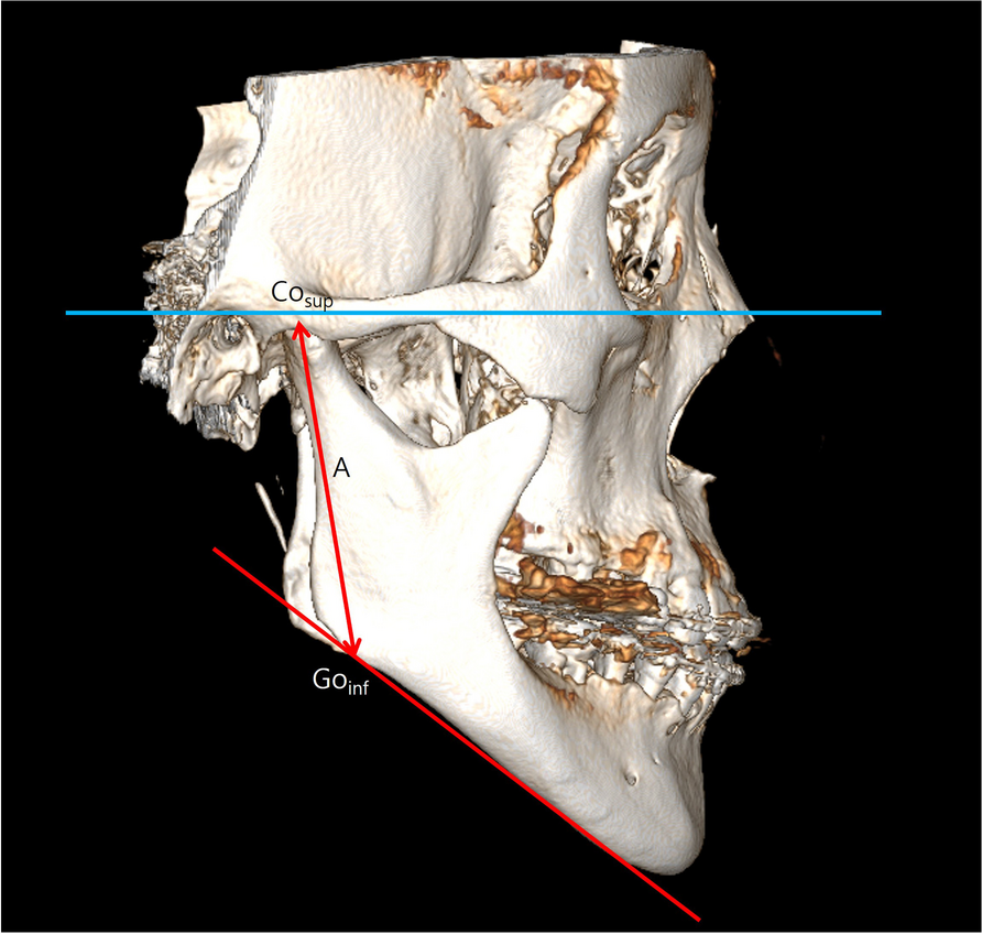
In our study, the ramus length was defined as the distance from the most superior point of the condyle to Go. To investigate the potential effect of the condylar head better, we measured the condylar height (Con_cranial to Con_dorsal) and ramal-proper height (Con_dorsal to Go) separately. In patients with skeletal class III asymmetry, the ramus length of the two sides are significantly different [4,5,6, 24]. Data presented in Table 3 show that asymmetric prognathism also exhibited a pattern similar to that observed in previous studies. The differences in ramal and body lengths in group B showed similar pattern to the other reports [5, 6].
However, it was interesting that ramus-proper height, excluding the condylar height, did not show inter-group differences. At the same time, the deviated side in asymmetric prognathism and the contralateral side in asymmetric retrognathism showed similar condylar width and height, ramus-proper height, and ramus height. Goto and Langenbach [19] reported that the condylar height (Con_cranial to Con_dorsal) of the deviated side in the asymmetric group was similar to the control group, whereas the contralateral side was significantly longer than the control (symmetry) group and deviated side. They suggested that the overgrowth of the condylar process on the contralateral side would result in an asymmetric mandible. Other study group also suggested that 3D asymmetry in the ramus would be attributed to the asymmetric spatial orientation of the Gonion rather than the 3D position of the condyle [24]. Although the chin deviation was significantly correlated with the ramal and condylar lengths in mandibular prognathism and retrognathism, no statistically significant correlation with the ramus proper height was found in both groups in our study (Table 5). This result is supported by those of previous study [19]. Furthermore, the results imply that an over-growth of the condylar process on the contralateral side in prognathism and an under-growth of the short side in retrognathism would be an important factor in the development of facial asymmetry. Data concerning condylar volume would provide more insight in the effect of condylar growth on condylar volume and subsequent mandibular asymmetry.
Our results showed that there were significant body length differences in both groups in a similar pattern, which is in agreement with results from previous studies [4, 22, 23]. Therefore, our result and those of others indicate that the body of the mandible follows a similar growth pattern of ramus length in both retrognathic and prognathic asymmetry.
There were no marked differences in right and left 3D condylar positions in patients with asymmetry with prognathism. It was in agreement with previous data [24, 25]. In asymmetric retrognathism, we could find bilateral difference in condyle position on sagittal direction. However, the bilateral difference was smaller than the inter-group difference. The left and right condyles of asymmetric retrognathic patients were positioned more posteriorly, inferiorly and medially compared to asymmetric prognathic patients. This suggested that chin asymmetry did not readily mean an asymmetric condylar position. Even if there is asymmetry in 3D condylar head position, this position would be more affected by the horizontal position of the mandible (retrognathic or prognathic) rather than the magnitude of mandibular asymmetry within each group. The result of correlation analysis in Table 5 also emphasized that there was no significant correlation between the degree of chin asymmetry and condylar position. Even though the difference in transverse condylar position significantly correlated with the degree of chin deviation in asymmetric retrognathism, the bilateral difference was not very great (1.2 ± 4.1 mm).
At the same time, the magnitude of the asymmetric 3D spatial orientation of the condyle in both groups of our study was remarkably smaller than that of Gonion in both group. As also shown in previous studies [24, 25], the results suggests that asymmetric 3D position of condyle is less severe than asymmetric displacement of the gonion point.
In both groups, the mandibular angle (Go) of the contralateral side was shifted more medially, and the mandibular angle of the deviated side was located more laterally, posteriorly and superiorly. At the same time, asymmetric 3D position of the Go also significantly correlated with mandibular asymmetry in prognathism patients, particularly in the vertical and transverse direction. A larger chin deviation in the horizontal direction seemed to also increase the ramus length on the contralateral side, displacing the mandibular angle downward and causing more posterior vertical asymmetry of the mandible. Thus, the spatial position of Go would be the more influential factor in facial asymmetry rather than the 3D condylar position, and surgical procedures to correct this bony asymmetry at the angle of the mandible may be required to obtain an optimal treatment result especially for deviated mandibular prognathism.
In previous reports [4, 6, 24], the contralateral side of the ramus was shifted medially to the side of chin deviation and was more anteriorly inclined in deviated mandibular prognathism. Our results showed a similar pattern of the sagittal ramal inclination reported in previous studies.
Frontal ramal inclination also showed similar pattern but was not statistically significant because of the large individual variations (Table 4). The ramus was in fact more upright or even a little posteriorly inclined in the retrognathic mandible. The bilateral difference in ramus orientation in prognathic patients was more significant than in retrognathic patients, but the absolute left and right differences were smaller than inter-group differences. Therefore, surgeons should consider the correction of the right-left difference in the frontal ramus inclination according to the individual bases. At the same time, the axial rotation of the ramus should not be the critical concern in the correction of mandibular asymmetry in both groups. These bilateral angular measurements of axial, frontal and lateral ramal inclinations exhibited large individual variations, and the degree of chin deviation was not proportional to the asymmetry of the ramus inclination or condylar axis. Therefore, the results suggest that a surgical correction of ramal asymmetry needs to be performed with individualized surgical planning to optimize posterior mandibular symmetry.
The limitation of this study was the lack of control data on normal occlusion from the two different institutes. Even though the demographic variables were not statistically different between the groups in this study, there can be the potential difference of shape and size of the different ethnic groups. Nevertheless, this study has shown different skeletal characteristics of asymmetry by using a large sample of 3D CT data from mandibular prognathism and retrognathism with asymmetry.


Comments (0)