Mandibular setback surgery can be performed as an independent procedure to achieve posterior movement by the mandible or as part of a combined orthognathic surgery involving maxillary surgery. Among the complications, skeletal stability after orthognathic surgery is crucial for achieving satisfactory long-term results. Various studies are actively investigating factors for predicting relapse, including maxillary surgery, the need for presurgical orthodontic treatment, direction and amount of mandibular movement, surgical technique, and the duration of intermaxillary fixation. However, conflicting results are still being reported in the literature [13,14,15].
Kim et al. attributed early relapse (within 6 months post-surgery) to mandibular condyle position changes and bone cut slippage [16]. In contrast, Tong Xi et al. linked late relapse (6–12 months post-surgery) to factors such as surgical extent, technique, and mandibular condyle resorption [17].
Valls-Ontañón et al. described skeletal stability after orthognathic surgery as a state without relapse, indicating no undesired movements in the sagittal, transverse, and vertical directions at the measurement points on the 3D radiographic images [18].
Conversely, Habets et al. defined vertical asymmetry of the mandible in panoramic radiographs as a significant difference between the right and left sides exceeding 6% [9]. McCrea et al. defined an AI exceeding 3% as indicative of a structural difference of 6% or more in mandibular length, establishing 3% as the threshold for detecting asymmetry in mandibular length [19].
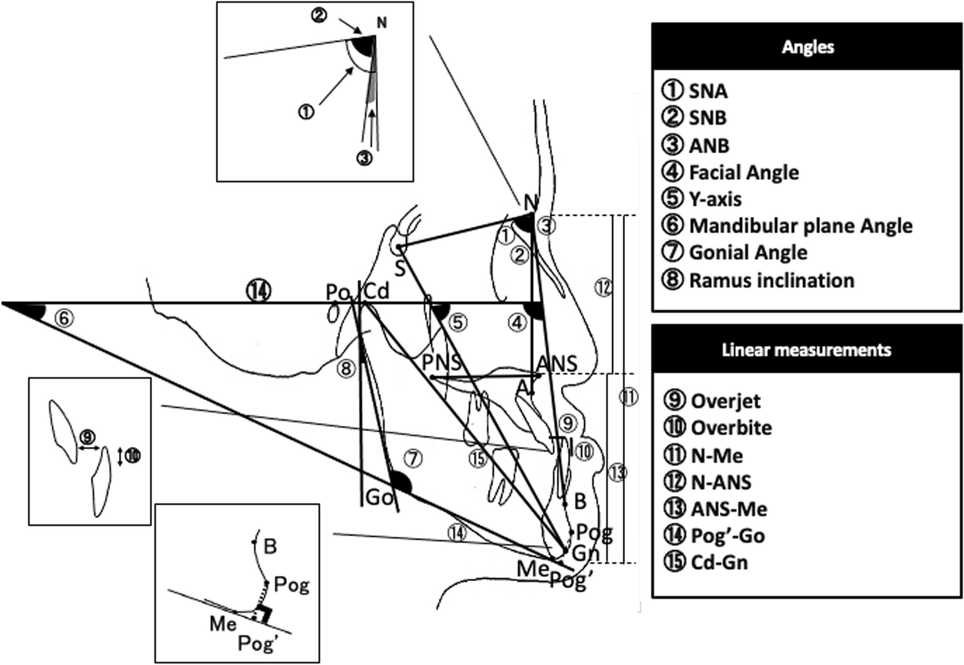
Many studies on post-orthognathic surgery relapse have relied on 2D lateral cephalometric radiographs for evaluation. However, these measurements can be prone to errors due to variations in X-ray beam angles, challenges in replicating head position, and inaccuracies in distance measurements. In contrast, 3D analysis enables assessment of skeletal changes across the x-axis, y-axis, and z-axis with minimal distance measurement errors [20, 21]. While many post-orthognathic surgical relapse studies have primarily focused on the sagittal plane, it is widely acknowledged that relapse can also occur in the vertical and transverse planes [22].
For CT overlap in 3D analysis, Rania et al. suggested that voxel-based image registration is an accurate and reproducible semiautomatic 3D-CBCT overlap method commonly used for overlapping regions such as the anterior cranial base or zygomatic arch [23]. This study conducted a 3D analysis using CBCT scans overlapping the anterior cranial base region. Additionally, to minimize the impact during surgery, bilateral mental foramina, a region minimally influenced during surgery, were measured as reference points to analyze changes in B point and rotational movements.
The purpose of this study was to evaluate the stability after mandibular orthognathic surgery in patients with significant vertical asymmetry of the mandible. Three-dimensional CT superimpositions were used to assess the postoperative stability of mandibular movement in three dimensions, as well as yaw and roll movements.
The results indicated that, in the x-axis direction of lateral movement, an AI of > 6% in Group 4 exhibited significantly greater lateral movement than Group 1 during the 6–12 months post-surgery period. No significant differences were observed between the four groups in the y-axis, z-axis, and 3D movement of the B point. When comparing mandibular roll direction rotational movement, Group 4 showed a significantly larger mandibular roll movement compared to Group 1 during the 6–12 months post-surgery period. This suggests that in cases where the AI indicates a bilateral mandibular asymmetry of 6% or higher, significant lateral and mandibular roll movements may occur post-surgery, signifying potential decreased skeletal stability in those directions.
The correlation analysis between the vertical asymmetry index of bilateral mandibles and the movement of the B point in the x, y, and z axes, as well as the yaw and roll rotational movements of the mandible, revealed a significant positive correlation between the AI and the increase in the x-axis movement of the B point. Additionally, a significant positive correlation was observed between the AI and the magnitude of the mandibular roll rotational movement. This indicates that as the asymmetry in the bilateral mandibles becomes more pronounced, there is a decrease in postoperative skeletal stability, particularly in lateral movement and mandibular roll rotational stability.
The potential cause of these results is the interference difference in the centrically based bone segment during surgery due to the asymmetry in the mandibles. During orthognathic surgery, the inevitable interference between the centrically based bone segment and the postoperative position of the mandible's lateral movement occurs. To minimize this interference and enhance postoperative skeletal stability, the surgical approach involves removing the interfering bone segment to allow for passive contact during surgery. However, the asymmetry in the mandibles may result in different interference amounts between the left and right sides.
In other literature, various factors contributing to the relapse of menton deviation in patients with facial asymmetry have been reported in different studies. Commonly recognized factors include differences in muscular activity due to asymmetrical amounts of set-back on the left and right sides, as well as condylar deviation [24, 25]. To prevent these factors, minimizing interference between the proximal and distal segments to prevent condylar deviation is crucial [26]. Additionally, it is believed that counterclockwise rotation of the mandible's proximal segment, as well as reducing the lengths of the masseter and pterygoid muscles, can prevent relapse, as previously known. Moreover, it is thought that securely detaching the pterygomasseteric muscle from the bone or lingual short cut technique could also prevent relapse.
In this study, patients with severe asymmetry, such as those in group 4, also exhibited canting of the maxilla. However, 1-jaw surgery was performed based on patient preference, resulting in satisfactory outcomes postoperatively. Nevertheless, it is hypothesized that if 2-jaw surgery had been performed to correct the canting in severe asymmetry cases, it might have yielded different results in terms of postoperative stability.
This study focused solely on assessing postoperative changes within a one-year. Therefore, further research is necessary to investigate long-term stability and potential impacts of factors like mandibular setback extent and inclusion of bimaxillary surgery on postoperative outcomes. Another limitation is the small sample size of 24 cases, which constrained the study's ability to divide groups effectively. Additionally, the lack of prior research on postoperative instability based on the asymmetry index limited the study, as the index was arbitrarily classified into four groups. Future studies with larger sample sizes are required to address these limitations comprehensively.


Comments (0)