
Remember me


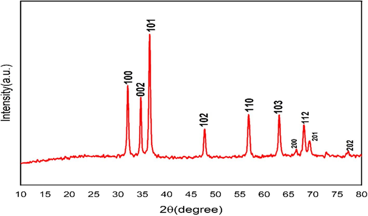


X-ray diffraction analysis provides insights into the crystal structure of the samples. The XRD pattern of ZnO nanoparticles is depicted in Fig. 1. The diffraction peaks for ZnO nanoparticles were more intense and narrower, indicating that these nanostructures exhibit good crystallinity. The peaks observed at scattering angles (2θ) of 31.98°, 34.66°, 36.48°, 47.78°, 56.78°, 63.08°, 66.6°, 68.14°, 69.3°, and 77.08° correspond to the (100), (002), (101), (102), (110), (103), (200), (112), (201), and (202) crystal planes, respectively [41].
Fig. 1 Transmission electron microscopy (TEM)
A supplementary morphological description is obtained using the (HR-TEM) analysis. The (HR-TEM) image of ZnO NPs with a mean size of 22.94 ± 4.77 nm is shown in Fig. 2. The image demonstrates that the particles are spherical [42].
Fig. 2 Dynamic light scattering (DLS)
The particle size and zeta potential of the synthesized ZnO nanoparticles are presented in Fig. 3a, b, respectively. The mean particle size was 24 ± 3.06 nm, with a narrow distribution as indicated by a low polydispersity index (PDI) of 0.324. Zeta potential measurements were conducted to evaluate the surface charge and colloidal stability of the ZnO-NPs. The average zeta potential was – 21 ± 2.40 mV, indicating a moderately negative surface charge (Fig. 3b). This level of surface charge is generally associated with good dispersion stability in aqueous environments [34, 43]. It may also influence cellular interactions, potentially enhancing cellular uptake and selective internalization in cancer cells due to electrostatic interactions with the cell membrane [44]. These properties are important when considering the biological behavior and cytotoxic effects of nanoparticles in subsequent assays. In comparison with previous studies, similar zeta potential values ranging from − 20 to −30 mV have been linked to enhanced nanoparticle uptake and biological activity in various cancer models. For example, ZnO nanoparticles with a zeta potential of approximately − 22.4 mV showed increased cytotoxicity in HepG2 cells [43], while those with a surface charge near − 25.7 mV exhibited improved internalization in MCF-7 breast cancer cells [45]. Such zeta potential values are generally considered optimal for promoting cellular uptake while maintaining colloidal stability, suggesting that the zeta potential measured in the present study falls within an effective range for biological applications.
Fig. 3
a Size distribution by the intensity of ZnO NPs. b Zeta potential for ZnO NP
Scanning electron microscopy (SEM) and energy dispersive X-ray analysis (EDX)SEM images were used to examine the surface morphology of ZnO nanoparticles, as shown in Fig. 5. The findings demonstrated the spherical form of ZnO NPs. The elemental composition of ZnO NPs was determined by EDX analysis. Agglomeration is responsible for the appearance of some larger ZnO NPs in SEM scans [46]. Figure 4 displays the EDX spectra of samples of ZnO nanoparticles. The label for the ZnO sample displays the element names and percentages. Since zinc and oxygen are the sample’s primary ingredients [47], no traces of contaminants were detected within the EDX detection limit. Figure 5 and Table 1 illustrate the weight percentages of ZnO NPs, which were 75.64% Zn and 24.36% O.
Fig. 4 Fig. 5
 Table 1 The EDX of ZnO NPsDifferential scanning calorimetry (DSC)
The DSC technique was employed to investigate the oxidized structure and isothermal oxidation behavior of ZnO nanoparticles in ambient air at temperatures ranging from 50 to 600 °C. The DSC curves of ZnO nanoparticles are displayed in Fig. 6. The low-temperature endothermic peak at 155.7 °C can be attributed to the loss of volatile surfactant molecules adsorbed on the surface of ZnO nanoparticles during synthesis. A second thermal event near 261.2 °C can be attributed to the decomposition of intermediate zinc hydroxide phases, leading to the formation of ZnO nanoparticles. A broader endothermic peak at approximately 401.2 °C may result from internal structural rearrangements or the elimination of residual organic content. No indication of ZnO reduction to metallic zinc is observed under these oxidative conditions, as ZnO remains thermally stable in air at these temperatures [48].
Fig. 6
DSC thermogram of synthesized ZnO nanoparticles
Cytotoxic activity on (A549) and (WI-38) by MTT assayThe A549 lung cancer cell Line demonstrated considerable resistance to gamma radiation at 5 to 15 Gy doses. Even at the highest dose of 15 Gy, cell viability remained above 70%, indicating that the rate of cell death did not exceed 30% (Fig. 7). While 5 Gy of gamma radiation induced noticeable toxicity in A549 cells, its cytotoxic impact was less pronounced than 10 and 15 Gy doses. This resistance implies that A549 cells possess efficient cellular mechanisms that protect against radiation-induced damage and enable recovery [49, 50]. Viability assessments conducted 48 h post-irradiation showed sustained metabolic activity in A549 cells, with survival rates frequently exceeding 80% even after exposure to 15 Gy. Clonogenic assays corroborated these findings by revealing that A549 cells are less susceptible to radiation-induced reproductive death compared to other, more radiosensitive lung cancer lines [51]. This inherent radioresistance is largely attributed to proficient DNA repair systems. Upon exposure to ionizing radiation, A549 cells rapidly initiate pathways to repair double-strand breaks (DSBs), a critical form of lethal DNA damage caused by radiation [49, 50]. Additionally, radiation generates reactive oxygen species (ROS) that inflict oxidative damage on cellular components, including DNA and lipids [52]. Gamma radiation remains a cornerstone in lung cancer radiotherapy by inducing DNA lesions that trigger cancer cell death; however, its efficacy is often compromised by the tumor cells’ radioresistance and collateral damage to healthy lung tissue [53]. Our findings confirm that higher radiation doses amplify the toxic effects of ZnO nanoparticles on lung cancer cells, highlighting their potential as radiosensitizers to improve radiotherapy outcomes. This strategy could also enable reduced radiation doses, minimizing harm to normal tissues while maintaining therapeutic efficacy.
Fig. 7
The cytotoxic effect of different doses of gamma radiation 5, 10, 15 Gy, on the A549 cell Line for 48 h
When A549 cell lines were exposed to various concentrations of ZnO nanoparticles (ZnO NPs) combined with different γ-radiation doses (5, 10, and 15 Gy), a significant, dose-dependent decrease in cell viability was observed. This effect was more pronounced in the combination treatment compared to ZnO NPs or γ-radiation alone after 48 h, as illustrated in Fig. 8.
Fig. 8
The cytotoxic effect of different concentrations of ZnO NPs combined with different doses of radiation, 5, 10, 15 Gy (γ-radiation) on the A549 cell Line for 48 h
Our findings demonstrate that the IC₅₀ value for ZnO NPs alone was 26.78 ± 0.44 μg/mL, which is consistent with earlier studies reporting IC₅₀ values between 25 and 30 μg/mL for A549 cells under similar in vitro conditions. However, this value dropped significantly to 17.97 ± 0.45 μg/mL upon combining ZnO NPs with 15 Gy γ-radiation, indicating a clear synergistic effect (p < 0.001). This enhanced cytotoxicity is likely driven by an increase in reactive oxygen species (ROS) generated by both ZnO NPs and γ-radiation [54, 55]. This represents a 32.8% reduction in IC₅₀, which appears more substantial than effects reported in other cancer cell types. For comparison, studies in PC-3 prostate cancer cells showed only a 20% reduction in IC₅₀ (from 28.3 to 22.6 μg/mL) when combining ZnO NPs with 10 Gy radiation [56]. Similar cytotoxic responses to ZnO NPs have been documented in various cancer cell types, including MCF-7 breast cancer cells (IC₅₀ of 27.3 μg/mL) [57], HeLa cervical cancer cells (~ 30.1 μg/mL) [58], and HepG2 liver cancer cells (~25.6 μg/mL) [59]. While less data is available on combination treatments with radiation, the pronounced reduction in IC₅₀ observed in this study highlights the potential of ZnO NPs to act as radiosensitizers across different tumor models (Fig. 9).
Fig. 9
A schematic mechanism by which nano zinc oxide can potentially cause cancer cells to become toxic or undergo apoptosis
ROS accumulation leads to oxidative stress that damages DNA, proteins, and mitochondria, ultimately triggering apoptosis. Cancer cells, including A549, are particularly sensitive to oxidative stress due to their high metabolic rates and deficient antioxidant defenses [19, 60, 61]. Gamma radiation further amplifies ROS production through water radiolysis [61], and ZnO NPs are known for their photocatalytic properties that intensify ROS formation under irradiation [62, 63]. Similar synergistic effects have been reported in other studies where ZnO NPs enhanced radiation-induced cytotoxicity in lung cancer cells [23, 64]. Some studies suggest that ZnO NPs may impair DNA repair pathways in cancer cells, particularly after radiation-induced damage. By interfering with repair mechanisms such as homologous recombination or non-homologous end joining, ZnO NPs may promote the accumulation of unrepaired DNA breaks, pushing cells toward apoptosis. This potential mechanism is supported by findings in which ZnO NPs enhanced the effect of chemotherapy agents and reduced radioresistance in A549 cells by disrupting redox balance and inhibiting cellular recovery after stress [21, 22, 24]. Altogether, these findings reinforce the potential of ZnO NPs as effective radiosensitizers. Their combination with γ-radiation not only increases cancer cell death but may also allow for the use of lower radiation doses, thus minimizing adverse effects on surrounding healthy tissues. Further in vivo and mechanistic studies are warranted to explore long-term safety and therapeutic potential.
The results presented in Table 2 and Fig. 10 provide the half maximal inhibitory concentration (IC50 μg/mL) values of ZnO NPs in combination with different doses of γ-radiation.
Table 2 IC50 values (μg/mL) of ZnO NPs on human cancer cell line (A549) and in combination with different doses of γ radiation after 48 hFig. 10
IC50 values of ZnO NPs with various doses of γ-radiation on the A549 cell line. ***p value < 0.001 refers to that there is a statistically significant difference as compared to ZnO NPs treatment
In the current study, the cytotoxic effects of zinc oxide nanoparticles (ZnO NPs) on normal lung fibroblast cells (WI-38) were evaluated both independently and in combination with gamma radiation as illustrated in fig. 11. The findings revealed a clear concentration-dependent reduction in cell viability, with higher concentrations (500 μg/mL) reducing viability to approximately 31% (p < 0.001) compared to control). While lower doses did not induce significant cytotoxicity (p > 0.001), the threshold for toxicity appears to intensify beyond 31.2 μg/mL, where cell viability was significantly decreased (p < 0.001). Notably, when ZnO NPs were combined with 15 Gy of gamma radiation, cell viability decreased even further, showing a statistically significant reduction compared to either treatment alone (p < 0.001). This synergistic effect was quantitatively reflected in a significant decrease in the half-maximal inhibitory concentration (IC₅₀) from 81.91 ± 0.96 μg/mL (without radiation) to 49.88 ± 0.65 μg/mL under irradiation (p < 0.001), as shown in Table 3. These observations align with several prior investigations demonstrating the ability of nanoparticles to sensitize cells to radiation. ZnO NPs, in particular, have been shown to elevate radiosensitivity through mechanisms involving enhanced oxidative stress leading to DNA damage and disruption of repair processes [65,66,67]. Moreover, the combined exposure to ZnO NPs and ionizing radiation has been linked to increased genetic damage and programmed cell death in both malignant and healthy cell lines, corroborating the mechanisms suggested by our findings [68,69,70,71]. Additionally, [72] showed that green-synthesized tin nanoparticles exhibited strong biological activity, including anti-haemolytic effects, underscoring the therapeutic potential of eco-friendly nanoparticle production for biomedical uses. Their results support the idea that nanoparticles, beyond targeting cancer, may also provide protective or restorative functions in biological systems, which is crucial when considering toxicity to normal cells. Supporting these insights, [73] found that silver nanoparticles made from Anethum graveolens extract significantly boosted radiotherapy effectiveness in colon cancer models, illustrating the benefits of combining metal nanoparticles with natural extracts. Similarly, [74] created iron-based nanoparticles that improved chemotherapy delivery and efficacy against lung cancer cells, demonstrating the multifunctionality of such nanosystems. [75] also showed that green-synthesized Cu₂O nanoparticles from Camellia sinensis leaves have notable antioxidant and anticancer activity, highlighting the advantages of eco-friendly synthesis methods. In addition, [76] developed composite nanofibers made of polyvinyl alcohol, gum tragacanth, and graphene oxide, which enabled targeted antibiotic release, exemplifying nanomaterials’ versatility in drug delivery. Likewise, [77] demonstrated that silver nanoparticles coated on chitosan-alginate magnetite not only catalyzed chemical reactions but also protected lung cells, indicating a combined therapeutic and protective effect of nanoparticle formulations.
Table 3 IC50 values (µg/mL) of ZnO NPs on normal lung cells (WI-38) and in combination with 15 Gy of γ-radiation after 48 hFig. 11
The cytotoxic effect of different concentrations of ZnO NPs alone or combined with 15 Gy (γ-radiation) on the WI-38 cells for 48 h
Despite promising therapeutic implications, the observed cytotoxicity in normal lung fibroblast cells highlights the necessity for carefully delineating safe dosing parameters to minimize damage to healthy tissues. Additionally, it should be noted that the single high dose of gamma radiation (15 Gy) used in this study differs from the fractionated dosing regimens typically employed in clinical radiotherapy. Therefore, subsequent studies should focus on fractionated radiation schedules combined with ZnO NPs to better replicate clinical conditions and optimize the therapeutic index.
However, it is important to note that the single high radiation dose (15 Gy) used in this study does not reflect standard clinical radiotherapy practices. Future experiments should incorporate fractionated dosing schemes to more effectively assess radiosensitizing effects under clinically relevant conditions. While a single supra-clinical dose of 15 Gy was used in this study, we acknowledge that a fractionated dosing regimen (e.g., 2 Gy × 5 fractions) may better reflect clinical radiotherapy practices. Future studies should explore the effects of such fractionated protocols to enhance clinical relevance [78] (Fig. 11).
Apoptosis analysis by flow cytometryThis work analyzed the cellular apoptosis and necrosis of ZnO NPs on lung cancer cell line (A549) without or with gamma radiation at a dose of 15 Gy for 48 h using flow cytometry with an annexin V/PI double staining. The treatment with the IC50 (μg/mL) concentration of ZnO NPs increases apoptosis percentages in A549 cells as compared to the untreated cells. However, compared to ZnO NPs or gamma radiation alone, the rates of apoptosis in the combination of ZnO NPs and 15 Gy of gamma radiation were noticeably higher Fig. 12 and Table 4.
Fig. 12
Apoptosis induction by ZnO NPs treatment combined with or without 15 Gy of γ-radiation after 48 h on the A549 cell line
Table 4 Assessment of A549 cell apoptosis following 48 h of ZnO NPs applied alone or in combination with 15 GyThe present study investigated the anticancer efficacy of zinc oxide nanoparticles (ZnO NPs), both alone and in combination with gamma (γ) radiation, against A549 lung carcinoma cells. ZnO NPs have garnered considerable interest in cancer therapy due to their ability to generate reactive oxygen species (ROS), particularly when activated by ionizing radiation. This leads to elevated oxidative stress, DNA damage, and ultimately, the induction of apoptosis in cancer cells. The radiosensitizing effect of ZnO NPs is mainly attributed to the catalytic activity of their surfaces under radiation, which amplifies oxidative stress levels within tumor cells.
As a result of these mechanisms, ZnO NPs activate apoptosis pathways in cancer cells. In particular, nano ZnO mediates ROS production through the p53 pathway. The p53 protein becomes activated in response to oxidative stress–induced DNA damage, thereby initiating programmed cell death [64].
The findings of the present study align with these mechanisms. ZnO NPs exhibited notable cytotoxicity against the A549 lung carcinoma cell line. Furthermore, when combined with gamma (γ) radiation, a synergistic cytotoxic effect was observed. This combined treatment led to a significant reduction in cell viability and a pronounced increase in apoptosis compared to either treatment alone [68].
Furthermore, supporting our findings, earlier research demonstrated that ZnO nanoparticles induce apoptosis in various cancer cell Lines. Notably, one study reported approximately 40% total apoptosis in MCF-7 breast cancer cells after 48 h of treatment with ZnO NPs [79]. Similarly, around 38% apoptosis was observed in HepG2 liver cancer cells through ROS-mediated mitochondrial pathways [80]. Comparable apoptotic effects (~ 36%) were also reported in A549 lung carcinoma cells [81]. Our findings of 29.6% total apoptosis in A549 cells treated with ZnO NPs alone fall within this range, supporting the broad cytotoxic potential of ZnO nanoparticles across different cancer types.
Additionally, it is crucial to highlight that both apoptosis and necrosis can result from elevated intracellular Zn2⁺ levels. Numerous studies have indicated that Zn2⁺ increases occur due to the dissolution of ZnO nanoparticles (NPs) in lysosomes after cellular uptake, or when ZnO NPs dissolve in the extracellular space, facilitating the transport of Zn2⁺ ions into the cell. This leads to a rise in intracellular Zn2⁺ concentrations that surpasses the capabilities of the Zn2⁺ homeostatic system. Consequently, the toxic levels of Zn2⁺ disrupt the mitochondrial membrane potential, prompting the generation of reactive oxygen species (ROS) and DNA fragmentation, activating caspases and resulting in apoptosis. When Zn2⁺ concentrations escalate further, necrosis becomes the predominant mode of cell death [70].
Comments (0)