
Remember me
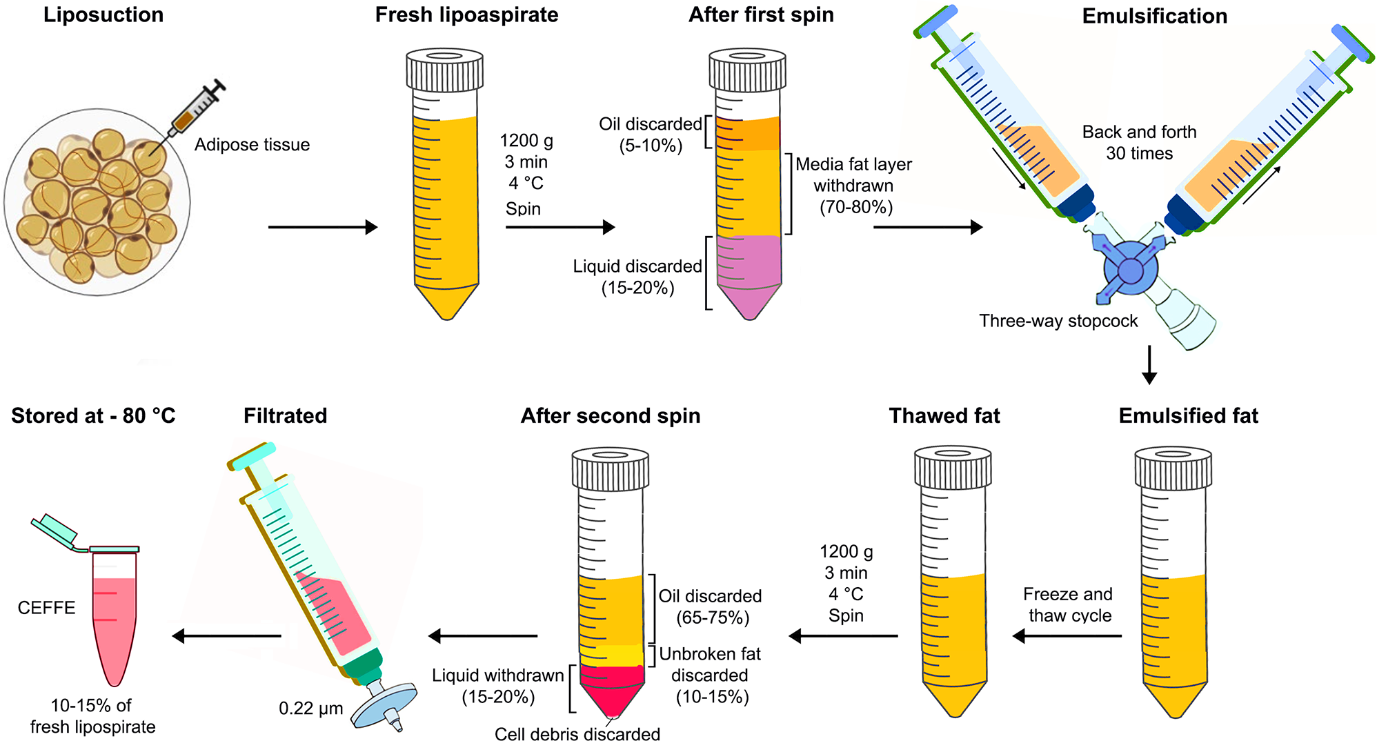


The total protein concentration of CEFFE was measured by BCA, and the concentration of various factors (bFGF, TGF-β1, HGF, VGEF, BDNF, PDGF, EGF and NT-3) was measured by ELISA (Fig. 3). The total CEFFE protein concentration was found to be 4.855 ± 0.751 µg/mL. Besides, the results of ELISA indicated that bFGF was the most abundant growth factor in CEFFE, with a concentration of approximately 33.5 ng/mL. TGF-β1 was also present in significant amounts, showing a concentration of roughly 29.4 ng/mL. The concentration of HGF was observed to be around 15.2 ng/mL, while VEGF was present at approximately 12 ng/mL. Other growth factors including, BDNF, PDGF, EGF and NT-3 were found in lower concentrations.
Fig. 3
The concentration of various growth factors in CEFFE detected by ELISA (n = 3)
The successful extraction and characterization of CEFFE from fresh adipose tissues represent a crucial step towards harnessing its therapeutic potential for tissue regeneration. Our study demonstrates that CEFFE is rich in various growth factors essential for tissue repair and regeneration.The substantial concentration of bFGF within CEFFE highlights its significance in promoting cellular proliferation, angiogenesis, and tissue remodeling [24]. Additionally, the presence of TGF-β1, a key regulator of extracellular matrix synthesis and chondrogenesis, underscores the potential of CEFFE in promoting articular cartilage regeneration [25]. HGF, known for its mitogenic and anti-apoptotic properties, further enhances the regenerative capacity of CEFFE by promoting cell survival and tissue repair.
Moreover, the presence of angiogenic factors such as VEGF within CEFFE suggests its potential to stimulate neovascularization, facilitating the delivery of nutrients and oxygen to the regenerating tissue [26]. This angiogenic activity is crucial for the establishment of a conducive microenvironment for tissue regeneration, particularly in avascular regions such as articular cartilage.
Although PDGF and EGF were found in relatively lower concentrations compared to other growth factors, their presence is noteworthy due to their roles in promoting cell migration, proliferation, and wound healing [24]. Despite their lower abundance, PDGF and EGF contribute to the overall regenerative potential of CEFFE, complementing the actions of other growth factors. On the other hands, BDNF and NT-3 are neurotrophic factors that support the survival and differentiation of neurons. Their presence, although in lower concentrations, indicates that CEFFE might also have a role in nerve regeneration, which can be beneficial in wounds involving nerve damage.
Fabrication and physicochemical characterizationThe concentration of 2% w/w nZnO in PLCL was selected based on previous studies that demonstrated effective antibacterial activity and biocompatibility at this level. Research has shown that ZnO nanoparticles at concentrations between 1 and 3% in polymer matrices provide enhanced surface roughness and bioactivity without compromising the structural integrity of the fibers [27, 28]. Furthermore, higher concentrations of ZnO nanoparticles tend to lead to excessive particle aggregation, which could negatively impact fiber morphology, as well as reduce the mechanical properties of the electrospun mats. By keeping the concentration at 2%, we aimed to balance the antimicrobial benefits of nZnO while minimizing aggregation and maintaining the fiber’s mechanical and structural properties. Besides, the 7:3 ratio of CEFFE to HA in the core solution was chosen based on preliminary studies and existing literature [29], which suggest that this ratio maximizes the biological activity of CEFFE while ensuring sufficient viscosity and electrospinnability of the solution. CEFFE contains bioactive components such as lipids, proteins, and growth factors, which are crucial for promoting tissue regeneration and wound healing. However, an excessive amount of CEFFE can decrease solution stability and impede the electrospinning process. The addition of 5% HA solution serves as a stabilizing agent that aids in maintaining solution viscosity, facilitating smoother fiber formation during electrospinning. This ratio has been optimized to ensure both successful coaxial electrospinning and the biological efficacy of CEFFE.
FE-SEM was utilized to analyze the surface morphology of the fabricated nanofibrous membranes (Fig. 4A), revealing significant differences among the four types: PLCL, nZnO/PLCL, PLCL-CEFFE/HA, and nZnO/PLCL-CEFFE/HA. The FE-SEM images illustrate a web-like structure across all samples, characteristic of electrospun nanofibers. However, the incorporation of nZnO has a marked impact on surface morphology and fiber diameter.
The PLCL and PLCL-CEFFE/HA membranes exhibit smooth surfaces, indicating uniform fiber formation during electrospinning. In contrast, the nZnO/PLCL and nZnO/PLCL-CEFFE/HA membranes display visible particle aggregation on the fiber surface, resulting in a rougher texture. This roughness can be attributed to the presence of nZnO, which tend to aggregate, as corroborated by previous studies [30, 31], where nanoparticle incorporation led to similar surface modifications.
The frequency histograms of fiber diameter distributions further underscore the impact of nZnO (Fig. 4B). PLCL and PLCL-CEFFE/HA membranes show relatively narrow diameter distributions centered around 400–500 nm. In contrast, the presence of nZnO in nZnO/PLCL and nZnO/PLCL-CEFFE/HA results in broader distributions with higher average diameters, extending up to 700 nm and 800 nm, respectively. These observations align with the findings of previous works [32, 33], where nanoparticle addition generally increased fiber diameters due to alterations in solution viscosity and electrospinning dynamics.
TEM provided insights into the internal fiber morphology and nanoparticle distribution (Fig. 4C). TEM images confirm the successful encapsulation of nZnO within the fiber matrix and on the surface, appearing both as individual particles and clusters. The core-shell structure in PLCL-CEFFE/HA and nZnO/PLCL-CEFFE/HA is clearly visible, indicating effective co-axial electrospinning and successful loading of CEFFE within the core, consistent with similar coaxial electrospinning studies [32, 34].
Fig. 4
Morphology and size characterization of nanofibers. (A) FE-SEM images with (B) diameter distribution of PLCL, nZnO/PLCL, PLCL-CEFFE/HA, and nZnO/PLCL-CEFFE/HA. (C) TEM images of the fabricated fibers
XRDThe XRD patterns of HA, nZnO, and electrospun fiber membranes composed of PLCL, nZnO, HA, and CEFFE are depicted in the Fig. 5A. A detailed analysis of the XRD patterns provides valuable insights into the structural properties and interactions of these materials within the composite membranes. The XRD pattern of pure HA shows a broad peak around 28° indicating its semi-crystalline nature. This broad peak is characteristic of the amorphous regions within the polymer matrix, suggesting a lower degree of crystallinity typical for biopolymers like HA. nZnO exhibit a highly crystalline structure as evidenced by the presence of multiple sharp peaks in the XRD pattern. The prominent peaks at 3°, 34°, and 36° correspond to the (100), (002), and (101) planes, respectively, of the hexagonal wurtzite structure of ZnO. These peaks confirm the high crystallinity and purity of the nZnO used in this study. The XRD pattern of PLCL shows characteristic peaks at 16° and 22°, indicative of its semi-crystalline nature. These peaks correspond to the (110) and (200) planes of PLCL, respectively. The relatively broad peaks suggest a lower degree of crystallinity, which is typical for this type of polymer. The XRD pattern of the nZnO/PLCL composite shows characteristic peaks of both PLCL and nZnO. The peaks of nZnO are visible at 31°, 34°, and 36°, indicating successful loading of nZnO into the PLCL matrix. The absence of new peaks suggests that the interaction between nZnO and PLCL is primarily physical, likely through hydrogen bonding, rather than chemical interaction. In the PLCL-HA composite, the XRD pattern displays the characteristic peaks of PLCL and a broad peak of HA around 28°. This indicates the presence of HA in the composite without forming new crystalline phases, suggesting a physical mixture of HA and PLCL. Finally, he XRD pattern of the nZnO/PLCL-CEFFE/HA composite shows peaks corresponding to PLCL, nZnO, and the broad peak of HA. Notably, the representative peak of PLCL at 22° is more intensified, which can be attributed to the presence of CEFFE. This intensified peak suggests that CEFFE is effectively incorporated into the composite, enhancing the crystallinity of PLCL. The absence of new peaks indicates that there is no chemical interaction among PLCL, nZnO, and CEFFE, further supporting the hypothesis of physical interaction.
Fig. 5
Physicochemical characterization of electrospun nanofibers. (A) the XRD patterns of HA, nZnO, and electrospun fiber membranes composed of PLCL, nZnO, HA, and CEFFE, (B) Water contact angle of the various electrospun fibers, and (C) typical stress-strain curve of various electrospun nanofibers. n = 3, *p < 0.05 vs. Control
Water contact angleThe hydrophilicity of the fabricated membranes was evaluated by measuring the water contact angle (Fig. 5B). The PLCL membrane exhibited a hydrophobic nature with a contact angle of approximately 121.73°. The incorporation of nZnO into the PLCL membrane (nZnO/PLCL) resulted in a further increase in the contact angle to 132.06°, indicating enhanced hydrophobicity, although this increase was not statistically significant (p > 0.05). In contrast, the core-shell membrane containing CEFFE and HA (PLCL-CEFFE/HA) demonstrated a drastic reduction in the contact angle to 21.85°, reflecting a highly hydrophilic surface. This significant decrease can be attributed to the presence of HA in the core, which is known for its hydrophilic properties. The addition of nZnO to the shell of the core-shell membrane (nZnO/PLCL-CEFFE/HA) resulted in a slight increase in the contact angle to 59.92°. Despite this increase, the membrane remained within the hydrophilic range, indicating that the presence of nZnO does not significantly hinder the hydrophilicity imparted by HA and CEFFE in the core.
The evaluation of hydrophilicity through contact angle measurements provides critical insights into the surface properties of the fabricated membranes. The contact angle data indicates that the incorporation of nZnO into PLCL membranes increases their hydrophobicity, while the inclusion of hydrophilic components such as HA and CEFFE significantly enhances hydrophilicity. Comparing these findings with existing literature, it is evident that the integration of hydrophilic substances like HA can effectively reduce the water contact angle and improve surface wettability. For instance, Liu et al. demonstrated that HA-modified membranes exhibited superior hydrophilicity with contact angles below 30°, consistent with our observations for the PLCL-CEFFE/HA membrane [35]. Additionally, the role of nZnO in modulating surface properties has been well-documented. Studies by Li et al. reported that nZnO enhance hydrophobicity due to their inherent surface characteristics [36], aligning with the increased contact angle observed in our nZnO/PLCL membranes. The slight increase in contact angle upon incorporating nZnO into the core-shell membrane (nZnO/PLCL-CEFFE/HA) suggests a balancing effect between the hydrophilic core and the hydrophobic nature of nZnO, which is crucial for optimizing membrane performance in biomedical applications [37]. These findings have significant implications for the design of innovative core-shell nanofiber wound dressings. The hydrophilic nature of PLCL-CEFFE/HA membranes ensures better wound exudate management, while the inclusion of nZnO provides the necessary antibacterial properties without significantly compromising hydrophilicity.
Mchanical propertiesThe mechanical properties of the fabricated nanofibrous scaffolds, including PLCL, nZnO/PLCL, PLCL-CEFFE/HA, and nZnO/PLCL-CEFFE/HA, were evaluated through tensile strength tests, elongation at break measurements, and Young’s modulus calculations (Fig. 5C) (Table 1). The pure PLCL scaffold exhibited the highest tensile strength of 5.30 MPa, showcasing its inherent mechanical robustness. In contrast, the addition of nZnO in the nZnO/PLCL scaffold reduced the tensile strength to 4.50 MPa, likely due to the brittle nature of nZnO, which introduces stress concentration points leading to early failure. The PLCL-CEFFE/HA scaffold displayed a tensile strength of 5.10 MPa, slightly lower than pure PLCL, indicating that CEFFE/HA did not significantly compromise the mechanical integrity of PLCL. However, the combination of nZnO and CEFFE/HA in the nZnO/PLCL-CEFFE/HA scaffold resulted in the lowest tensile strength of 3.90 MPa, suggesting a compounded effect of both modifications on the mechanical properties.
Table 1 The mechanical properties of various nanofibrous scaffoldsThe elongation at break data followed a trend consistent with the tensile strength results. Pure PLCL demonstrated the highest elongation at break of 320%, indicative of its excellent ductility. The incorporation of nZnO resulted in a notable decrease in elongation to 250%, which can be attributed to the rigid nature of nZnO. The PLCL-CEFFE/HA scaffold showed an elongation at break of 290%, suggesting that while CEFFE/HA impacts flexibility, it does so less drastically than nZnO. The nZnO/PLCL-CEFFE/HA scaffold exhibited the lowest elongation at break of 240%, reinforcing the observation that the combination of nZnO and CEFFE/HA impairs mechanical flexibility.
Young’s modulus results indicated that the addition of CEFFE/HA to PLCL increased the stiffness of the scaffold to 2.60 MPa, likely due to the reinforcing effect of CEFFE/HA. In contrast, the addition of nZnO decreased the Young’s modulus to 1.80 MPa, indicating reduced stiffness, possibly due to the introduction of stress concentrators within the polymer matrix. The nZnO/PLCL-CEFFE/HA scaffold demonstrated a moderate Young’s modulus of 2.00 MPa, reflecting a balance between the stiffening effect of CEFFE/HA and the softening effect of nZnO.
In conclusion, the mechanical properties of the nZnO/PLCL-CEFFE/HA core-shell nanofibers suggest that these modifications can tailor the mechanical behavior to meet specific needs in wound healing and dressing applications. While pure PLCL and PLCL-CEFFE/HA scaffolds are suitable for applications requiring high mechanical strength and flexibility, nZnO/PLCL-CEFFE/HA scaffolds offer a balance of mechanical properties that may be beneficial for applications where moderate stiffness and additional functionalities provided by nZnO are desired.
In vitro release of nZnO and CEFFEThe release of Zn²⁺ ions from the nZnO/PLCL-CEFFE/HA core-shell electrospun nanofibers was studied using inductively coupled plasma atomic emission spectrometry (ICP-AES) (Fig. 6A). The release profile demonstrated a biphasic pattern, consisting of an initial burst release followed by a sustained release phase.
For nZnO/PLCL-CEFFE/HA core-shell nanofibers, in the first 8 h, a burst release of Zn²⁺ ions up to 630 µg was observed. This rapid release can be attributed to the presence of nZnO on the surface of the PLCL shell, which readily dissolve into the release medium. Following the initial burst, a sustained release of Zn²⁺ ions amounting to 853 µg occurred over the next 72 h. This phase likely results from the gradual degradation of the PLCL polymer matrix and the controlled diffusion of Zn²⁺ ions from the nZnO/PLCL-CEFFE/HA core-shell nanofibers. The total cumulative release of Zn²⁺ ions over a period of 96 h reached approximately 930 µg.
Fig. 6
Cumulative release of (A) Zn ion from nZnO/PLCL and nZnO/PLCL-CEFFE/HA, and (B) release of bFGF, TGF-β1, and VEGF from nZnO/PLCL-CEFFE/HA core-shell nanofibers. The data are presented as mean ± SD (n = 3)
For the nZnO/PLCL nanofibers, a burst release of Zn²⁺ ions amounting to 330 µg/mL was noted within the initial 8 h. This is indicative of the rapid dissolution of nZnO directly exposed to the release medium. The subsequent cumulative release over the next 24 h reached 490 µg. The shorter duration of sustained release in nZnO/PLCL, compared to nZnO/PLCL-CEFFE/HA, can be attributed to the absence of the CEFFE/HA core, which provides an additional barrier and modulation effect on the Zn²⁺ release.
The biphasic release pattern observed in the nZnO/PLCL-CEFFE/HA nanofibers is advantageous for biomedical applications requiring an initial burst of antimicrobial activity followed by a prolonged release for sustained therapeutic effects. The initial burst ensures rapid disinfection, while the sustained release phase maintains a therapeutic level of Zn²⁺ ions over an extended period, which is beneficial for wound healing and tissue regeneration.
The enhanced release profile in nZnO/PLCL-CEFFE/HA compared to nZnO/PLCL highlights the significance of the core-shell structure. The CEFFE/HA core not only provides a secondary phase of Zn²⁺ release but also introduces bioactive molecules from the CEFFE, potentially offering synergistic effects in promoting tissue repair and regeneration.
The cumulative release of three key growth factors, namely bFGF, TGF-β1, and VEGF, from nZnO/PLCL-CEFFE/HA core-shell nanofibers was monitored over a period of 96 h (Fig. 6B). The release of bFGF exhibited a rapid increase during the initial 24 h, reaching approximately 150 pg/mL. This was followed by a more gradual release, achieving a maximum cumulative release of approximately 500 pg/mL by the end of the 96-hour period. The release of TGF-β1 followed a similar trend to that of bFGF but at a lower rate. Within the first 24 h, the release reached around 100 pg/mL, with a continued steady increase over time, peaking at approximately 400 pg/mL by 96 h. Besides, the release of VEGF was slower compared to bFGF and TGF-β1. By 24 h, the release was around 75 pg/mL. The cumulative release then increased gradually, reaching a maximum of approximately 300 pg/mL by the end of the study period.
The observed release pattern of growth factors from the nZnO/PLCL-CEFFE/HA core-shell nanofibers is consistent with the expected release behavior from core-shell nanofiber systems. The slight initial burst release followed by a sustained release phase aligns well with typical core-shell nanofiber release mechanisms, confirming the effectiveness of the core-shell design in achieving controlled and prolonged delivery of growth factors.
On the other hands, the differential release rates of the growth factors are significant for tissue engineering applications. bFGF is known for its role in cell proliferation and differentiation, making its higher release rate beneficial for the initial stages of tissue regeneration. TGF-β1, with its role in matrix production and remodeling, showed a moderate release, which aligns with its function in later stages of tissue repair. VEGF, crucial for angiogenesis, exhibited the slowest release, ensuring a sustained supply for vascularization over the extended period [38].
Antibacterial activityThe antibacterial efficacy of the fabricated electrospun nanofibers was assessed against two bacterial strains: E. coli and S. aureus. The OD measurements at 600 nm after 24 h of incubation provide insight into the antibacterial properties of various nanofiber compositions.
As shown in the Fig. 7, the nZnO/PLCL and nZnO/PLCL-CEFFE/HA nanofibers demonstrated significantly lower OD values compared to the control, indicating substantial antibacterial activity. The antibacterial efficacy calculated using equation (i) shows that these nanofibers reduced bacterial growth by approximately 40% compared to the control. The presence of nZnO in these fibers likely contributed to the observed antibacterial effect, as nZnO are known for their antibacterial properties. In the case of S. aureus, similar to E. coli, the nZnO/PLCL and nZnO/PLCL-CEFFE/HA nanofibers showed a significant reduction in OD values, indicating a strong antibacterial effect. The reduction in bacterial growth was approximately 60% and 50% for nZnO/PLCL and nZnO/PLCL-CEFFE/HA, respectively. This further confirms the enhanced antibacterial activity imparted by the incorporation of nZnO.
Fig. 7
Antibacterial activity against (A) E.coli and (B) S. aureus measured through turbidity method. The data are presented as mean ± SD (n = 3), *p < 0.05 vs. control was considered significant
The results demonstrate that the incorporation of nZnO into PLCL nanofibers significantly enhances their antibacterial properties against both gram-negative (E. coli) and gram-positive (S. aureus) bacteria. In the case of nZnO/PLCL-CEFFE/HA nanofibers, the presence of nZnO in the shell layer is particularly effective in reducing bacterial growth. nZnO are known to induce oxidative stress and disrupt bacterial cell membranes, leading to bacterial cell death. Additionally, the use of CEFFE/HA in the core may provide further benefits for wound healing applications, potentially promoting tissue regeneration while simultaneously providing antibacterial protection.
The turbidity method is a widely recognized and reliable quantitative assay for measuring bacterial growth and inhibition, particularly in liquid cultures where planktonic bacteria are the primary focus. This method directly quantifies bacterial cell density by measuring OD at 600 nm, which is proportional to bacterial concentration. In the context of our study, where we aimed to evaluate the overall antibacterial efficacy of the nanofiber dressings against planktonic bacteria in a liquid environment, the turbidity assay provided a straightforward and accurate measure of bacterial growth inhibition. Studies have shown that turbidity assays are particularly suitable for evaluating nanoparticle-containing materials, as they allow continuous monitoring of bacterial growth in the presence of test materials, thus offering dynamic insight into the antibacterial properties over time [39].
The spread plate technique, while highly effective for assessing zone of inhibition formation on solid media, is often more suitable for materials that release antibacterial agents into the surrounding medium. In our study, the core-shell nanofiber dressings with nZnO and CEFFE were designed to maintain controlled release and localized activity. Since the antibacterial mechanism of these materials relies on the sustained release of nZnO and the activity of the CEFFE in a wound-like environment, the liquid-based turbidity assay was chosen to simulate conditions closer to in vivo situations where bacteria exist in a planktonic state rather than as colonies on solid surfaces. Furthermore, some studies suggest that the spread plate method may not fully capture the bactericidal activity of certain nanomaterials due to the limited diffusion of nanoparticles across the agar surface [40].
In addition, our materials include PLCL loaded with nZnO and HA loaded with CEFFE, which are designed for prolonged activity and biocompatibility in moist environments, such as wound dressings. The turbidity assay allows us to evaluate their antimicrobial efficacy over an extended period under conditions that mimic wound exudates, where bacteria are likely to remain in suspension rather than form solid colonies.
By focusing on turbidity measurements, we ensured a quantitative and reproducible assessment of the antibacterial activity across all test samples. However, future studies may explore the inclusion of additional assays, such as the spread plate technique for a comprehensive evaluation of both planktonic and biofilm-forming bacteria on solid surfaces, as seen in other comparative studies [41].
MTTThe MTT assay results depicted in the graph demonstrate the proliferation of NIH-3T3 cells on different fabricated fiber membranes over a period of 7 days (Fig. 8A). The experimental groups included control, PLCL, nZnO/PLCL, PLCL-CEFFE/HA, and nZnO/PLCL-CEFFE/HA. The data indicate a notable variation in cell growth among the different groups.
On Day 1, the OD values for all groups were relatively low, indicating minimal cell proliferation at the initial stage. By day 3, there was a significant increase in cell proliferation across all groups, with the control and PLCL groups showing higher OD values compared to the nZnO/PLCL group, suggesting that the presence of nZnO initially inhibited cell growth.
By day 7, a substantial increase in cell proliferation was observed in all groups, with the PLCL-CEFFE/HA group exhibiting the highest OD value, significantly surpassing the control, PLCL, and nZnO/PLCL groups. This indicates that the controlled release of CEFFE from the core-shell structure of PLCL-CEFFE/HA nanofibers significantly enhanced cell proliferation.
The release of growth factors from CEFFE is a key factor contributing to the increased cell proliferation observed in the PLCL-CEFFE/HA and nZnO/PLCL-CEFFE/HA groups. Growth factors such as VEGF, PDGF, and FGF present in CEFFE promote cell growth, differentiation, and migration, essential for wound healing and tissue regeneration. The controlled release of these growth factors from the core of the nanofibers provides a sustained supply, creating an optimal environment for cell proliferation over the 7-day period.
Fig. 8
Cell viability and adhesion on the various electrospun nanofibers. (A) graph of MTT results showing the absorbance proportional to the viability of NIH-3T3 cells, and (B) FE-SEM images, showing the growth of NIH-3T3 cells on fiber membrane, taken at day 4 and 7 (scale bar = 30 μm)., The data are presented as mean ± SD (n = 3), *p < 0.05
The nZnO/PLCL group consistently showed the lowest cell proliferation among the fabricated fiber membranes across all time points, suggesting that the incorporation of nZnO reduced the proliferation of NIH-3T3 cells. However, the continuous growth of cells over the 7-day period indicates that the membranes were only slightly cytotoxic, and the used concentration of nZnO did not inhibit cell proliferation entirely. In the nZnO/PLCL-CEFFE/HA group, while ZnO might initially reduce cell proliferation, the presence of growth factors from CEFFE likely mitigates this effect, resulting in a net increase in cell proliferation over time.
The observed trend in cell growth can be correlated to the hydrophilicity of the membrane and the presence of nZnO. The PLCL-CEFFE/HA group’s superior performance is likely due to the hydrophilic nature of CEFFE and its controlled release from the core of the nanofibers, promoting better cell adhesion and growth. Conversely, the hydrophobic nature of nZnO/PLCL may have contributed to the reduced cell proliferation observed in the nZnO/PLCL group.
FE-SEM analysisThe morphology and adhesion of NIH-3T3 cells seeded on the scaffolds were visualized using FE-SEM. The FE-SEM images captured after 4 and 7 days of culture provided insights into the cell-surface interactions (Fig. 8B). The NIH-3T3 cells showed initial adhesion and spreading on the scaffolds. The PLCL-CEFFE/HA group displayed a more extensive cell network and better cell morphology compared to other groups, indicating that the controlled release of growth factors promoted initial cell attachment and proliferation. The nZnO/PLCL group showed less cell spreading and fewer cell extensions, supporting the MTT assay results that nZnO initially inhibited cell growth. Besides, there was a noticeable increase in cell density and spreading across all groups. The PLCL-CEFFE/HA group exhibited the most significant cell proliferation, with cells forming a dense and interconnected network. This suggests that the sustained release of growth factors from the CEFFE/HA nanofibers continued to support cell growth and proliferation. The nZnO/PLCL group, while showing improved cell adhesion compared to day 4, still had the lowest cell density among the groups, aligning with the MTT results indicating lower proliferation rates. The FE-SEM images corroborate the MTT assay results, showing that the PLCL-CEFFE/HA nanofibers provide an optimal environment for cell proliferation through the sustained release of growth factors, while the nZnO/PLCL nanofibers, despite being slightly cytotoxic, do not entirely inhibit cell growth.
In vitro scratch assayThe in vitro scratch assay was employed to evaluate the impact of various nanofiber formulations on the migration of NIH-3T3 cells. The wound area percentages were calculated after 48 h of incubation for each treatment group. The results are presented in the Fig. 9. The PLCL group exhibited a wound area percentage of approximately 60%, indicating moderate cell migration. The nZnO/PLCL group showed a higher wound area percentage of around 80%, suggesting a lower efficacy in promoting cell migration compared to PLCL alone. The PLCL-CEFFE/HA group demonstrated a significantly reduced wound area percentage of about 40%, highlighting its superior performance in facilitating cell migration and wound healing. Finally, the nZnO/PLCL-CEFFE/HA group displayed a wound area percentage of approximately 50%, which, while better than nZnO/PLCL, did not outperform the PLCL-CEFFE/HA formulation. The in vitro scratch assay results provide insightful data regarding the wound healing efficacy of different nanofiber formulations. The PLCL-CEFFE/HA nanofiber showed the most promising results, with the lowest wound area percentage, indicating the highest level of cell migration and potential wound healing efficacy. The combination of CEFFE loaded in HA within the core appears to significantly enhance cell migration compared to the other formulations.
Fig. 9
Representation of cell migration performed by the scratch assay. (A) Scratch results showing the migration of cells co-cultured with the various electrospun nanofibers as analyzed by taken images at 0 h and 48 h. (B) The quantification results of the cell migration images by using the Image J software. * p < 0.05 vs. control was considered significant. Results are mean ± SD (n = 3)
The released CEFFE with a variety of growth factors, which are crucial for cell proliferation and migration, likely contribute to the observed superior performance of the PLCL-CEFFE/HA formulation in promoting cell migration and wound healing. Conversely, the nZnO/PLCL formulation, which integrates nZnO into the PLCL polymer, exhibited the highest wound area percentage, suggesting that the presence of nZnO may inhibit cell migration to some extent. This finding aligns with previous studies indicating that while nZnO have antibacterial properties, their high concentration may be detrimental to cell viability and migration. The nZnO/PLCL-CEFFE/HA nanofibers, while demonstrating better performance than nZnO/PLCL, did not achieve the same efficacy as PLCL-CEFFE/HA. This suggests that the beneficial effects of CEFFE/HA on cell migration may be partially counteracted by the presence of nZnO.
The novel nZnO/PLCL-CEFFE/HA core-shell nanofibers offer significant advantages over traditional wound dressings like AgNPs and hydrogel-based systems. Antimicrobial efficacy is comparable between nZnO and AgNP dressings. Both exhibit strong antibacterial activity, disrupting bacterial membranes and generating ROS. However, nZnO has lower cytotoxicity and reduced risk of bacterial resistance, making it safer for long-term use in chronic wound care. AgNPs, though effective, pose concerns regarding toxicity and resistance development with extended exposure [7, 8].
Tissue regeneration is where the core-shell nanofibers truly excel compared to AgNPs and hydrogel-based systems. Hydrogels are biocompatible and provide moisture retention, but they lack bioactive components that actively promote tissue healing [4]. By incorporating CEFFE, rich in growth factors such as bFGF and TGF-β1, the core-shell nanofibers enhance fibroblast proliferation, ECM deposition, and wound healing. The study’s results demonstrated superior cell migration and wound closure with CEFFE-loaded nanofibers, far exceeding hydrogel-based systems’ passive role in wound care.
Mechanical properties of hydrogels can limit their application in large or irregular wounds due to poor structural integrity [42]. The PLCL matrix in the core-shell nanofibers balances mechanical strength and flexibility, making them more durable than hydrogel alternatives, even with the slight reduction in mechanical properties from nZnO incorporation.
A key advantage of the nZnO/PLCL-CEFFE/HA nanofibers is their biphasic release of zinc ions and growth factors, providing sustained antimicrobial activity and regenerative effects. This controlled release is more beneficial than AgNP dressings, which may overexpose tissues to silver, and hydrogel systems, which lack active bioactive delivery mechanisms.
Comments (0)