Over than 14 million people in the United States (US), and more than 200 million people globally have osteoporosis, a condition characterized by reduced bone mineral density. The primary causes of bone loss include obesity, genetic disorders, accidents, and aging [98]. While autografts are the standard treatment for bone injuries, their use is limited due to accessibility and risks. As a result, new approaches such as tissue engineering are gaining traction in bone repair. Tissue engineering focuses on creating scaffolds to support bone cells and promote the regeneration of bone tissue [99]. From a histological point of view, bone tissue is a natural mixture of bioceramic and polymeric phases, mainly apatite and collagen [100].
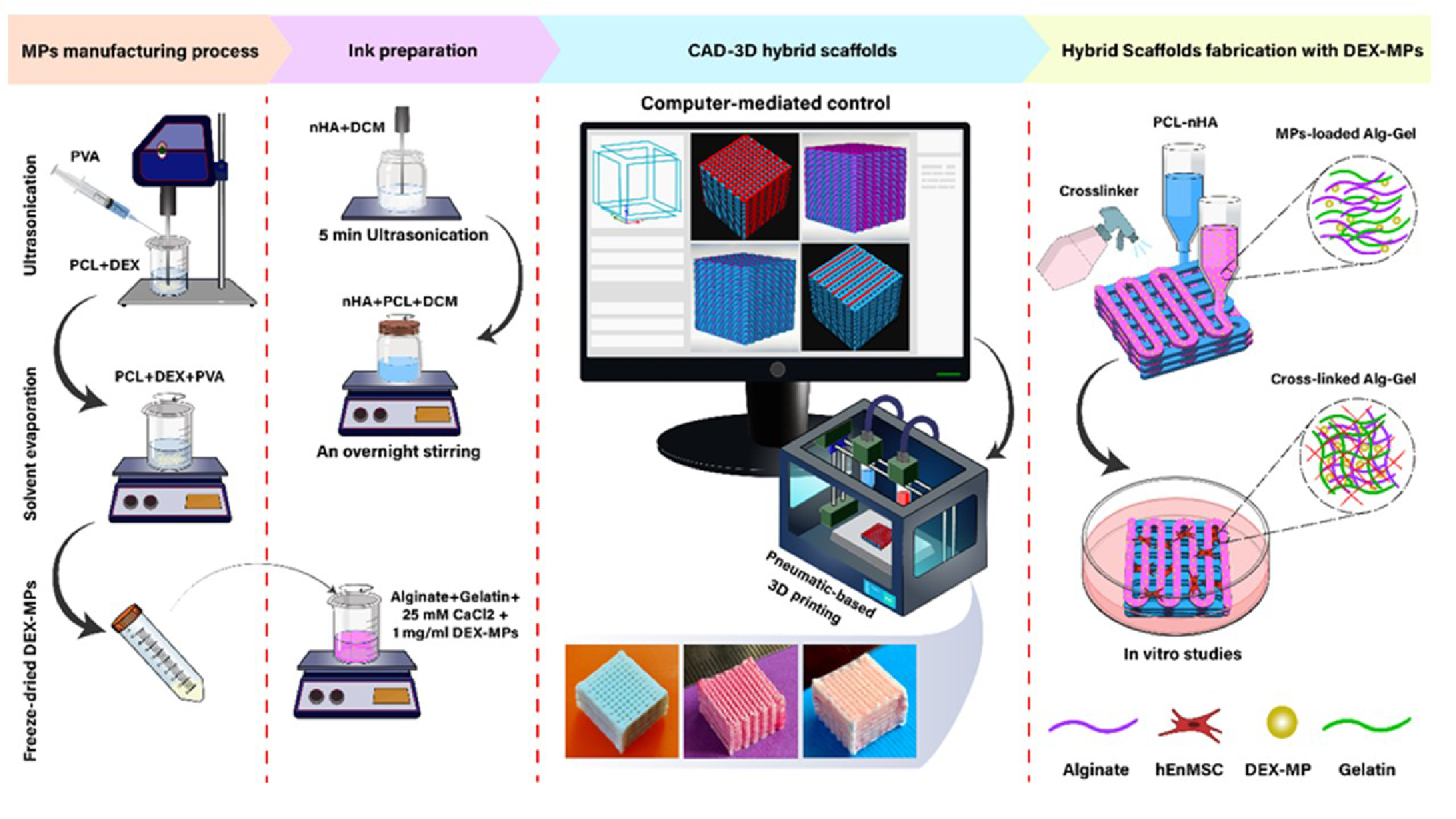
Hence, scaffolds which consist of organic-inorganic hybrid polymeric composites containing hydroxyapatite nanoparticles, show promise for bone tissue engineering. Specifically, 3D-printed porous polymer nanocomposite scaffolds, including those made from polycaprolactone, are suitable for supporting various types of cells due to their mechanical properties, cost-effectiveness, biocompatibility, and printability [101, 102]. Moreover, recent approaches in bone tissue engineering focus on creating biomimetic scaffolds using hydrogels, such as alginate-gelatin, to induce biomineralization and drug delivery [103]. Sustained release of drugs, including microcarriers and nanocarriers, has been shown to support tissue healing [104]. Amongst, Dexamethasone, a drug for inducing bone differentiation, has been used in microparticles within the scaffold structure [105]. The current study involves fabricating biomimetic nanocomposite scaffolds with sustained drug release using 3D printing, optimizing for biological and mechanical performance to promote osteogenesis in endometrial mesenchymal stem cells. The development of hybrid systems, combining osteoinductive 3D-printed scaffolds and cell-laden hydrogels, has shown great potential for bone tissue engineering and the treatment of bone defects based on active tissue regeneration.
Basically, the main aim of the study was to develop hybrid scaffolds with sustained release of DEX, which mimicked the extracellular matrix (ECM) while enhancing the differentiation potential of hEnMSCs into osteoblasts, thus facilitating bone regeneration. One of the key aspects of this study was the similarity of the hybrid scaffolds, composed of nHA, gelatin, and alginate, to the ECM of bone tissue which promotes osteoconductivity and creates a biomimetic microenvironment. Given the self-renewal nature of MSCs and their inherent potential for osteogenic differentiation [106], these scaffolds have proven effective in enhancing hEnMSCs adhesion, migration, proliferation, and ultimately, bone formation by guiding the behavior of MSCs.
The homogenous incorporation of DEX-MPs within the hydrogel, entrapped in the hybrid scaffolds, allowed precise control over sustained drug release (Fig. 3). Furthermore, the use of 3D printing technology played a pivotal role in designing the hybrid scaffolds as it provided precise control over dimensions, structure, and porosity, ensuring optimal structural integrity (Fig. 2). These scaffolds exhibited favorable mechanical properties, ensuring sufficient stability and support for hEnMSCs colonization and osteogenesis. By combining the structural, mechanical, and bioactive properties of the hybrid scaffolds with controlled release of DEX, a synergistic approach for BTE was sought to be achieved.
Overview data of (Table 2) demonstrated that increasing the initial DEX percentage compared to the polymer led to a corresponding rise in DEX encapsulation within the MPs. This trend could be explained by considering that the drug content in these MPs is influenced by the interactions between the drug and polymer, with higher drug mixing resulting in greater drug incorporation [107]. Furthermore, the percentage of drug loading closely mirrors the initial drug concentration applied during fabrication. This suggests that, regardless of the initial drug concentration, a significant amount of drug is consistently loaded into the MPs. In this study, encapsulation and drug loading efficiencies of DEX align with previous reports [75, 82].
Also, the results from DLS indicate that the size of microparticles increases with higher initial drug concentrations. Given that only a fixed amount of drug can be accommodated within a specific quantity of polymer, the increased drug content leads to a more viscous dispersed phase, which in turn, contributes to larger microparticle dimensions [107]. Notably, the particle size distribution falls within the range of 0.24 to 5.58 μm (Fig. 1b), which is consistent with similar studies and is well-suited for the intended application of controlled drug release [74]. Moreover, FESEM images (Fig. 1a) reveal the smooth and uniform morphology of the MPs, consistent with the DLS results (Fig. 1b). These visual observations affirm the successful fabrication of MPs with the desired properties, reinforcing their potential for controlled drug release applications.
The zeta potential, a measure of surface charge, illustrates that drug-free MPs exhibit a zeta potential of -27.7, while drug-containing microparticles display a more negative value of approximately − 31.56 (Fig. 1c). This change can be attributed to the presence of the drug within the microparticles. Zeta potential values within the range of -15 to -30 mV are considered ideal for stabilizing microparticles [108, 109]. The negative charge plays a crucial role in preventing microparticle aggregation, maintaining their dispersed state, and ensuring the stability of the microparticle system, which are essential factors for controlled drug release and targeted delivery to hEnMSCs [110].
In this study, we successfully fabricated hybrid scaffolds with integrated and coherent structure using a layer-by-layer approach, which included Alg-Gel and PCL-nHA composites. Calcium chloride (CaCl2) and glutaraldehyde were applied as crosslinkers after each Alg-Gel layer printing and after the completion of the complete 3D scaffold printing. In a general sense, the polyanionic carboxylate groups (-COO−) in alginate [111] and the polycationic amine groups (NH2-) in gelatin [112] can potentially form weak electrostatic interactions between the two materials [88]. Simultaneously, the Ca2+ ions present in the CaCl2 solution infiltrate the alginate matrix, leading to an ion exchange with Na+ ions. This process facilitates the formation of ionic bridges between alginate’s carboxylate groups (-COO−) and the formation of a gel network [111]. This gelation process results in the creation of a coherent and stable structure of Alg-Gel, onto which the layer of PCL-nHA can be easily placed. Moreover, glutaraldehyde, with its aldehyde functional groups (-CHO), forms covalent imine bonds (C = N) with the amine groups (-NH2) of gelatin [113], thereby enhancing the structural integrity.
Additionally, there may be cross-links between the -OH group of nHA and the -CHO of glutaraldehyde [114]. Previous studies have also proved interactions between the phosphate functional groups (-PO43−) and Ca2+ ions in hydroxyapatite and the carboxyl and amine functional groups of gelatin-chitosan-alginate when combining these materials [114,115,116]. These findings support the hypothesis that interactions between calcium ions, phosphate groups, and carboxyl/amine groups within hydroxyapatite, gelatin, and alginate contribute to scaffold stability. However, the precise mechanisms underlying this phenomenon remain unclear. Furthermore, the textured and porous surface of the PCL-nHA composite layers allows for the penetration and interlocking of Alg-Gel layers. These physical interactions contribute to the mechanical stability and strong interface between the layers. Further research is still needed to fully elucidate the specific mechanisms and extent of these interactions.
SEM images (Fig. 2) revealed scaffold shrinkage following freeze-drying. This shrinkage varied among scaffold types, likely due to a combination of material properties, solvent type, and inter-component interactions. Alg-Gel scaffolds exhibited the highest shrinkage (35.99 ± 2.09%), potentially reflecting the hydrogel’s susceptibility to deformation during the drying process. In contrast, PCL-nHA scaffolds showed minimal shrinkage (5.37 ± 0.677%), possibly attributed to strong interactions between PCL and nHA [117] that maintained scaffold structural integrity. The substantial difference in shrinkage may also be linked to the solvents employed. DCM, a volatile organic compound (VOC) used for PCL, evaporates rapidly during 3D printing and drying [118], whereas water, used for the alginate and gelatin in Alg-Gel scaffolds [119], evaporates more slowly. The higher water content in Alg-Gel inks (resulting from a higher solvent-to-material ratio in ink preparation—see Methods section), combined with the hydrophilicity of alginate and gelatin [120], likely led to greater water loss and consequently higher shrinkage during drying [121]. This observation aligns with findings reported in other studies [121, 122].
Hyb-1 and Hyb-2 scaffolds, containing both PCL-nHA and Alg-Gel components, exhibited moderate shrinkage. Hyb-2 (9.93 ± 1.95% shrinkage) showed less shrinkage than Hyb-1 (14.07 ± 2.53%), likely due to differences in Alg-Gel distribution. In Hyb-2, Alg-Gel filaments were less numerous and more dispersed, between PCL-nHA filaments which provided greater structural support and reduced the Alg-Gel’s contribution to overall shrinkage compared to Hyb-1, where Alg-Gel and PCL-nHA layers are stacked.
Hyb-1 scaffolds exhibited pore size changes in addition to overall shrinkage. However, the scaffold’s overall dimensions remained largely unchanged due to interlayer crosslinking and the structural support provided by the PCL-nHA component. Subsequent immersion in aqueous media resulted in hydrogel swelling and pore size recovery to near-original dimensions, consistent with the findings of Luo Y et al. [121].
The change in pore dimensions in the Hyb-1 scaffold after drying and rehydration can be attributed to the different swelling properties of the PCL-nHA and Alg-Gel components. Drying resulted in greater Alg-Gel shrinkage than PCL-nHA shrinkage, leading to elongated pores. The observed variations in scaffold shrinkage and pore dimensions are likely due to the different solvents, rehydration, and swelling properties of the PCL-nHA and Alg-Gel components.
Previous studies conducted degradation tests within a timeframe of less than 30 days [76, 123, 124]. The present study was monitored scaffold degradation over a period of 12 weeks (Fig. 3a). Dorj et al. investigated the degradation rate of PCL-HA scaffolds with a 40:60 ratio using acetone and tetrahydrofuran (THF) solvent, which resulted in approximately 6% degradation after 14 days [123]. In our study, this composite achieved a similar degradation level after 12 weeks, indicating a slower degradation rate. Additionally, other studies assessed the degradation of PCL-13-93B3 composite with a 50:50 ratio in chloroform solvent, revealing a degradation rate of about 30% after 2 and 4 weeks [76, 124, 125]. Our study demonstrated that as the content of Alg-Gel hydrogel increased in the overall scaffold structure, the degradation rate increased. Due to their natural composition, hydrophilic nature, and weaker chemical bonds, Alg-Gel scaffolds are more susceptible to enzymatic decomposition, hydrolysis, and swelling, resulting in a higher degradation rate. This makes them suitable for drug delivery systems that require rapid degradation [40]. Similar research conducted by Kolan et al. evaluated hybrid scaffolds PCL-13-93B3 and Pluronic F127 hydrophilic and temperature-sensitive hydrogel. Their study reported a degradation rate of 20% after one week [77], whereas in our study, at the same time, Hyb-1 and Hyb-2 scaffolds exhibited degradation rates of only 2.5 ± 0.63 and 2.2 ± 0.68, respectively. Another study investigating the degradation behavior of PCL-GelMA hybrid scaffolds reported a degradation rate of 2% after three weeks [126], which corresponds with our findings for Hyb-1 (5.08 ± 0.35) and Hyb-2 (2.7 ± 0.62) after three weeks. As a result, the choice of solvent and temperature, material type and percentage, interactions and cross-linking characteristics, material distribution, presence of impurities, and scaffold design and layering can all influence the physicochemical properties of the scaffold, including the degradation rate [127, 128].
Moreover, the combination of PCL and nHA in the scaffold provided structural integrity and biocompatibility, while the addition of Alg-Gel improved mechanical performance. The PCL-nHA scaffolds exhibited brittle behavior, whereas the Hyb-1 scaffold with the inclusion of Alg-Gel showed advantages in terms of deformation capacity and stress distribution [129]. The hydrogels contributed to increased toughness and energy absorption, enabling the scaffold to withstand higher compressive loads without failure [130], as evidenced by the highest strain at failure (Fig. 3d). Although, the mechanical behavior of the Hyb-2 scaffold was slightly higher than Hyb-1, particularly the higher strength observed in Hyb-2, are relevant to lower Alg-Gel content, this study mainly emphasized investigating the biological efficiency of scaffolds on osteogenesis. Therefore, the Hyb-1 scaffold was chosen for drug loading and osteogenesis assays due to its higher cell interaction, porosity, and promising preliminary results in osteogenic differentiation.
Additionally, it can be claimed that the presence of Alg-Gel in the scaffold structure particularly in Hyb-1 scaffolds, can mimic the role of collagen as a soft phase in bone structure which leads to outstanding toughness of bone [128].
Overall, this study underscores the significance of meticulous material selection, optimization, and scaffold design in achieving desired mechanical properties for BTE. Processing parameters, such as solvent choice, temperature, material percentage, and interactions between composite components, can significantly influence the physicochemical properties and degradation rate of the scaffold. Thus, the combination of PCL-HA and Alg-Gel composites presents a promising approach for constructing scaffolds with favorable mechanical behaviors for bone regeneration and drug delivery purposes.
The results of the release curves fitting with mathematical models showed different release kinetics for free MPs (MPs-DEX) and the MPs-DEX entrapped in the Hyb-1 (Hyb-1-MPs-DEX) (see Table 4). The release of DEX from the free MPs follows a primarily Fickian diffusion-controlled pattern with a high release rate, whereas the release from the Hyb-1 scaffolds exhibits a non-Fickian Super Case-II transport mechanism dominated by polymer relaxation and degradation, showing a slower and potentially more controlled release [97]. The structural components of the Hyb-1 matrix seem to be involved in sustained DEX release [97]. This drug-release kinetic model has also been previously reported in systems consisting of hydrogels [131, 132]. The release rate constants were also lower for Hyb-1-MPs-DEX in comparison with MPs-DEX, indicating a reduced release rate for the Hyb-1 scaffold system. The MPs-DEX do not present any physical barrier to diffusion, contrary to the porous structure of the Hyb-1, in which the drug needs to diffuse through an additional barrier in addition to the MPs itself.
The slow and sustained release profiles observed from the scaffold-encapsulated microparticles hold promise for controlled drug delivery. Sustained release allows long-term drug availability, improving therapeutic efficacy and reducing dosing frequency [133]. The initial burst release is due to weakly attached DEX molecules on the microparticle surface or poorly encapsulated regions in the scaffold [134]. This burst release is reduced in scaffold-encapsulated microparticles compared to free MPs, thanks to an additional diffusion barrier created by the hybrid scaffold matrix.
Interestingly, we observed a strong correlation between the DEX release and the in vitro degradation profiles of the Hyb-1 (Fig. 3). The degradation test showed a slow weight loss during the first 3 weeks, after which the degradation rate accelerated. Similarly, the initial stage and then the gradual increase in drug release from days 16 to 30 align with the degradation pattern of Hyb-1 (Fig. 3). This strong correlation suggests that scaffold degradation plays a crucial role in controlling the drug release from Hyb-1. The initial slow release may be attributed to the low initial degradation and the need for the drug to diffuse through the matrix. The increased degradation rate of Hyb-1 creates larger pores [131], causing incorporated MPs to lose their structural support, which in turn facilitates the release of more microparticles and dexamethasone. Changes in hydrogel cross-linking density and dexamethasone diffusion characteristics contribute to this phenomenon [135]. This is consistent with the zero-order fit, which suggests that the DEX release rate is approximately constant after an initial phase that overcomes the immediate-release first-order systems and results in long-term maintenance of drug concentrations in the therapeutic range [95]. The release mechanism is thus influenced by a combination of polymer erosion, and possibly diffusion with zero-order model. So if initially the release mechanism may be accompanied by diffusion, matrix degradation becomes the rate-limiting factor. Overall, both polymer relaxation, as suggested by the Korsmeyer-Peppas model, and subsequent matrix degradation contribute to the sustained and non-Fickian release of DEX in Hyb-1.
In the scaffold-entrapped DEX-MPs, we observed an initial burst release of about 2 µg, followed by a slower, more uniform release. This controlled release can inhibit inflammatory reactions in the first days since a concentration of DEX between 0.5 and 5 µg/ml is known to inhibit the inflammatory response of macrophages [136]. In previous studies, the effective dose of DEX for osteogenic differentiation has been reported between 4 and 400 ng/ml [137,138,139]. In our study, the average drug release from day 1 to day 16 was approximately 133 ng from each scaffold, providing insights into drug release kinetics relevant to bone differentiation, and biomineralization (Fig. 3f). Sustained release of DEX from scaffold-entrapped DEX-MPs, especially in the effective concentration range, benefits osteogenic differentiation, demonstrating successful drug delivery for BTE applications [25, 140, 141].
The evaluation of osteogenic differentiation, using gene expression analysis of RUNX2, COL1A1, and OST at days 14 and 21, revealed a complex interplay between DEX-MPs, the biomaterial properties of the scaffold, and intrinsic cellular responses. In line with previous research, our study demonstrates that scaffolds containing controlled-release DEX-MPs enhance the differentiation of hEnMSCs into osteoblasts compared to scaffolds without the DEX [136, 142, 143]. Gene expression analysis revealed significant upregulation of COL1A1 and OST, key markers of late-stage osteoblast differentiation and matrix maturation [144], in the Hyb-1-DEX compared to Hyb-1 and PCL-nHA on days 14 and 21, confirming the beneficial effect of the DEX release system on osteoblast activity and bone formation (Fig. 6). These findings align with our visual and quantitative assessments from the ICC test, which reveal stronger and more extensive green fluorescence in the Hyb-1-DEX, in comparison to both PCL-nHA and Hyb-1 on days 14 and 21 (Fig. 6e). Moreover, the higher level of OST protein in the Hyb-1, compared to the PCL-nHA, suggests that the natural polymer composition, along with HA, provides a conducive environment for promoting bone differentiation and facilitating increased expression of OST.
RUNX2, Known as a key transcription factor responsible for regulating osteoblast differentiation [145, 146], exhibited increased expression in all groups, highlighting its critical role in governing osteoblast differentiation. However, the absence of a significant difference in RUNX2 expression between the groups on day 14, and only a slight difference against PCL-nHA on day 21 (p < 0.01), suggests the involvement of additional factors that contribute to the observed differentiation effects caused by the DEX-MPs (Fig. 6c). This trend, which is that the presence of a component can significantly increase only one or two bone biomarkers but has no significant effect on the others, has also been seen in the work of other researchers [3]. This indicates that the DEX-MPs may primarily influence later stages of osteogenic differentiation, consistent with studies showing enhancement of downstream signaling pathways involved in ECM protein synthesis, rather than directly affecting the initial transcriptional activation mediated by RUNX2 [147,148,149]. Despite observed improvements in bone formation, the lack of a statistically significant difference in RUNX2 expression likely reflects the complex interplay of multiple factors influencing osteogenic differentiation. Firstly, the limitations in experimental design might not have fully captured the dynamic expression patterns of RUNX2, highlighting the need for future studies employing a more comprehensive temporal analysis. Secondly, the complexity of osteogenesis suggests compensatory mechanisms may be at play. While RUNX2 is a crucial transcription factor, bone formation is regulated through multiple interconnected pathways, such as TGF-β and Wnt signaling [148, 149]. The controlled release of DEX influences osteogenesis via diverse pathways, including the induction of mineralization, secretion of BMP2, and activation of the hedgehog pathway [25, 150,151,152]. Furthermore, DEX has been shown to stimulate osteoblast-derived extracellular vesicle secretion, promoting bone differentiation [153]. These alternatives signaling pathways could compensate for subtle changes in RUNX2 expression, resulting in enhanced bone formation despite the non-significant change in RUNX2 levels. Finally, inherent biological variability must also be considered, including intrinsic cellular heterogeneity, variations in MSC responsiveness to DEX, and the unique biophysical properties of scaffold and paracrine signaling within the 3D microenvironment [154,155,156]. Further research is necessary to fully elucidate the contribution of each of these factors to the observed effects and clarify the complex interplay between RUNX2 and other signaling pathways in the context of the controlled DEX release system.
The higher ALP activity in the Hyb-1-DEX compared to the two other groups over time, especially the significantly higher ALP activity on day 7 in the Hyb-1-DEX compared to the PCL-nHA (p < 0.05), can be partially attributed to the presence of the DEX-MPs within the scaffolds. However, the absence of significant differences between the Hyb-1-DEX and the other two groups on days 14 and 21 requires further analysis. Scaffold structural components, including alginate and gelatin, in both the Hyb-1-DEX and Hyb-1, may have influenced ALP activity [157,158,159]. The presence of these biopolymers in Hyb-1 likely mitigates the long-term impact of DEX, leading to similar ALP activity levels between groups by days 14 and 21. As shown in a study, the potential of chitosan addition to the culture medium for osteogenic differentiation of DPSCs was almost similar to that of DEX by evaluating markers such as ALP and RUNX-2 [160]. Also, in another study, Amjadian et al. did not observe a significant difference in ALP activity on day 7 in the PLLA, nHA, DEX/gela


Comments (0)