HHV-6 is a rare cause of infectious uveitis, which can present in various forms, including corneal endotheliitis, posterior uveitis (more frequently as acute retinal necrosis), panuveitis, or optic neuritis [6,7,8,9,10,11,12,13,14,15].
In our report, we describe a case of unilateral retinitis in a splenectomised patient, hospitalized due to invasive pneumococcal disease.
Before this episode, our patient did not present any risk factors for infection other than being splenectomised. Following splenectomy, individuals have an elevated risk of infection to encapsulated bacteria, such as Streptococcus pneumoniae, Neisseria meningitidis, and Haemophilus influenzae type b, Gram-negative pathogens, and intra-erythrocyte parasites [16].
Vaccination against Streptococcus pneumoniae, Neisseria meningitidis, and Haemophilus influenzae type b can help prevent overwhelming post-splenectomy infection by establishing immunological memory. Initial vaccinations should be administered 14 days before a planned splenectomy or 14 days after an urgent splenectomy to ensure an adequate response [16].
Splenectomy is not associated with an increased risk of viral reactivation; however, in this case, the presence of the invasive meningococcal disease may have triggered its reactivation. In fact, the neurotropism of HHV-6 is currently well known. HHV-6 can invade the brain during acute infection, when there is disruption of the blood-brain barrier, and can cause febrile seizures. Moreover, its genome has been shown to persist in the CNS, even in healthy adults without symptoms, as shown by PCR exams of tissues obtained at postmortem examination [4]. Therefore, in the face of a serious CNS infection, the reactivation of the virus will likely occur more easily. Furthermore, the presence of bacteremia and the prescription of intravenous corticosteroids may have contributed to a reduction in overall immunity.
Regarding the diagnosis, it was inferred from the results of PCR analysis of both serum and CSF. Similarly, in Schallhorn CS et al. diagnosis was also made based on PCR analysis of serum and CSF [15].
In our case, given the presence of active and concurrent CNS disease, the positive PCR analysis of both serum and CSF for HHV-6, and the retinal lesion characteristics suggestive of a viral etiology, we concluded that the ocular disease was most likely caused by HHV-6. A posteriori, the good response to treatment, was also a point in favor of this diagnosis. The absence of ocular hypertension, while not supporting a herpetic etiology, did not exclude it either.
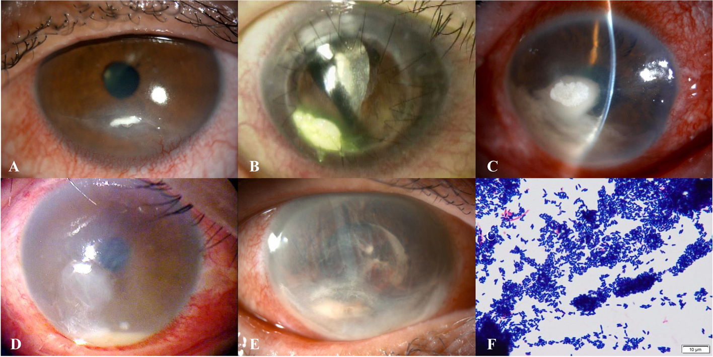
Streptococcus pneumoniae should be considered as a cause of endophthalmitis, especially in patients with meningitis and bacteriemia associated with the microorganism. The authors did not consider this a pneumococcal endogenous endophthalmitis because the patient did not present with ocular pain, had only mild ciliary injection and mild vitritis, and had been receiving treatment with intravenous ceftriaxone since the first day of hospitalization.
Retinitis without significant vitreous infiltrate is more suggestive of a neurotropic infection such as those associated with the herpes family of viruses, rather than bacterial endophthalmitis. In the case of atypical or unfavorable progression, immediate collection of an aqueous or vitreous sample should have been performed. In our case, since no ocular sample was collected, the diagnosis remained presumptive.
The treatment of this type of infection is still a matter of debate. Similar to CMV, in vitro studies have shown that ganciclovir and foscarnet can effectively inhibit HHV-6 replication, while acyclovir has been found to be less effective. Insufficient response to acyclovir has been observed in clinical cases reported by Keorochana N. et al., Schallhorn SC. et al., and Maslin J. et al., where treatment was switched to ganciclovir/foscarnet, resulting in a successful outcome. However, there are also reports in the literature in which the infection was resolved with acyclovir/valacyclovir, as in Malamos P. et al [4, 6, 8, 10, 15].
The most frequent treatment scheme is intravenous ganciclovir 5 mg/kg/dose every 12 h for 2 weeks followed by oral valganciclovir 900 mg twice daily as a maintenance therapy. Several studies had variable maintenance periods, ranging from 4 weeks to 8 months [10, 11, 14, 15]. In our case, given the favorable clinical progression and the observed healing of the retinal lesion, we decided to discontinue oral therapy after 4 weeks.
In Maslin J. et al. the treatment was stopped on day 45 as PCR analysis for HHV-6 on a second CSF and aqueous humour samples were negative [8]. The decision to suspend treatment based on the PCR result must be cautious, as chromosomal integration may occur. Chromosomal integration signifies that the entire viral genome is incorporated into the host’s DNA. Typically, HHV-6 DNA is not present in the serum and/or plasma of healthy individuals, non-infected by HHV-6. Thus, when HHV-6 DNA is detected in plasma or serum, one might assume that there is active viral replication. However, in individuals with HHV-6 chromosomal integration, whose cells contain at least one copy of the HHV-6 genome, HHV-6 DNA loads of 10⁴ to 10⁵ copies/mL of plasma can be detected, as the simple act of drawing blood causes a certain amount of cellular lysis. If chromosomal integration happens, PCR becomes an unreliable method for monitoring treatment, as the number of DNA copies may not correlate with active viral replication. This event should be suspected if, given a good clinical evolution, the copy number remains high. This was a point that was raised in the clinical case of Bajric J. et al. [13, 18].


Comments (0)