1)
Phantom study of [177Lu]Lu-PSMA-617 administration
For these experiments, a [177Lu]Lu-PSMA-617 infusion was administered using a peristaltic infusion system (Plum 360® pump, ICU Medical, San Clemente, CA, United States), and a hybrid titanium and polyoxymethylene TIVAP (Polysite 3007ISP®) equipped with a silicone catheter was used (Perouse Medical, Ivry-le-Temple, France).
An experimental scheme was built to reproduce an infusion of [177Lu]Lu-PSMA-617 solution through a TIVAP with peristaltic pump, as per clinical conditions.
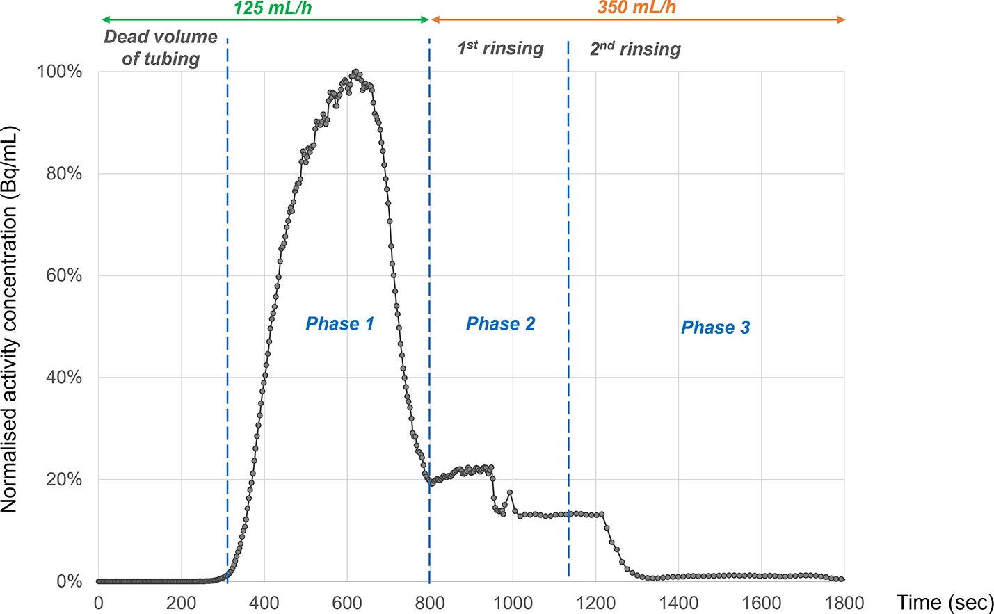
NaCl 0.9% was added to each vial of [177Lu]Lu-PSMA-617 to obtain a total of 10mL and these were infused through TIVAP. The infusion scheme was performed in 3 phases: (i) 125 mL/h injection rate for administration of 10mL of [177Lu]Lu-PSMA-617 solution and 5mL of NaCl 0.9% for rinsing the line; (ii) 15 mL of NaCl 0.9% was transferred by the peristaltic pump into the [177Lu]Lu-PSMA-617 vial for the first vial rinsing with injection rate of 350 mL/h, (iii) a last vial rinsing with 15 mL of NaCl 0.9% at 350 mL/h injection rate, as per usual clinical conditions in our department. During the administration of [177Lu]Lu-PSMA-617, the injected solution was collected in 3 fractions of 15 mL, one after each phase just described. The experiments were repeated 3 times to assess the repeatability of the results. The total duration of each administration is 30 min.
Each activity was measured using a dose calibrator (CRC-55tR Capintec®). The initial [177Lu]Lu-PSMA-617 activities were measured, as well as the three collected fractions obtained after the administration procedure. Residual activities within materials used for the administration (implantable chamber, TIVAP catheter, line from the [177Lu]Lu-PSMA-617 vial to the implantable chamber and the empty [177Lu]Lu-PSMA-617 vial) were also measured.
During administration, a dynamic 3D Single Photon Emission Computed Tomography/Computed Tomography (SPECT/CT) acquisition was performed on a 360° CZT camera (VERITON-CT® 400 series, Spectrum Dynamics, Caesarea, Israel). CT was acquired first with 120 kV, 80 mAs, 0.625 mm slice thickness followed by a 30-minute dynamic 3D SPECT recording. Consecutive 3-sec SPECT images were reconstructed with 2 iterations, 8 subsets, a median post-filter and 4.92 mm3 voxel size.
A 4mL sphere volume of interest (VOI) was drawn on the chamber of the TIVAP on the dynamic 3D SPECT images to analyze the kinetics of [177Lu]Lu-PSMA-617 through it during the injection procedure.
2)
Clinical [177Lu]Lu-PSMA-617 SPECT analysis
We retrospectively included consecutive patients from our department with progressive mCPRC who received [177Lu]Lu-PSMA-617 treatment on TIVAP between February 2022 and October 2023. The functionality of the TIVAP was assessed prior to each administration by verifying blood return on aspiration to ensure the absence of any relevant blockage or thrombus. The study protocol was registered on ClinicalTrials.gov (identifier NCT06858995).
Whole body SPECT/CT acquisitions were recorded 4 h after [177Lu]Lu-PSMA-617 infusion using the same parameters as those previously published [7].
A 4 mL sphere VOI was manually drawn on the TIVAP chamber on each SPECT image (VOITIVAP). Blood background was determined by a VOI on the right atrium (VOIRA). The VOIRA was automatically delineated on the CT using TotalSegmentator deep learning model [8]. Maximum Standardized Uptake Values (SUVmax), SUVpeak and SUVmean were extracted from the VOIs. Unilateral comparisons of superiority of VOIRA values over VOITIVAP values were performed using Wilcoxon tests. A qualitative analysis along the TIVAP catheter was also performed and considered positive if an uptake was superior to the blood background or otherwise considered negative.


Comments (0)