Animals
Seven-week-old male rats were purchased from Central Laboratory Animal, Inc. (Seoul, Korea). The rats were housed three per cage in a room with temperature control (21℃–22℃) and a 12 h/12 h light/dark cycle. All animals had access to sterile food and water. The rats were randomized into four groups of six rats each. All of the study procedures were approved by the Animal Research Ethics Committee of The Catholic University of Korea (2018-0174-04).
MIA-induced OA and treatment
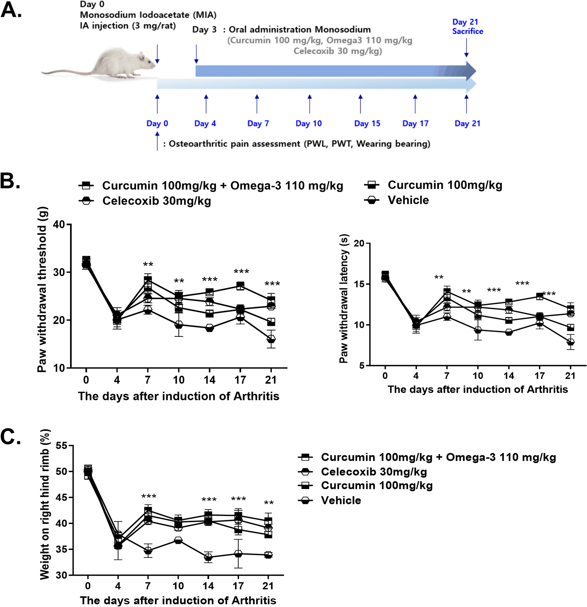
Rats were randomly assigned to treatment groups (curcumin, curcumin + omega-3, or celecoxib group) or the control (vehicle) group before the study began. After anesthetization with isoflurane, the rats (n = 6 per group) were injected with 3 mg MIA (Sigma-Aldrich, St. Louis, MO, USA) in the intra-articular space of the right knee through the patellar ligament, using a 26.5-G needle. Celecoxib was provided by Hanlim Pharm (Seoul, Korea). Curcumin (100 mg/kg), omega-3 (110 mg/kg), and celecoxib (30 mg/kg) were administered orally every day starting from day 3 after MIA induction and continued for 21 days. The control rats were given an equivalent of corn oil. The rats were sacrificed at day 21 from MIA injection.
Osteoarthritis pain assessment
Mechanical sensitivity was used to assess pain, as described previously [19, 20]. Following MIA injection, a dynamic plantar aesthesiometer (Ugo Basile, Gemonio, VA, Italy) was used to evaluate the response. Von Frey hair was used for mechanical sensitivity assessment. Pain was scored based on previous stimulation of the masseter, using rigid von Frey filaments and a force transducer (model 2290; Electronic von Frey, IITC, Inc., Woodland Hills, CA, USA). The force required to elicit hind-paw withdrawal was recorded three times following stimulations at 1-min intervals. The meaning of the three values was used for analysis.
Weight balance assessment
Weight bearing was evaluated using an incapacitance tester (Linton Instrumentation, Norfolk, UK) that included a dual-channel weight mean value. The rats were positioned in a plastic chamber. The strength applied by an individual hind limb was averaged over more than a 3-s time period. Individual data points were the average of three measurements. The percentage of weight on the handled (ipsilateral) hind limb was calculated utilizing the following equation: (weight on right leg/weight on right and left legs) × 100.
Histopathological analysis
Histological changes were analyzed to determine the effects of curcumin, and the combination treatment of curcumin + omega-3 administration in the knee joints of rats. The rats were perfused via the ascending aorta with 10% neutral buffered formalin. The knee joints, including the patella and joint capsule, were resected and maintained in the same fixative for an additional 48 h at 4 °C. The fixed specimens were decalcified with 5% formic acid decalcifier for 6 days at 4 °C. After decalcification, the specimens were embedded in paraffin. Standardized 7-mm serial sections were obtained at the medial and lateral midcondylar level in the sagittal plane and stained with hematoxylin and eosin (H&E). A modified Mankin’s score was used to classify histological injury of the articular cartilage, as follows: = normal; 1 = irregular surface, including fissures into the radial layer; 2 = pannus; 3 = absence of superficial cartilage layers; 4 = slight disorganization (cellular row absent, some small superficial clusters); 5 = fissure into the calcified cartilage layer; and 6 = disorganization (chaotic structure, clusters, and osteoclast activity). Cellular abnormalities were scored on a scale ranging from 0 to 3, where 0 = normal; 1 = hypercellularity, including small superficial clusters; 2 = clusters; and 3 = hypocellularity. Matrix staining was scored on a scale ranging from 0 to 4, where 0 = normal/slight reduction in staining; 1 = staining reduced in the radial layer; 2 = staining reduced in the interterritorial matrix; 3 = staining present only in the pericellular matrix; and 4 = staining absent. The joint space width was estimated based on the sum of the nearest distance of the medial and lateral tibiofemoral joints. Histological evaluations were performed independently by two experienced researchers who were blinded to the study groups.
Immunohistochemistry
Immunohistochemistry slides were deparaffinized and rehydrated using a graded ethanol series. The sections were incubated overnight at 4 °C with antibodies to COX IV (Santa Cruz Biotechnology, Santa Cruz, CA, USA) and Tomm20 (Abcam, Cambridge, UK). The slides were then treated with secondary antibodies and biotinylated anti-mouse IgG for 20 min, conjugated to a streptavidin peroxidase complex (Vector Laboratories, Burlingame, CA, USA) for 1 h, and then treated with 3,30-dia-minobenzidine (Dako, Glostrup, Denmark). The slides were counterstained with Mayer’s hematoxylin and photographed using a photomicroscope (Olympus, Tokyo, Japan).
In-vivo micro-computed tomography imaging and analysis
Micro-CT imaging and analysis were performed using a bench-top cone-beam-type in-vivo animal scanner (mCT 35; SCANCO Medical, Bruttisellen, Switzerland). The animals were imaged at settings of 70 kVp and 141 µA using an aluminum 0.5-mm-thick filter. The pixel size was 8.0 μm, and the rotation step was 0.4 °C. Cross-sectional images were reconstructed using a filtered back-projection algorithm (NRecon software, Bruker Micro CT, Kontich, Belgium). For each scan, a stack of 286 cross-sections were reconstructed at 2000 × 1335 pixels. The bone volume and surface were analyzed at the femur.
Primary culture and treatment of OA chondrocytes
All relevant protocols were approved by the Institutional Review Board of Uijeongbu St. Mary’s Hospital (HC14TISI0071) and performed in accordance with the Declaration of Helsinki. All patients provided written informed consent. OA was diagnosed using the American College of Rheumatology criteria [21]. Isolation of human chondrocytes was performed as described previously [12]. Briefly, chondrocytes were isolated from the cartilage of patients. Cartilage was digested with 0.5 mg/mL hyaluronidase, 5 mg/mL protease type XIV, and 2 mg/mL collagenase type V. Finally, chondrocytes were incubated in Dulbecco’s modified Eagle medium (DMEM), including 10% fetal bovine serum. The isolated human OA chondrocytes of passage 3 were cultured in the presence or absence of IL-1β (20 ng/mL), curcumin (100 μm), DHA (Docosahexaenoic acid), (50 μm), and celecoxib (10 μm).
Enzyme-linked immunosorbent assay
The concentrations of MCP-1 in culture supernatants were measured using a DuoSet enzyme-linked immunosorbent assay (ELISA) kit (R&D Systems, Minneapolis, MN, USA). The 96-well plates (Nunc, Roskilde, Denmark) were coated with capture antibodies for anti-human monocyte chemoattractant protein-1 (MCP-1, R&D Systems) and incubated overnight at 4 °C. After the overnight incubation, the plates were blocked with phosphate-buffered saline containing 1% bovine serum albumin and 0.05% Tween 20 for 2 h at room temperature. Cell culture supernatants were added to the plates and incubated at room temperature for 2 h. Subsequently, the plates were washed, detection antibodies were then added, and the reaction mixtures were incubated for 2 h at room temperature. The plates were washed again and then incubated with streptavidin-horseradish peroxidase for 20 min. Following an additional wash step, the substrate solution was added for incubation for 20 min. The stop solution was then added. The results were analyzed by determining the absorption at 405 nm (A405).
Real-time polymerase chain reaction
Total RNA was extracted using TRI Reagent (Molecular Research Center, Cincinnati, OH, USA) according to the manufacturer’s instructions. Complementary DNA (cDNA) was prepared by reverse transcription of single-stranded RNA using a high-capacity cDNA reverse transcription kit (Applied Biosystems, Foster City, CA, USA), according to the manufacturer’s instructions. Polymerase chain reaction (PCR) amplification was performed using a LightCycler 2.0 instrument (software version 4.0; Roche Diagnostics, Indianapolis, IN, USA). All reactions were conducted using LightCycler FastStart DNA Master SYBR Green I (TaKaRa, Shiga, Japan), according to the manufacturer’s instructions. The primer pairs used were as follows: control human gene β-actin, 5′-GGA CTT CGA GCA AGA GAT GG-3′ (sense) and 5′-TGT GTT GGC GTA CAG GTC TTT G-3′ (antisense); human MMP-1, 5′-CTG AAG GTG ATG AAG CAG CC-3′ (sense) and 5′-AGT CCA AGA GAA TGG CCG AG-3′ (antisense); MMP-3, 5′-CTC ACA GAC CTG ACT CGG TT-3′ (sense) and 5′-CAC GCC TGA AGG AAG AGA TG-3′ (antisense); MMP-13, 5′-CTA TGG TCC AGG AGA TGA AG-3′ (sense) and 5′-AGA GTC TTG CCT GTA TCC TC-3′ (antisense). All expression values were normalized to that of β-actin mRNA. PCR amplification and analysis were performed using a LightCycler real-time PCR system (Roche Holding AG, Basel, Switzerland).
Statistical analysis
Statistical analyses were performed using the nonparametric Mann–Whitney U-test for comparisons between two groups, and one-way analysis of variance with the Bonferroni post-hoc test for multiple comparisons. GraphPad Prism (ver. 9.2.0; GraphPad Software Inc., San Diego, CA, USA) was used for all analyses. The data are presented as the mean ± standard deviation. For all comparisons, P < 0.05 was taken to indicate statistical significance.


Comments (0)