Mice
C57BL/6 J (Cat. No. 000664), Casp1−/− (Cat. No. 016621) [31] and Nlrp3−/− (Cat. No. 021302) [32] mice were purchased from The Jackson Laboratory. Casp1−/− and Nlrp3−/− mice were backcrossed onto the C57BL/6 J background for an additional five generations before use. All mice were bred and housed in specific-pathogen-free conditions under a 12-h light/dark cycle. Drinking water and food were available ad libitum. All methods were carried out in accordance with relevant local and University of Wisconsin guidelines and regulations. All animals were handled in accordance with the Animal Research: Reporting of in vivo Experiments (ARRIVE) guidelines and the University of Wisconsin's Institutional Animal Care and Use Committee policies and our approved protocols.
Primary cell culture
BMDM were prepared as previously described [33]. Briefly, tibias and femurs were removed from mice, clipped on each end, and flushed with RPMI 1640 (Gibco, Cat. No. 11875–093) supplemented with Glutamax (1X) (Gibco, Cat. No. 35050–061), non-essential amino acids (1X) (Gibco, Cat. No. 11140–050), sodium pyruvate (1X) (Gibco, Cat. No. 11360–070), penicillin/streptomycin (1X), and 10% heat-inactivated fetal bovine serum (FBS) (Cytvia HyClone, Cat. No. SH30071.03HI) (complete RPMI media). Bone marrow was cultured in petri plates with complete RPMI media supplemented with 20% L-cell conditioned medium (LCCM). Cells were allowed to differentiate for 5–7 days prior to use.
Screening FDA-approved drugs for NLRP3 inflammasome inhibitors
A drug library containing 875 FDA-approved drugs was purchased from Cayman Chemical Company (Cat. No. 23538; Batch No. 0592679). For the primary screen, 25,000 BMDM/well were seeded in a 96-well tissue culture plate in glucose-free DMEM (Gibco, Cat. No. 11966–025) supplemented with glucose (3 mM) (Gibco, Cat. No. A24940-01) and recombinant murine macrophage colony stimulating factor (MCSF) (10 ng/ml) (PeproTech, Cat. No. 315–02) (plating media) (0.1 ml/well). BMDM were stimulated with LPS (50 ng/ml) from Escherichia coli O26:B6 (Sigma, Cat. No. L2654) for 4 h at 37℃, 5% CO2. BMDM were then incubated with drug (1 µM in DMSO) or DMSO (control) for 15 min followed by stimulation with the NLRP3 inflammasome activator, ATP (Research Products International, Cat. No. A300305) for 1 h. Following incubation, supernatants were collected and tested for IL-1β secretion by ELISA as described below. The primary screen was performed once with one technical replicate for each drug and four technical replicates for the DMSO control. A drug was considered to be a NLRP3 inflammasome inhibitor if the drug reduced IL-1β levels by ≥ 33% when compared to the DMSO control average.
Drugs identified as NLRP3 inflammasome inhibitors during the primary screen were subjected to a secondary screen. BMDM were seeded in a 24-well plate at a cell density of 400,000 cells/well in plating media (0.5 ml/well). BMDM were incubated with LPS, drug/DMSO, and ATP as described above for the primary screen. Following incubation, supernatants were collected and assayed for secreted IL-1β by ELISA as described below. The secondary screen was performed once with three technical replicates for each drug and the DMSO control. A drug was considered to be a NLRP3 inflammasome inhibitor if the drug significantly reduced IL-1β levels (P < 0.05) when compared to the DMSO control. Following the secondary screen, a literature review was performed in PubMed using the pharmaceutical drug name and “NLRP3” to determine if they had previously been reported to influence NLRP3 activity. Drugs were considered to be an unidentified NLRP3 inhibitor if the number of NLRP3 related papers was ≤ 2.
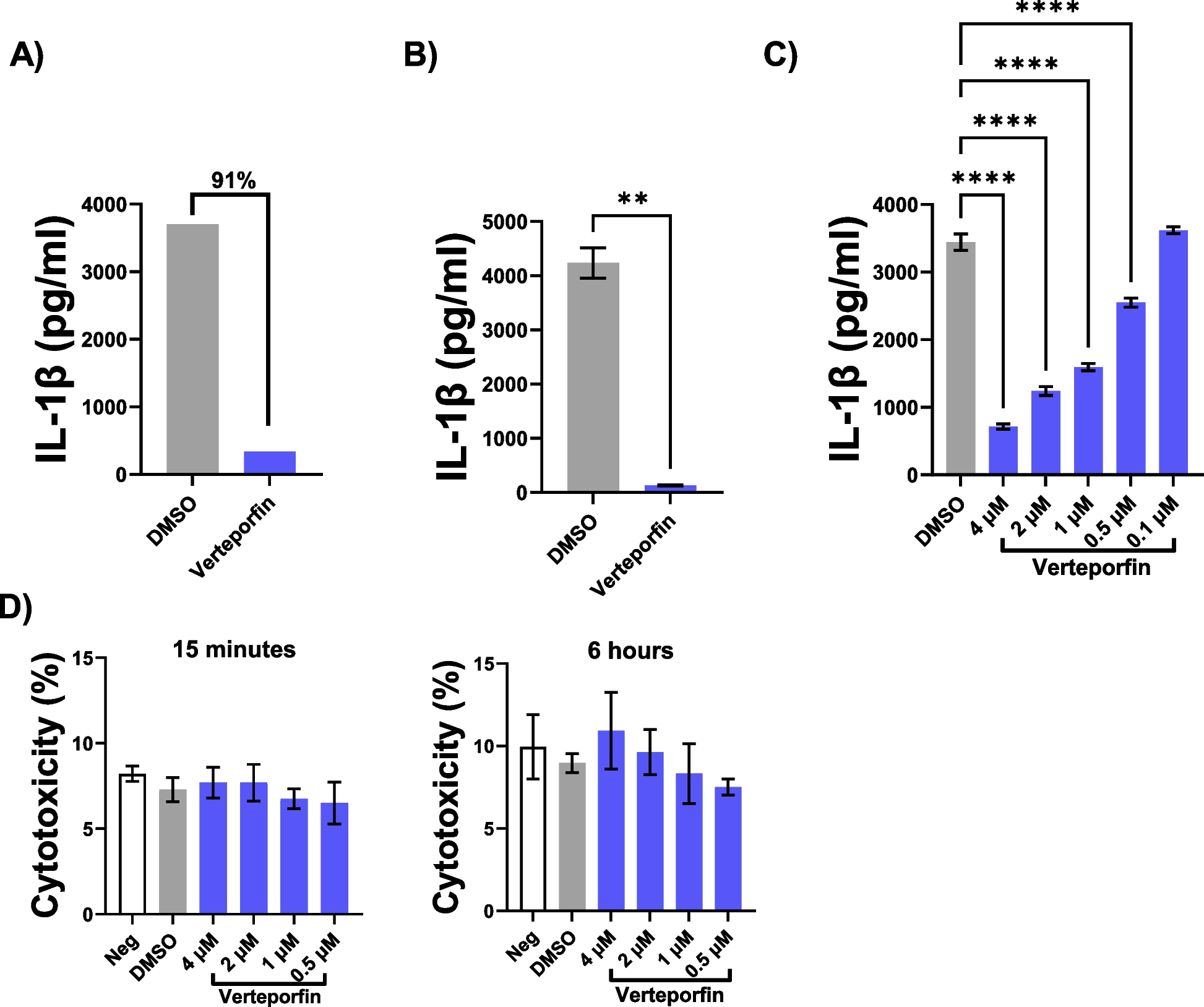
Drugs found to inhibit IL-1β secretion were prepared at concentrations of 4 μM, 2 μM, 1 μM, 0.5 μM, and 0.1 μM to determine the optimal in vitro drug concentration for IL-1β inhibition. BMDM (400,000 cells/well) were seeded in a 24-well plate and incubated with LPS, drug/DMSO, and ATP as described above for the primary screen. Following incubation, supernatants were collected and tested for secreted IL-1β by ELISA as described below. The dose curve was performed three times with three technical replicates for each drug and the DMSO control. Drug concentrations that significantly reduced IL-1β levels (P < 0.05) when compared to the DMSO control were then subjected to toxicity testing. BMDM were seeded in a 96-well tissue culture plate at a concentration of 20,000 cells/well in phenol red-free RPMI 1640 (Gibco, Cat. No. 11835–030) supplemented with 5% heat-inactivated FBS and MCSF (10 ng/ml). Cells were incubated with media, drug, and DMSO for 15 min or 6 h at 37 °C, 5% CO2. Drug toxicity to BMDM was determined by levels of lactate dehydrogenase (LDH) present in cell culture supernatants using the Cytotox 96® Non-Radioactive Cytotoxicity Assay (Promega, Cat. No. G1780) following manufacturer’s instructions. The experiment was repeated three times with three technical replicates per group. A drug concentration was considered toxic if the LDH release was significantly greater (P < 0.05) than the DMSO control. Overall, a drug was considered an NLRP3 inflammasome inhibitor if the drug had a concentration that significantly inhibited IL-1β secretion (P < 0.05) when compared to the DMSO control in the dose curve that did not cause toxicity to BMDM at the same concentration in the cytotoxicity assay.
Verteporfin NLRP3 inflammasome cell culture assays
Verteporfin was purchased from Cayman Chemical Company (Cat. No. 17334) and dissolved in DMSO (10 mM) to create a stock solution identical to the drug screen library. Following a confirmatory dose curve and cytotoxicity assay as described above, the optimal in vitro concentration of verteporfin (4 µM) was used to determine its ability to inhibit the NLRP3 inflammasome in BMDM. Briefly, WT, Casp1−/−, and Nlrp3−/− BMDM were seeded in a 24-well plate (400,000 cells/well) and incubated with LPS, verteporfin (4 µM)/DMSO, and ATP as described above. Following treatment, BMDM were lysed with Radio-Immunoprecipitation Assay (RIPA) buffer (Sigma, Cat. No. R0278) containing phenylmethylsulfonyl fluoride (PMSF), sodium orthovanadate, and Halt™ Protease Inhibitor Single-Use Cocktail (Thermo Fisher Scientific, Cat. No. 78425). Supernatants and lysates were flash frozen on dry ice and stored at − 80 °C until use. The experiment was repeated five times with three technical replicates per group.
NLRP3 inflammasome inhibition by verteporfin was also evaluated in WT BMDM using the NLRP3 inflammasome activator, MSU. BMDM (400,000 cells/well) were seeded in a 24-well plate and stimulated with LPS (50 ng/ml) for 4 h. BMDM were incubated with verteporfin (4 µM)/DMSO for 15 min followed by stimulation with MSU crystals (InvivoGen, Cat. No. tlrl-msu-25) (150 µg/ml) for 16 h. Following treatment, supernatants were flash frozen on dry ice and stored at − 80 °C until use. The experiment was repeated three times with three technical replicates per group.
Verteporfin inflammation cell culture assays
Assays were performed to determine if verteporfin inhibits general inflammation in BMDM. Briefly, WT BMDM (400,000 cells/well) were seeded in a 24-well plate and treated with verteporfin (4 µM) or DMSO for 15 min. Following incubation, cells were stimulated with LPS (50 ng/ml) for 6 h. Following stimulation, supernatants were flash frozen on dry ice and stored at − 80 °C until use. The experiment was repeated three times with three technical replicates per group.
ELISAs
For the drug screening, BMDM supernatants were assayed for IL-1β using the Mouse IL-1β/IL-1F2 DuoSet ELISA (R&D Systems, Cat. No. DY401) following manufacturer’s instructions.
BMDM supernatants from the verteporfin NLRP3 inflammasome cell culture experiments were assayed for IL-1β using the Mouse IL-1β/IL-1F2 DuoSet ELISA (R&D Systems, Cat. No. DY401) and IL-18 using the Mouse IL-18 DuoSet ELISA (R&D Systems, Cat. No. DY625) following manufacturer’s instructions. The WT BMDM supernatants were also assayed for IL-1α, IL-1β, IL-2, IL-3, IL-4, IL-5, IL-6, IL-10, IL-12, IL-13, IL-17, MCP-1, IFN-γ, TNF-α, MIP-1α, RANTES, GM-CSF, Eotaxin, CXCL1, MDC, and TARC using a multiplex assay (Quansys Biosciences).
WT BMDM supernatants from the verteporfin inflammation cell culture experiments were assayed for IL-6 using the Mouse IL-6 DuoSet ELISA (R&D Systems, Cat. No. DY406), TNF-α using the Mouse TNF-α DuoSet ELISA (R&D Systems, Cat. No. DY410), MCP-1 using the Mouse CCL2/JE/MCP-1 DuoSet ELISA (R&D Systems, Cat. No. DY479), CXCL1 using the Mouse CXCL1/KC DuoSet ELISA (R&D Systems, Cat. No. DY453), RANTES using the Mouse CCL5/RANTES DuoSet ELISA (R&D Systems, Cat. No. DY478), and MIP-1α using the Mouse CCL3/MIP-1α (R&D Systems, Cat. No. DY450) following manufacturer’s instructions.
Mouse paw homogenates were assayed for IL-1β using the Mouse IL-1β/IL-1F2 DuoSet ELISA (R&D Systems, Cat. No. DY401), IL-18 using the Mouse IL-18 DuoSet ELISA (R&D Systems, Cat. No. DY625), IL-6 using the Mouse IL-6 DuoSet ELISA (R&D Systems, Cat. No. DY406) and TNF-α using the Mouse TNF-α DuoSet ELISA (R&D Systems, Cat. No. DY410) following manufacturer’s instructions. Mouse paw homogenates were also assayed for MPO using the Mouse Myeloperoxidase DuoSet ELISA (R&D Systems, Cat. No. DY3667) following manufacturer’s instructions.
Caspase-Glo® 1 Inflammasome Assay
Caspase-1 activity in the supernatants was determined in the WT BMDM supernatants from the verteporfin NLRP3 inflammasome cell culture assays using the Caspase-Glo® 1 Inflammasome Assay (Promega, Cat. No. G9951) following the manufacturer’s instructions. Luminescence was recorded at 60, 90, and 120 min to ensure the luminescent signal was stabilized. A no-cell control was used to measure the background luminescence attributed to the cell culture media and Caspase-Glo® 1 Reagent. Final values for each treatment group were calculated by subtracting the Caspase-Glo® 1 YVAD-CHO Reagent values from the corresponding Caspase-Glo® 1 Reagent value. The experiment was repeated three times with three technical replicates per group.
Immunoblotting
Immunoblots were performed as previously described [33]. Briefly, BMDM lysates from the verteporfin NLRP3 inflammasome cell culture assays were prepared in NuPAGE LDS sample buffer (Invitrogen) and 10% β-mercaptoethanol and heated for 10 min at 85 °C. Samples were run on NuPAGE 4–12% Bis–Tris precast gels (Invitrogen), transferred to nitrocellulose membranes, and blocked with 5% milk in PBS plus 0.05% Tween 20 (PBST) for 1 h at room temperature. Membranes were probed with anti-Caspase-1 (p20) (mouse), mAb (Casper-1) (Adipogen, Cat. No. AG-20B-0042) in 5% milk in PBST overnight at 4 °C. Membranes were subsequently washed and incubated with IR Dye® 800 CW goat anti-mouse antibody (LI-COR, Cat. No. 926–32,210) for 2 h at room temperature, washed, and scanned with the Odyssey CLx (LI-COR) imaging system. Membranes were washed and stripped using Restore™ Western Blot Stripping Buffer (Thermo Fisher Scientific). Membranes were subsequently reprobed with β-actin antibody (Cell Signaling Technology, Cat. No. 4970), incubated with IR Dye® 800 CW goat anti-rabbit antibody (LI-COR, Cat. No. 926–32,211) and imaged as described above. The densities of the caspase-1 bands were normalized to the β-actin loading control using Fiji/ImageJ software [34, 35].
MSU-induced acute gout arthritis mouse model
Male C57BL/6 J mice (10 weeks old) were administered verteporfin (75 mg/kg) (n = 8) or sterile PBS (200 µl) (n = 8) by intraperitoneal (ip) injection for two consecutive days. One hour following the second injection, 2 mg of MSU crystals (InvivoGen, Cat. No. tlrl-msu-25) were subcutaneously injected into the plantar aponeurosis of the right hind paw [36]. Sterile PBS (50 µl) was subcutaneously injected into the plantar aponeurosis of the left hind paw. Mouse paw edema was measured with a digital caliper (Fisher Scientific, Cat. No. 15–077-957) at 0 (baseline) and 24 h post-MSU injection. These time points were chosen based on a pilot study where the 1-, 2-, and 6-h time points showed little to no edema in the MSU-injected mouse paws. At 24-h post MSU injection, mice were euthanized, and the paws were surgically removed and frozen with liquid nitrogen. Paws were then homogenized in RIPA buffer (100 mg/ml), centrifuged at 12,000 rpm for 15 min at 4℃, and the supernatants were used to determine MPO and pro-inflammatory cytokine/chemokine levels by ELISA.
Statistical analyses
Statistical analyses were performed using Prism (GraphPad, v. 10.0.2). Data are presented as mean ± standard error of the mean (SEM). Comparison between two groups was performed using a Student’s t-test. Comparison between multiple groups was performed using one-way analysis of variance (ANOVA) with a Dunnett’s multiple comparisons test and two-way ANOVA with a Tukey’s multiple comparisons test. A P-value < 0.05 (*P < 0.05, **P < 0.01, ***P < 0.001 and ****P < 0.0001) was used as the significance cutoff.


Comments (0)