
Remember me
The experimental scheme and implementation procedure have been approved by the Experimental Animal Ethics Committee of Guilin Medical University (GLMC-IACUC-2021014). Thirty-two healthy adult SPF male SD rats, 8–10 weeks old and weighing 240-260 g, were purchased from Hunan Shrek Jingda Experimental Animal Co., Ltd. (SCXK (Hunan) 2019–0004). The rats were raised in the Animal Experimental Center of Guilin Medical University at room temperature 22–25 ℃, relative humidity 35–60%, alternate light:dark cycle for 12 h and free diet. After a week of adaptive feeding, rats were divided into four groups by the method of random number table (n = 8): sham operation group (sham group), AOLT group (I/R group), AOLT + ferroptosis inhibitor ferrostain-1 group (I/R + Fer-1 group), AOLT + iron chelator deferoxamine (DFO) group (I/R + DFO group). In the sham group, only laparotomy was performed and the abdomen was closed after the corresponding blood vessels were dissociated. The AOLT model was established in the other three groups. In the I/R + Fer-1 group, 5 mg/kg of ferrostain-1 (batch number: HY-100579,MCE Company) was injected intraperitoneally 30 min before operation and dissolved with DMSO 2 ml. In the I/R + DFO group, DFO (batch number: HY-B0988,MCE) 100 mg/kg was injected intraperitoneally 1 h before operation and 12 h after operation.
The rats were euthanized before the liver samples were taken out. Necessary measures were used to minimize the animals’ suffering during the experiment.
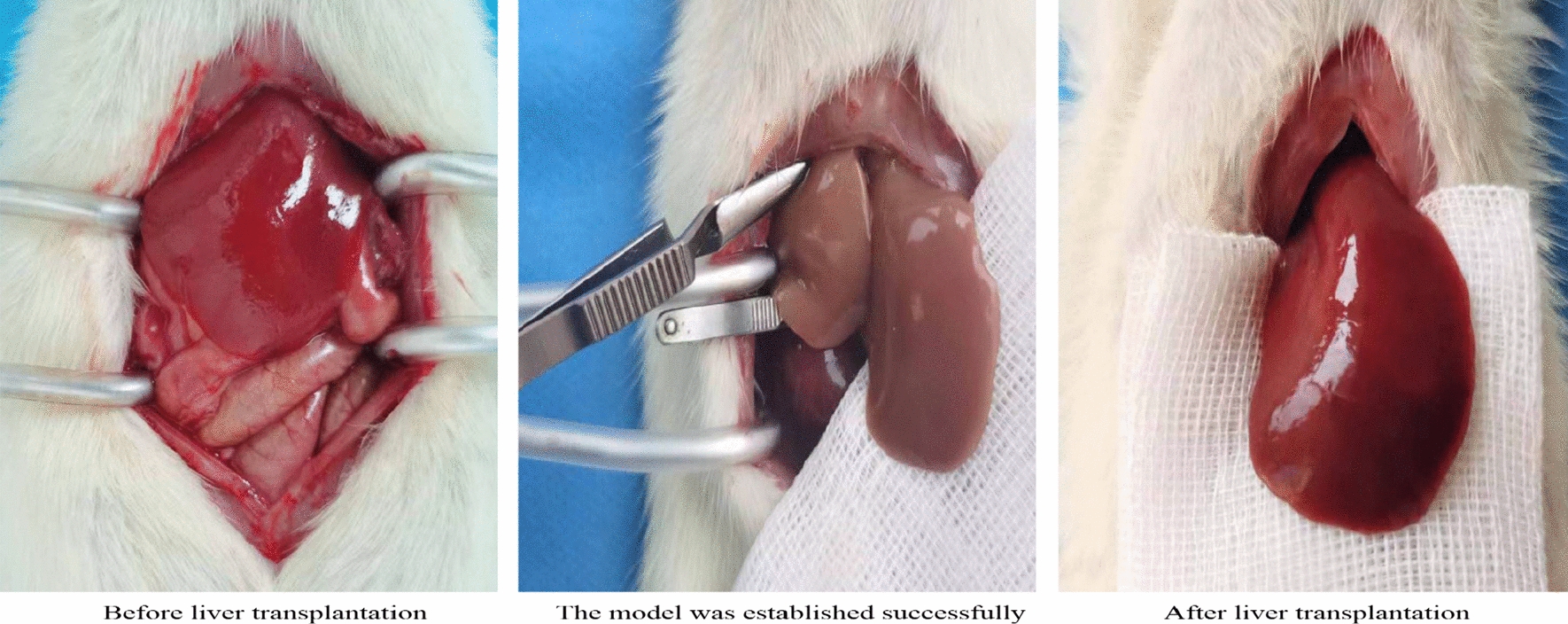
Model establishmentFor establishment of the AOLT model [8], The rats were fasted for 12 h before surgery and drank water freely, until 2 h before surgery. Rats were injected intraperitoneally with 3% sodium pentobarbital (0.2 ml/100 g) for anesthesia maintenance before surgery, and 0.005% fentanyl (0.16 ml/100 g) was injected intraperitoneally to relieve pain, After the supine position was taken and fixed, the femoral artery was punctured under skin incision, and the transducer was connected to continuously monitor the arterial blood pressure and observe the respiration of the rats. The abdomen depilates and prepares the skin, carries on the disinfection with the iodophor, spreads the aseptic hole towel, cuts along the abdominal midline. After laparotomy, the left triangular ligament and hepatic falciform ligament were dissociated, and the inferior hepatic vena cava (IHVC) and superior hepatic vena cava (SHVC) were carefully and bluntly separated, so that they were fully exposed. The hepatoduodenal ligament was cut open to separate the proper hepatic artery and hepatic portal vein (PV). IHVC, PV and proper hepatic artery were blocked with a non-invasive hemostatic clamp. Above the blocking site of PV, 1 ml of saline containing 30U heparin was infused after puncturing the scalp with a needle. The blood in the liver was allowed to enter the systemic circulation through SHVC, then the SHVC was blocked with a hemostatic clamp. After starting the anhepatic phase and time, normal saline was continuously infused at 4 ℃ through the PV. A small opening was cut above the blocking site of the IHVC as an outflow channel, and the fluid circulating in the liver discharged at a perfusion time of 30 ± 1 min. After perfusion, the liver gradually changed from dark red to khaki. After perfusion, the outflow tract in IHVC and the puncture hole in PV were sutured with 8–0 non-invasive vascular suture, The proper hepatic artery, PV, IHVC and SHVC were opened successively, and the vascular reflow was made to restore the liver color to bright red. After the operation, the abdominal cavity was rinsed with 36℃ normal saline and carefully sutured layer by layer (Fig. 1).
Fig. 1
Color changes of rat liver in three different periods
Specimen collection24 h after the end of ALOT in the rat, the rat was anesthetized again and the abdominal cavity opened. 2 ml of inferior vena cava blood was taken and placed in a BD tube. A high-speed centrifuge was used to prepare serum samples, and which were stored in a refrigerator at-80℃ for later use. After successful collection of blood from the inferior vena cava, 50 ml of heparinized physiological saline at 4 °C was slowly injected into the left ventricle of the rat, and a small opening was cut in the right atrial appendage to serve as the outflow tract. By fully perfusing heparinized physiological saline at 4 °C, residual blood in each organ was washed as thoroughly as possible, and then the liver tissue of the rat was taken out and stored in a refrigerator at-80° C for later use.
Details of testing indicatorsThe pathological changes of the liver of rats in each group were observed under high magnificationPartial rat liver tissue was fixed with 4% paraformaldehyde solution for over 24 h. After embedding the liver tissue specimen in paraffin, the liver tissue specimen was sectioned (with a thickness of 5 μm) using a microtome and gradually stained with H&E. The pathological changes of liver tissue sections were observed under a high-power light microscope (EVOS M5000, Thermo Fisher Scientific, USA) (× 200). The operation was carried out by two pathology professionals from the Department of Pathology of the Affiliated Hospital of Guilin Medical University.
Detection of serum ALT and AST levels in ratsThe serum was thawed and the levels of AST and ALT in serum of mice were detected by the AST and ALT detection kits, respectively, according to the instructions of the kit.
Determination of serum IL-6 levels in ratsIL-6 ELISA KIT (item number: SEKR-0005, Beijing Solebo Biotechnology Co., Ltd.) was used to detect the level of inflammatory cytokines (serum IL-6) in rat serum.
Analysis of MDA content in liver tissue and serumHe maximum absorption peak was at 532 nm, and the peroxide product MDA combined with TBA to form a red product. An instrument was used to measure the OD value of the sample, and then the MDA content calculated. The degree of lipid oxidation was assessed by determining the MDA concentration. In short, 0.1 g of rat liver tissue was collected, and the homogenate was prepared and centrifuged at 8000 g at 4 °C for 10 min. The supernatant was collected and placed on crushed ice for testing. After centrifuging the anticoagulated blood, the upper serum was collected, and the MDA content in liver tissue and serum was measured using a lipid diamine peroxide content detection kit (catalog no.: BC0025, Beijing Solarbio Science & Technology Co., Ltd.).
Determination of SOD activitySuperoxide dismutase (SOD) is the main enzyme of antioxidation in the body, which can protect the cells from oxidative damage by scavenging ·O2–. The instructions of the SOD activity test kit (item number: BC0170, Beijing Solebao Biotechnology Co., Ltd.) was strictly followed for testing.
Determination of Fe 2+content in liver tissue0.1 g of liver tissue and 1 ml of extract were mixed and homogenized in an ice bath, centrifuged at 4000 g for 10 min at 4 °C and the supernatant collected. The Fe2+ content in the supernatant of liver tissue samples was detected using a tissue iron content detection kit (Beijing Solarbio Science & Technology Co., Ltd., catalog number: BC4355).
GPX4 activity in liver tissue0.1 g of rat liver tissue stored at -80° C was completely ground using a fully automatic rapid sample grinder, and 1 ml of extract was added to make tissue homogenate. After centrifugation, the supernatant was collected and stored at low temperature for later use. The instructions of the GPX4 detection kit (catalog no.: K003083P Beijing Solarbio Science & Technology Co., Ltd.) were strictly followed to detect the GPX4 content in liver tissue. Then, the OD value of the sample was obtained at a wavelength of 450 nm, and the results were analyzed to calculate the GPX4 content.
The GSH content in liver tissue (1)0.1 g of frozen liver tissue was weighed and fully ground using an automatic sample rapid grinder (Shanghai Jingxin, China), and 1 ml of extract was added to prepare liver tissue homogenate. The samples were then centrifuged at 8000 g for 10 min at 4℃; the supernatant was taken and stored at 4 °C for subsequent analysis.
(2)Anticoagulated blood was centrifuged at 600 g at 4 °C for 10 min. The upper plasma was transferred to another PE tube and centrifuged at 8000 g for 10 min at 4 °C. The supernatant was transferred to a new tube and stored at 4 °C. GSH content in liver tissue and serum was detected using reduced GSH content detection kit (catalog no.: BC1175, Beijing Solarbio Science & Technology Co., Ltd.).
Statistical analysisA database was established and SPSS26.0 used for statistical analysis. Data conforming a normal distribution are expressed as mean and standard deviation(‾x ± s). Repeated measures analysis of variance was used to compare differences between groups, and then t tests were used to compare differences between the two groups. P < 0.05 was considered statistically significant.
Comments (0)