Details of materials and methods see supplementary material.
Test material
Test material consisted of gas exchange fibres (GF) and heat exchange membranes (HE) from various commercially available ECMO oxygenators with different non-thrombotic surface coatings [PLS, Bioline® coating (Getinge, Goteborg, Sweden); Hilite 7000LT, X.ELLENCE® coating (Fresenius, Bad Homburg, Germany); Nautilus, Balance® coating (Medtronic, Dublin, Ireland); EOS, PH.I.S.I.O® coating (LivaNova, London, UK); Table S1]. Uncoated-GFs membrane mats (reference material) were kindly provided by Getinge. Uncoated-GFs and GFs from all oxygenator types consisted of PMP. HEs were made of different material (Table S1). One naïve oxygenator of each manufacturer was analyzed.
For preparation of the test material a band saw was used to open the oxygenator housing. The GF/HE membrane core was sterile-packed. GF and HE membrane mats were separated without local assignment, cut to size (“samples”, length x width, 25 mm x 5 mm, 10 fibres connected via wrap threads), fixed in sterilized stainless steel inserts and placed in sterile Petri dishes (diameter, 35 mm; Thermo Fischer, Waltham, Massachusetts, USA) (Figure S1). The samples were coated with 3 mL HTP buffer + 30% AB serum (12 h, gentle movement, room temperature), dried, and used for PMN adhesion/activation within 24 h.
Poly-L-lysine-coated (0.01%, Sigma-Aldrich, St. Louis, USA) glass slides served as control samples to prove PMN adhesion and induction of NETosis.
Isolation of granulocytes
Granulocytes were isolated from lithium-heparin (16 IU/mL) anticoagulated blood samples of healthy volunteers (n = 6) using double density gradient centrifugation. Isolated PMNs from each volunteer were incubated with all test materials (one sample from each oxygenator type) in six independent experiments. Blood donors gave informed consent for inclusion before participation in the study. The study protocol was approved by the Ethics Committee of the University of Regensburg (vote no. 16-101-0322).
The cell density was counted using an analysis system (CASY, OMNI Life Science, Bremen, Germany) and adjusted to 2.5 * 105 cells / mL with the resuspension buffer.
For each oxygenator type, a negative control (adhesion of granulocytes and incubation in buffer) and a positive control (adhesion of granulocytes and incubation in 200 nM phorbol-12-myristate-13-acetate (PMA)) were always analysed to identify NETotic nuclei [16].
Contact of granulocytes with test materials
Dried samples were wetted with HTP-BSA-Ca/Mg (3 mL, 10 min, RT). After aspiration, 1 mL of freshly HTP-BSA-Ca/Mg was transferred below the clamped samples and 2 mL of the cell suspension was deposited on top of the samples. Petri dishes were capped and incubation was carried out for 3 h at 37 °C with gentle movement (shaking incubator, Kuhner Kelvin, Basel, Switzerland). The surfaces of the samples were analyzed for granulocyte adhesion and NET formation. The supernatants were collected to detect material-induced cell activation via flow cytometry (FACS).
Granulocyte adhesion and NET formation: immunolabeling
After washing twice with HTP buffer, the samples were stained with 4′,6-diamidino-2-phenylindole (DAPI, 500 ng/mL) (60 min, RT, dark), washed with 2 mL HTP buffer and covered with Fluoromount-GTM mounting medium (Thermo Fisher). Adherent cells were fixed with paraformaldehyde (4% in PBS, 3 mL, 10 min, room temperature). The samples were located between two coverslips and cured in the refrigerator for 3 days. DAPI-stained nuclei were visualized using a fluorescence microscope (BZ-8100E Keyence, Neu-Isenburg, Germany) with an integrated camera system (magnification, 16x to 80x).
Adherent granulocytes (on glass slides and GF samples) were stimulated with PMA (200 nM) for 30 min after a 3 h adhesion phase (positive control). Similar incubation with HTP-BSA-Ca/Mg was used as a negative control.
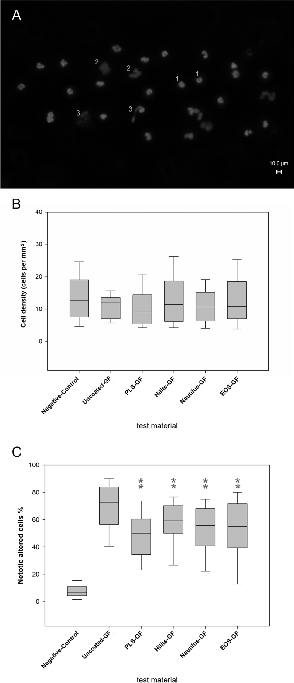
DAPI-staining allowed the quantification of cell density and the differentiation of nuclear morphologies to identify NETs [16]. Granulocytes without morphological changes in the form of nuclear deformations or protrusions are termed “normal”. Swollen nuclei and ruptured nuclei with ejected DNA were identified as NETs. A cell was considered swollen if its original outline was clearly deformed, with expansions and bulges that were more than half the size of the nucleus. The ratio of NETotic nuclei relative to the total cell number was examined.
ImageJ, an open platform for scientific image analysis was used for processing and analysing the fluorescence microscope images. The cell count was determined for each sample (detection area, 4.1 mm2; magnification, 40x) using the previously validated (Figure S2) cell automatically counting function (Figure S3). Ten randomly selected non-overlapping microscopic images from each sample (GF surfaces without wrap threads) were evaluated. In total, 300 microscopic images (10 images/sample, 6 blood donors, 5 different GF-coatings) were used for detection of cell counts. Additional 180 microscopic images (6 images/sample, 6 blood donors, 5 different GF-coatings) were recorded (magnification, 80x) and NETotic nuclei were manually counted by an experienced specialist. The ratio of NETotic nuclei (Figure S4) relative to the automatically detected cell count was determined per defined area of interest (2.05 mm2; Figure S4A–E).
NET degradation using DNAse treatment
DNAse is essential for NET degradation and was used to identify NETs under in vitro conditions [17]. Therefore, PMA-stimulated (and non-stimulated) granulocytes (seeded on poly-L-lysine-coated glass slides) were treated with DNase-1 (deoxyribonuclease I, Roche, Merck, Darmstadt, Germany) (10 µg/mL, in HTP-BSA-Ca/Mg). The removal of extracellular NET-structures and the retention of non-altered nuclei indicated the existence of NETs.
Material-induced activation of granulocytes: FACS analysis
Non-adherent cells in the supernatants were collected to detect material-induced cell activation via flow cytometry (FACS). This included the formation of ROS (reactive oxygen species) (oxidative burst) and the expression of activation markers (CD11b, CD62L (L-Selectin)). Details of sample preparation and gating strategy see Figure S5 and supplementary material.
Statistical analysis
The SigmaPlot 13.0 software programme (Systat, Erkrath) was used for statistical analysis of the experiments. The quantitative comparison of the neutrophil accumulations and the NETotic changes on the differently coated ECMO membranes was carried out using one-factorial ANOVA due to the non-normally distributed values. Statistical significance was assumed for a p value < 0.05.

Comments (0)