
Remember me
Mitochondrial-derived peptides (MDPs) are bioactive micro peptides encoded by short-open reading frames (sORF) in mitochondrial DNA and are known to exert myriad metabolic and cytoprotective effects [1]. Among the eight known members of MDPs, the mitochondrial open reading frame of the 12S rRNA-c (MOTS-c) is a 16 amino acid long peptide well known to increase exercise performance, improve insulin sensitivity, and reduce obesity [2, 3]. It is induced in response to exercise and metabolic stress and travels to the nucleus in a retrograde fashion to control the gene expression of many stress-responsive and anti-oxidant-responsive genes [3]. While the exercise benefits of MOTS-c peptide made it to the list of prohibited substances of the World Anti-Doping Agency (WADA), its metabolic benefits have been efficiently harnessed for therapeutic benefits in various metabolic and age-related diseases [4]. It is observed that exercise can induce the release of endogenous MOTS-c, chronic stress, and aging negatively influence the physiological levels of MOTS-c, making its external supplementation a potential therapeutic strategy for many cardiovascular diseases [4].
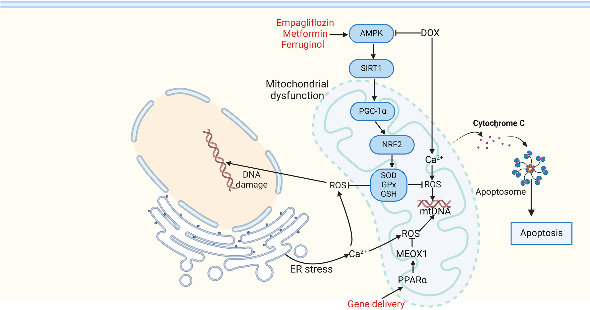
Diabetic cardiomyopathy is a known complication of diabetes characterized by dysregulated metabolism and impaired mitochondrial function. Since MOTS-c is known to promote glucose metabolism and mitochondrial function, it has been tested for its therapeutic utility in animal models of diabetic cardiomyopathy and found to exert cardio protection [5, 6]. While most of these studies were done in type 2 diabetes models, N Wu et al. recently examined the therapeutic effects of MOTS-c peptide in an animal model of type I diabetes-induced cardiomyopathy [7]. MOTS-c peptide was exogenously administered to streptozotocin (STZ)-induced type I diabetic mice for 3 months, followed by cardiac functional, histological, and molecular assessments. Systolic functional measures such as left ventricular ejection fraction (LVEF) and left ventricular fractional shortening (LVFS) were reduced in diabetic mice but were significantly improved by MOTS-c administration [7]. Similarly, while there are elevated cardiac chamber dimensions, left ventricular internal diameter at systole and diastole (LVIDs, LVIDd) in diabetic mice, these changes were attenuated by the MOTS-c administration. Further, MOTS-c also attenuated cardiac hypertrophy, fibrosis, and apoptosis in the diabetic mouse hearts as assessed by wheat germ agglutinin assay (WGA) staining, Picrosirius red and terminal deoxynucleotidyl transferase dUTP nick end labeling (TUNEL) staining, respectively [7]. At the molecular level, Wu et al. have identified significant activation of adenosine monophosphate-activated protein kinase (AMPK) (phosphorylation at Thr172) and reduction in myocardial inflammation (tumor necrosis factor-α, Interleukin-1β) by MOTS-c in diabetic mouse hearts, explaining its beneficial outcomes [7]. Although the findings of this study are of broad interest to the cardiovascular community, some concerns/limitations warrant further discussion (as highlighted in Fig. 1).
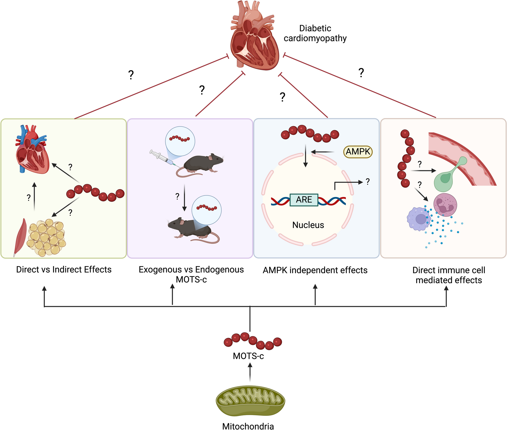
Fig. 1
Graphical summary of research gaps in the MOTS-c effects in diabetic cardiomyopathy. Abbreviations: AMPK adenosine monophosphate kinase, ARE antioxidant response element, MOTS-c mitochondrial open reading frame of the 12S rRNA-c. Created in BioRender. Yerra, V. (2025) https://BioRender.com/e90y553
The authors measured the terminal fasting blood glucose levels, HBA1c, and plasma insulin levels but reported no differences between the MOTS-c-administered mice and diabetic mice. They concluded that the cardio protective effects observed in their study are not secondary to the glucose level regulation [7]. Given the established effects of MOTS-c on skeletal muscle glucose transporter 4 (GLUT4) upregulation and improved muscle sensitivity for glucose uptake and metabolism [3], the lack of changes in the glycemic parameters is perplexing. However, since diabetic mouse suffers from significant sarcopenia and muscle atrophy, whether lack of skeletal muscle mass nullified the systemic effects of MOTS-c on glucose metabolism is worth investigating for future studies. Also, previous studies have reported the opposite influence of STZ-induced diabetes and MOTS-c on muscle myostatin and muscle growth [8, 9]. Hence, a more comprehensive study needs to be conducted to examine the influence of MOTS-c on these parameters (muscle weight and myostatin expression) when STZ is co-administered to better understand the systemic influences of MOTS-c and their indirect influence on beneficial cardiac outcomes.
Another facet missing in the study by N Wu et al. is the failure to distinguish whether the observed effects are mediated by exogenously administered MOTS-c or endogenous MOTS-c stimulated in response to the exogenous MOTS-c. Previous studies have characterized that the latter case is possible, especially in the myocardium [10], and hence, it is not known if there is myocardial induction of MOTS-c in response to the exogenously administered MOTS-c or if the exogenously administered MOTS-c traversed through the systemic circulation into the myocardium. The mechanisms governing the uptake of exogenous MOTS-c through the plasma membrane are not very well characterized. Future studies should answer this conundrum by identifying the exact protein transporters responsible for the uptake of exogenous MOTS-c, using an inhibitor of the corresponding transporters, and studying the biological effects of exogenous MOTS-c.
Also in the current study, the authors have attributed the beneficial cardiac outcomes of MOTS-c to AMPK activation. AMPK activation may be critical for the nuclear translocation of MOTS-c and some of the metabolic effects of MOTS-c [1, 3]; the actual gene targets that the MOTS-c administration might influence may involve many other genes apart from the AMPK activation. For instance, studies in diet-induced animal models of diabetes have previously reported other targets such as nuclear factor E2-related factor 2 (Nrf2), thioredoxin-interacting protein (TXNIP), and nucleotide-binding oligomerization domain-like receptor protein 3 (NLRP3)13, neuregulin1 (NRG1) signalling [5], critical for cardio protection. Hence, the current study could have benefited from an unbiased sequencing approach in elucidating more specific molecular targets regulated by MOTS-c, particularly in myocardial dysfunction in diabetes. Moreover, N Wu et al. have not confirmed AMPK dependency of MOTS-c peptide effects by using AMPK siRNA or AMPK inhibitor (Compound C). Given the cardio toxic effects of AMPK knockout mice, conducting AMPK inhibition experiments under in vitro conditions to test the anti-hypertrophic, anti-fibrotic effects of MOTS-c will provide helpful information on whether the complete spectrum of MOTS-c benefits are exclusively mediated through AMPK and or other targets. Also, AMPK activation by MOTS-c involves inhibition of the folate-methionine cycle and endogenous accumulation of 5-aminoimidazole-4-carboxamide ribonucleotide (AICAR) [3]. Although AICAR is well-known for AMPK activation, several AMPK-independent effects of AICAR are also appreciated [11], and hence, it is important to understand the actual molecular targets of MOTS-c apart from AMPK activation.
Another interesting aspect missing from the study by N Wu et al. is whether the reduction in the inflammatory mediators is a direct effect of MOTS-c on the immune cells or AMPK-mediated indirect effect. Previous studies have identified that MOTS-c treatment facilitated the anti-inflammatory activity of macrophages by negatively regulating mitogen-activated protein kinases (MAPKs) and facilitating the aryl hydrocarbon receptor (AhR) and signal transducer and activator of transcriptional 3 (STAT3) signalling [12]. Since diabetic cardiomyopathy is also characterized by an abnormal immune response and accumulation of immune cells, it is important to distinguish the immune cell-specific effects of MOTS-c in diabetic cardiomyopathy by using immune cell-specific AMPK knockout mouse strains.
Barring the enlisted limitations, the study by N Wu et al. has laid a strong foundation along with previously reported studies of MOTS-c protective effects on diabetic cardiomyopathy. MOTS-c joins the elite list of AMPK-modulating biological molecules (Fibroblast growth factor 21 (fgf21), adiponectin, and irisin), which have also proved to be beneficial against diabetes-related myocardial dysfunction. Expanding the knowledge on this preliminary evidence and our understanding is important to gain better molecular insights and facilitate the clinical translation of MOTS-c and other AMPK-activating molecules to manage diabetic cardiomyopathy efficiently.
Comments (0)