
Remember me


For this narrative review we conducted an extensive literature search up to July 2024 in PubMed and Embase using the terms ((cerebellar injury) AND ((neonate) OR (preterm))); ((cerebellar hypoplasia) AND ((neonate) OR (preterm))) resulting in 1058 studies after removing 240 duplicates. After abstract and full text screening and adding studies from cross-references and peer review process, 111 original studies or meta-analyses have been included in this study.
Imaging Cerebellar Development and InjuryPremature birth, along with associated environmental and intrinsic factors, can disrupt the rapid and intricate development of the cerebellum. Recognizing impaired or altered cerebellar development is important, as it has implications for neurodevelopmental outcome. To detect injury and/or dysmaturation, early neuroimaging of the cerebellum in preterm infants is of particular importance, especially in the most immature infants. Ultrasound is the primary neuroimaging technique to monitor cerebellar growth and maturation because it is safe, can be performed bedside at any time and allows serial scans with minimal stress to the infant [21,22,23]. To properly examine the cerebellum, additional ultrasound windows should be included in routine scans. Scanning through the mastoid fontanel provides more reliable measurements of the transverse cerebellar diameter (TCD) and better detects cerebellar injury compared to scanning only through the anterior fontanel. This method should be the standard for assessing the cerebellum (Fig. 4) [24,25,26,27]. In very low birthweight infants, transnuchal ultrasound via the foramen magnum is feasible and offers the advantage of direct comparison of both cerebellar hemispheres at close range with a high-resolution linear probe; this helps to detect smaller cerebellar lesion and asymmetry (Fig. 5) [23]. Several studies have been published on nomograms for measurements of the TCD and vermis height and diameter [23, 28, 29].
Fig. 4
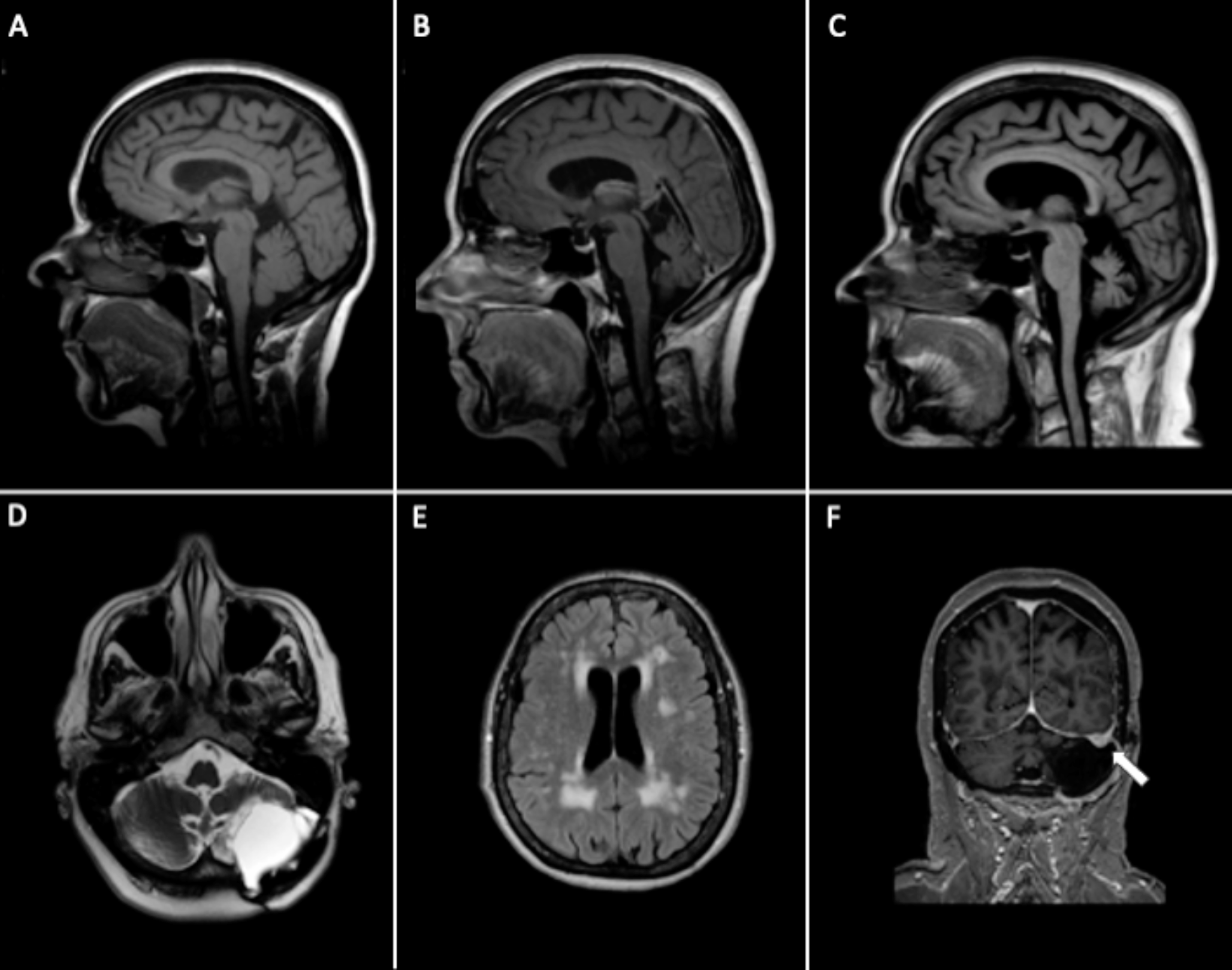
Unimpaired cerebellar growth in a preterm infant of 25 weeks gestational age. Serial coronal ultrasound scans (A-E) through the mastoid fontanel show a normal development with proceeding foliation representing the rapid increase in cerebellar surface. (F) T2 MRI scan at postmenstrual age of 33 weeks results in same transverse cerebellar diameter compared to corresponding ultrasound scan (D)
Fig. 5
Cerebellar hypoplasia of prematurity: Preterm infant of 25 4/7 weeks gestation age without supra- or infratentorial hemorrhage. Upper row shows serial transnuchal ultrasound scans, the lower row corresponding axial scans through the mastoid fontanel at day of life (DOL) 7 and 27, approximately postmenstrual age (PMA) 36 weeks and term equivalent age (TEA). A normal transverse cerebellar diameter (TCD) of 25-50th percentile according to Imamoglu et al. [29] was present at first scan with one week. Even without recognizable supra- or infratentorial hemorrhage in serial ultrasound scans a mild cerebellar hypoplasia developed during the unremarkable neonatal course with a TCD < 3rd percentile at PMA 36 weeks and TEA
MRI studies offer valuable insights into cerebellar development, connectivity, and the impact of premature birth. However, MRI is resource-intensive, and transporting infants to the scanner can be stressful and is restricted to those in stable condition. As a result, clinical MRI scans are typically performed only at term-equivalent age, with serial scans largely confined to specially designed research studies [10, 30,31,32]. Nonetheless, MRI has proven superior for the detection of cerebellar lesions compared to ultrasound, especially punctuate cerebellar hemorrhages (CBH), smaller than 4 mm, using susceptibility weighed imaging [27, 33, 34]. Furthermore, MRI offers several other advantages as volumetric measurements, parcellation of the different lobules, examination of cerebellar nuclei, and diffusion tensor imaging (DTI) to assess cerebellar white matter integrity and tractography [10, 30, 31, 35, 36]. Therefore, MRI and ultrasound should be seen as complementary neuroimaging modalities.
Pathophysiological Mechanisms of Disrupted Cerebellar DevelopmentDirect injury: Cerebellar Hemorrhage (CBH) (Figs. 6 and 7)Fig. 6
Limited cerebellar hemorrhage (CBH) left posterior-lateral hemisphere in a preterm infant of 29 weeks gestational age. A Coronal ultrasound scan through the mastoid fontanel, B corresponding transnuchal ultrasound scan on the fourth day of life shows the limited CBH (< 1/3 of the left hemisphere). C On the follow-up scan at postmenstrual age (PMA) of 34 5/7 weeks through the left mastoid fontanel, an asymmetrical appearance with smaller left hemisphere can be identified. D T2 weighted MRI scan at PMA 36 6/7 weeks and susceptibility weighted imaging (SWI) confirm the asymmetry with a hypoplastic left hemisphere and residual hemosiderin deposition after the CBH
Fig. 7
Massive cerebellar hemorrhage (CBH) of the right hemisphere in a preterm infant of 27 weeks gestational age. A Coronal scan via the anterior fontanel shows an increased echogenicity (CBH, arrow) in the right cerebellar hemisphere, which can be also seen in (B) the right paramedian sagittal scan (arrow). C the left cerebellar hemisphere appears normal (arrow). D Better visualization of the extend of the massive CBH (> 1/3 of the right hemisphere, arrowheads) via ultrasound scan through the mastoid fontanel with midline shift of the vermis and IVth ventricle. E Axial T2 MRI scan and paramedian sagittal T2 scans, F corresponding to (B) and (G) to (C), and susceptibility weighted imaging (H) proving the massive CBH of the right hemisphere
Infants at highest risk for CBH are often born extremely preterm (< 26–28 weeks) and are in overall worse general condition, e.g., high-frequency ventilation and inotropic support [37,38,39], therefore CBH is often accompanied by supratentorial hemorrhage [40, 41]. CBH occurs with an incidence of 10 to 38 percent [31, 42, 43]. Small punctate lesions represent around two thirds of CBHs while larger lesions are less common [44]. CBHs are localized most often in the inferior aspect of the posterior lobe as indicated by two MRI studies and a neuropathologic study [31, 41, 43]. The latter study of Haines et al. revealed that the origin of CBH in contrast to supratentorial hemorrhages is usually not the germinal matrix in the ventricular zone or external granular layer (EGL) but the paucicellular internal granular layer (IGL) where rapid vessel growth might cause an increased susceptibility for fluctuations in blood flow or blood pressure leading to rupture of these vessels [41]. Furthermore, histology showed that CBHs are often surrounded by smaller satellite hemorrhages and with evidence for acute and subacute changes which may indicate not only multilocal but also multitemporal onset of CBH [41]. However, as the results of this study are from postmortem investigations, this has to be interpreted with caution as postmortem changes on histologic level might have occurred.
Cerebellar growth and neurodevelopmental outcome are both negatively associated with the size of cerebellar lesions [31, 43,44,45] and therefore it is reasonable to use a classification system with respect to the size of the primary lesion [44]. The classification used by Boswinkel et al. described massive CBH as hemorrhage affecting more than 1/3 of the respective hemisphere; limited CBH affect less than 1/3 of the hemisphere but are still larger than 4 mm [44]. These massive or limited CBH are usually well detectable on cranial ultrasound via the mastoid fontanel [27, 33]. The punctate cerebellar lesions, less than 4 mm in size, usually require MRI e.g., with susceptibility weighed imaging (SWI) sequences, to be recognized [27, 33]. Neurodevelopmental outcome of patients with punctuate lesions was reported not to be different to infants without CBH in two studies [46, 47], whereas one study found increased odds for worse motor outcome [42] and another study impaired motor function, visuomotor integration and full scale IQ [43]. The CHOPin study compared infants with different sizes of CBH; infants with massive CBH had the worst outcomes even in absence of supratentorial injury [44]. Although not significant, rates of abnormal outcome were higher in infants with bilateral limited or massive CBH or if the vermis was involved. However, infants with limited CBH and punctate CBH had similar outcomes in this study [44]. Apart from the size of the hemorrhage, a separate notion should be made on the number of lesions, laterality, and whether the vermis is affected or not [44, 45, 48]. A sophisticated analysis of size and localization of cerebellar hemorrhages and associated outcomes has been performed by Garfinkle et al. [43]. Of 234 preterm infants born 24 to 32 weeks 36 (15.4%) had CBH, most of them in the inferior posterior lobe. Intriguingly no association of CBH and cognitive function could be shown in this study, but CBH size was independently associated with adverse motor outcomes, visuomotor integration and behavioral outcomes, e.g. externalizing and internalizing behavior. The authors calculated a voxel-wise probabilistic map of CBH and the associated outcomes showing that lesions extending more superiorly and affecting deeper aspects of the cerebellum were more likely to cause adverse outcomes compared to those affecting more superficial parts of inferior regions of the cerebellum [43].
A retrospective study assessed the impact of CBH on neurodevelopmental outcomes in preterm infants < 32 weeks with isolated CBH (identified on ultrasound, confirmed by MRI, n = 35) compared to age-matched controls (n = 35) and infants with CBH plus supratentorial parenchymal injury (n = 16) [48]. Infants with isolated CBH had lower scores for gross- and fine motor function, visual reception, language development, early learning composite as well as adverse behavioral outcomes and higher rate of positive screening for autism spectrum disorders [48]. Additional supratentorial parenchymal injury resulted in worse gross motor function but similar results in all the other evaluated domains. Bilateral CBH and CBH with vermis involvement were significantly associated with more profound disability [48]. Hortensius et al. performed a systematic review of eight studies on infants with isolated cerebellar injury (including the previous mentioned study) to evaluate the effect of isolated CBH on cognitive, motor, language and behavioral outcome [45]. Neurodevelopmental outcome of infants with punctate CBH was comparable to infants without brain injury. However, infants with larger CBH ≥ 4 mm had significantly worse cognitive outcomes (41% vs. 13% for punctate CBH) and motor outcomes (43% vs. 7%) and a severe combined impairment in 46–82%. Vermis involvement resulted in an even worse rate of severe combined impairment (87–93%) [45]. Furthermore, CBH including the vermis had a major impact on behavioral outcomes with severe behavioral impairment in 87% [45].
DTI studies showed the effect of CBH on several white matter tracts reflecting disrupted microstructure in the centrum semiovale, corpus callosum, posterior limb of the internal capsule and the superior and middle cerebellar peduncles [49]. These changes are due to remote transsynaptic effects of CBH on the cerebellar circuitry, leading to the so called crossed cerebello-cerebral diaschisis with to smaller contralateral regional cerebral cortex volumes [50] and retrograde degeneration of the pons [51]. These remote effects will be discussed in detail below.
A study using proton magnetic spectroscopy analyzed metabolite concentrations in the cerebellum on n = 53 very preterm infants [52]. Those with moderate-to-severe cerebellar injury had lower concentrations of N-acetylaspartate (NAA), choline (Cho) and creatine (Cr) compared to those with no or only mild cerebellar injury likely reflecting the neuronal loss [52]. Further studies on the metabolic profile may provide more insight, whether this advanced neuroimaging technique could be used as prognostic tool for the long-term outcome.
The presented studies clearly demonstrate the importance of direct cerebellar injury for further development, the necessity to scan the cerebellum and to provide long-term follow-up for those with cerebellar lesions.
Indirect Injury: Cerebellar UnderdevelopmentIndirect injury due to prematurity itself and its consequences can cause a disrupted cerebellar development or “cerebellar hypoplasia of prematurity” [8] (Fig. 5). The following sections summarize the current literature on the respective influencing factors.
PrematurityImmunohistochemical analysis in an animal model with moderate preterm (equal to 32 weeks in humans) compared to term pigs showed a reduction in granule cell precursors (GCP) and Bergmann glia fibers just by exposure to extrauterine environment [53]. The resulting reduced proliferation but normal differentiation of GCP caused a reduction of mature granule cells (GC) in the IGL [53]. A histopathologic study compared cerebella of preterm infants who died in the neonatal period and still born infants as age-matched “controls” [54]. Comparable to the animal study, deceased preterm infants had a reduced thickness of the EGL with a lower proliferation rate of GCP and decreased sonic-hedgehog (SHH) expression and also decreased Bergmann glia fibers. Maturation of Purkinje cells (PC) or thickness of the molecular layer were not affected by extrauterine environment, but the EGL and IGL showed an increased packing density of the cells [54]. These studies provide immunohistochemical evidence, that the complex programming of cerebellar development might be altered by exposure to the extrauterine environment. However, difficulties in translation of animal models to human (patho-)physiology must be considered.
A resting state functional MRI study of Herzmann et al. showed that corticocerebellar connectivity is already well established at term equivalent age, as well as the circuitry to subcortical or intrinsic networks [55]. Interestingly, infants show more functional connectivity structure within the cerebellum compared to adults, but to a lesser magnitude in preterm infants than in term-born infants [55].
A recent study by Basu et al. compared GABA-edited spectroscopy (MEscher-GArwood Point Resolved Spectroscopy, MEGA-PRESS) in 75 preterm infants without moderate-to-severe brain injury vs. 48 controls to analyze neurometabolic profile in cerebellum, basal ganglia and frontal lobe [56]. Lower concentrations of GABA + , glutamate and NAA in all three regions in moderate preterm infants might indicate a delayed or altered brain maturation due to stressors in the extrauterine environment [56]. However, as GABA + and glutamate concentrations in extremely preterm infants were not different from term infants, the underlying mechanism remains unclear and warrants further studies [56].
Hemosiderin or Blood Products (Fig. 8)The outer part of the EGL, where proliferation of the GCP takes place, is dir
Comments (0)