The study showed whether changing the irradiation method would have a bacteriostatic effect when implanted in a CV port. The experiment showed that 30- and 60-min irradiation of 680 nm wavelength red light showed significant inhibition of MRSA growth. Total ED for them was 340 J and 680 J. Lipovsky et al. have reported a power density of 400 mW/cm2, and a total energy dose of 120 J/cm2, which is much greater than this experiment. Sousa et al. [5] reported the growth inhibition of S. aureus was observed at 660 nm by irradiating more than 12 J/cm2 [5].
Sousa et al. achieved reduction of MRSA with irradiation doses ranging from 3 to 24 J/cm2 at 30 mW.
The observed irradiation dose per minute of this study was 14.21 W, while their study was 33.5 mW.
The need for equivalent or higher energy doses to achieve similar or better effects was anticipated. Experiments with such energy doses were planned and favorable results were obtained.
The most important difference between this experiment and theirs was the concentration of the light. The exposure area in Sousa’s [5] experiment was 0.02 cm2, which is quite concentrated beam irradiation; however, such irradiation is not realistic in the CVP chamber to suppress bacterial growth inside and outside. In this experiment, the light exposure area was large enough to cover all the bacterial suspension on it as nonconcentrated light irradiation was performed, which is not enabled by laser. The irradiation power in a unit area of this experiment was lower than that of Sousa et al. [5]; however, the bacterial growth suppression could be revealed in vitro; moreover, the temperature of the bacterial temperature was less than 37 °C, which is the temperature of the human deep body. The irradiation model in this experiment should be matched to a CVP that can induce PBM as the light from LEDs might diffusely irradiate in the chamber of the CVP, which cannot be achieved by the experimental modes of Sousa et al. Moreover, an implantable CVP with LEDs can be recognized when it is transduced via wireless energy transmission [10] as red light can penetrate into the dermis and epidermis more effectively compared with purple to yellow visible light. This novel CVP could contribute to medical safety in terms of antibacterial technology with PBM and avoid disjuncture via visualization. Sousa et al. achieved reduction of MRSA with irradiation doses ranging from 3 to 24 J/cm2 at 30 mW.
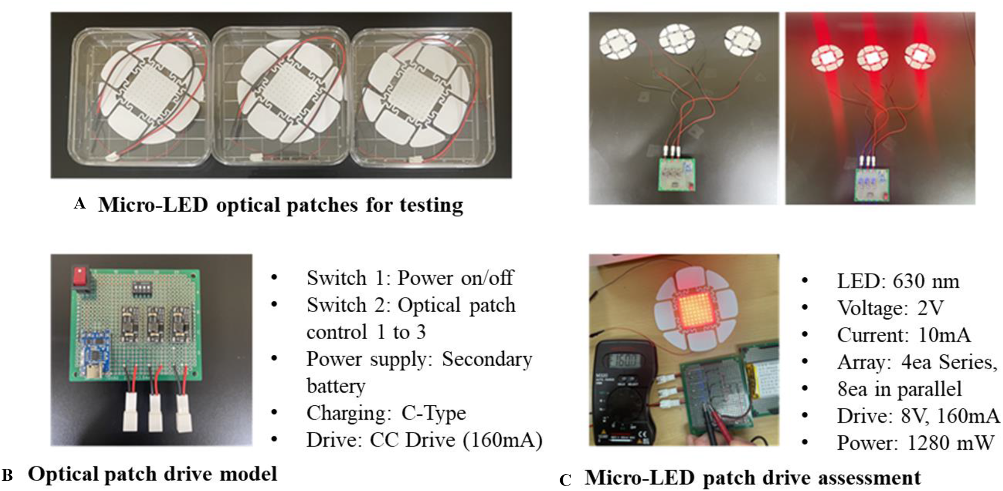
The wavelength selected in this study is based on the visibility of the CVP with LEDs under the skin. Compared with red light, other visible light wavelengths struggle to penetrate the skin. Previous reports indicate that light with a 660 nm wavelength can inhibit bacterial growth [11, 12]. It is suggested that the 680 nm wavelength falls within the permissible range of 660 nm. In this experiment, we used LEDs with a 680 nm wavelength, as this was the only LEDs suitable for embedding on the CVP produced by a 3D printer. Although there are reports that near-infrared light with wavelengths of 830–900 nm is more effective in suppressing bacterial growth [13], these wavelengths are not visible. Consequently, they are not applicable to this experiment, where the goal is to establish a visible and illuminated CVP even under the skin.
In addition, Ranjbar et al. reported the use of photobiomodulation therapy to surgical wounds in diabetic rats [16]. Implanting a CV port may be a potential treatment for SSI (surgical site infection).
PBM is a relatively new phenomenon, thus it is unclear what type of gene was expressed, how the intracellular signal system was activated, or why the microbe was prevented from growing. Comparing the overall quantity of irradiation dosage was assumed to be required, but that the irradiation dose rate should also be evaluated. Given irradiation dose was 14.21 mW, the irradiation dose of Sousa’s report was almost twice power [5]. This could be the difference in the total dose required to limit microbial growth. Various devices and reported targets in PBM include the following: a 410 nm LEDs effective against Enterococcus faecalis, MRSA, and Prevotella intermedia [14]; a 465 nm LEDs effective against Staphylococcus pseudintermedius or methicillin-resistant S. pseudintemedius [15], a 660 nm laser effective against Staphylococcus aureus [12], a 685 nm laser effective against S. aureus [16]; and 630, 660, 810, and 995 nm lasers effective against Pseudomonas aeruginosa, Escherichia coli, and S. aureus [17]. Walski et al. reported that near-infrared (NIR) and red-to-near-infrared (R/NIR) radiation exposure in immune cells typically involves the production of reactive oxygen species, nitrogen oxide, or interleukins. Several immune cell studies have demonstrated that exposure to R/NIR has an anti-inflammatory effect [4]. However, the results suggest that 680 nm wavelength light can inhibit the growth of S. aureus even without the immune system, as previously reported [5, 8, 16]. A 680 nm LEDs might be favorable for embedding on a CVP at risk of infection and beyond the reach of the immune system. Differences in transparency or absorption may result in varying impacts on bacteriostatic effect; however, this remains uncertain.
Kushibiki et al. reported that intracellular chromophores possess the ability to excite oxygen through the electron transport chain in eukaryotic cells [18]. Although intracellular porphyrin may contribute to the production of reactive oxygen species (ROS), the low q band absorption of porphyrin implies that it is not the primary source of ROS. Consequently, it is crucial explore candidate chromophores that react to 680 nm light and can induce toxic effects associated with oxidative stress, ultimately leading to cell death [19].
Within the best scope, there was no report of determining PBM by irradiating MRSA with red light. Some papers reported the PBM to S. aureus in vitro or in an animal model [5, 8, 16, 20]; however, all of them did not determine the sensitivity to methicillin so whether it is MRSA or methicillin-sensitive S. aureus (MSSA) was not determined. The next step in this experiment was to compare how PBM works in MSSA and MRSA.
Limitation
There were several limitations in this study. Only MRSA was used in this study as it was aimed to simply check up on how the low dose of red light works in suppressing the growth of MRSA, which is the most important microorganism in CVAP-BSI. The size and the number of LEDs which is assumed to be embedded in a CVP were different from the LEDs system in this experiment. The next step is to develop a CVP with LEDs to test its ability to induce PBM by infusing the MRSA suspension in the chamber and irradiating it using LEDs. Further clinical trials would be conducted by observing the infection rate of CVP under the skin by emitting the LEDs in a certain frequency.
In addition, the suspension has been irradiated, but the colonies themselves have not been; further experiments may be needed in the future.


Comments (0)