
Remember me
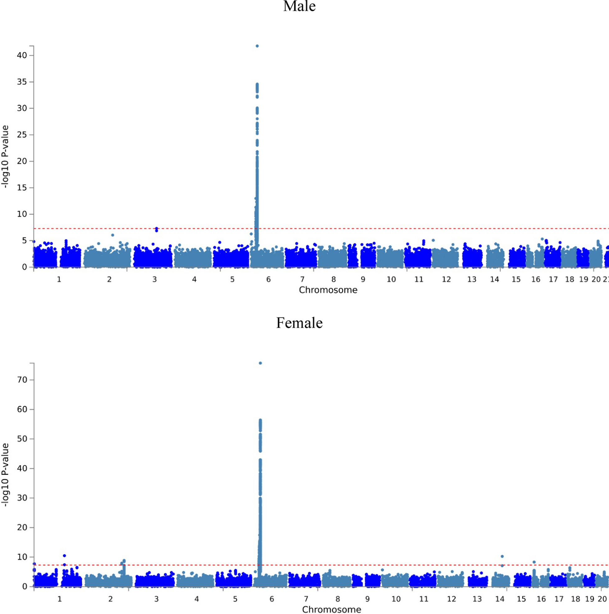


Table 1 showed the results of basic characteristics of participants among gender, HBV infection status and confounders. There were 23,178 men in the GWAS study, including 2656 HBV cases and 20,522 with no HBV infection subjects. Moreover, there were 48,874 women in the analysis which contained 4715 HBV cases and 44,159 healthy control subjects. HBV infection was significant between gender (P-value < 0.0001). All the covariates reached statistical significant difference between gender, including age (P-value = 0.0006), exercise (P-value < 0.0001), cigarette smoking (P-value < 0.0001), alcohol drinking (P-value < 0.0001), body mass index (P-value < 0.0001), hypertension (P-value < 0.0001), hyperlipidemia (P-value < 0.0001) and diabetes (P-value < 0.0001). In addition, all the variables were significant among genders with or without HBV infection. In age, mean age was 50.407 (± 0.079) years in male without HBV, 50.000 (± 0.194) years in male with HBV, 50.063 (± 0.050) years in female without HBV and 50.041 (± 0.136) years in female with HBV. The P-value of age was 0.0013. In exercise, a total of 11,651 (56.77%) no exercise and 8871 (43.23%) exercise in male without no HBV, 1613 (60.73%) no exercise and 1043 (39.27%) exercise in male with HBV. However, 26,665 (60.38%) no exercise and 17,494 (39.62%) exercise subjects were in female without HBV, 2885 (61.19%) no exercise and 1830 (38.81%) exercise in female with HBV. The P-value was < 0.0001.
Table 1 Basic characteristics of participants stratified by gender and HBV infection statusIn Table 2, genomic risk loci were different between men and women. In male, three risk loci (rs3732421, rs1884575 and Affx-28516147) were detected while eight risk loci (Affx-4564106, rs932745, rs7574865, rs34050244, rs77041685, rs107822, rs2296651 and rs12599402) were found in female.
Table 2 Genomic risk loci of HBV infection in men and womenIn Table 3, the differences in SNPs genotypes stratified by gender and HBV infection were presented. There were 11 SNPs significant among males or females in HBV infection in GWAS analysis. In rs3732421, there were 9217 (44.95%) AA, 8974 (43.76%) AG and 2314 (11.29%) in male without no HBV, 1048 (39.49%) AA, 1252 (47.17%) AG and 354 (13.34%) GG in male with HBV. Moreover, there were 19,644 (44.54%) AA, 19,595 (44.43%) AG and 4869 (11.04%) AG in females without HBV, 1984 (42.12%) AA, 2148 (45.61%) AG and 578 (12.27) GG in female with HBV. The P-value of the difference was < 0.0001. The only one SNP which was not significant was rs77041685 (P-value = 0.6100). There were 17,499 (85.33%) CC, 2894 (14.11%) CT and 115 (0.56%) TT in male without no HBV, 2254 (84.86%) CC, 387 (14.57%) CT and 15 (0.56%) in male with HBV. Moreover, there were 37,378 (84.71%) CC, 6479 (14.68%) CT and 268 (0.61%) in female without HBV and 3996 (84.84%) CC, 684 (14.52%) CT and 30 (0.64%) TT in female with HBV.
Table 3 Frequencies of HBV related SNPs stratified by gender and HBV infection statusIn Manhattan plot (Fig. 1), sex also presented different result. In females, the most significant SNPs gathered in chromosome 6. However, excepted for chromosome 6, significant HBV infection SNPs also could be found in chromosome 3 among males.
Fig. 1
Manhattan plot of HBV infection in men and women
Table 4 shows the results of gene to function analysis in FUMA and want to know the difference of reactome pre-Notch expression and processing between men and women. We found that POGLUT1 and HIST1H2BC only appeared in males but not in females.
Table 4 Result of Reactome pre notch expression and processing of HBV in men and women
Comments (0)