
Remember me
The evolution of breast cancer-related lymphedema (BCRL) management epitomizes decades of dedication, innovation, and persistence in improving patient outcomes. Initially emerging in surgical practice in the 1960s [1], early interventions for lymphedema were limited by technological constraints and lack of surgical experience, leading to inconsistent outcomes. However, these early endeavors formed the essential foundation from which contemporary lymphedema treatment has evolved dramatically, particularly in the past two decades.
Significant advancements began in the 1990s with vascularized lymph node transplantation (VLNT) [2,3,4], establishing a pathway toward reliable surgical success. The advent of supermicrosurgical techniques further refined lymphatic reconstruction, introducing procedures such as vascularized lymph vessel transplantation (VLVT) [5,6,7] and lymphaticovenular anastomosis (LVA) [8,9,10,11,12,13]. More recently, innovations such as lymph node-to-vein anastomosis (LNVA) [14, 15] and specialized debulking liposuction have expanded available treatment modalities, addressing both the fluid and solid disease components characteristic of BCRL.
These continuous improvements have sparked detailed evaluations of the optimal timing and combinations of interventions, fostering consensus and resolving many historical debates within the field. This paper presents a perspective informed by extensive clinical experience and ongoing research on the diagnosis, management, and future directions of BCRL treatment, integrating these innovations while highlighting the importance of understanding both primary and secondary lymphatic pathology.
Diagnostic ConsiderationsAccurate diagnosis of BCRL necessitates distinguishing between primary and secondary lymphatic conditions, recognizing their distinct management implications. Primary lymphedema is increasingly identified as systemic, often presenting asymptomatically until triggered by an acquired insult, such as axillary surgery or radiation in breast cancer patients. Comprehensive clinical evaluations and advanced imaging, specifically indocyanine green lymphography (ICGL), are utilized to detect subclinical primary dysfunction and accurately characterize lymphatic pathology [16]. This differentiation is vital to ensure tailored management, reducing potential donor-site complications associated with lymphatic tissue transfers [17,18,19,20].
Fluid Vs. Solid Disease ComponentsThe terminology “lymphedema” is somewhat misleading, as the condition encompasses more than just fluid accumulation. Chronic inflammation and disease progression drive pathological adipose tissue deposition and fibrosis, collectively referred to as solid components of the disease. Traditionally viewed as late-stage manifestations, these solid features are now understood to appear earlier in disease progression.
Clinically, patients describe solid-predominant disease as persistent swelling unresponsive to elevation or compression therapy. Physical examination of solid disease typically reveals edema-resistant tissue thickening, confirmed by imaging techniques such as ultrasound and MRI. Distinguishing fluid from solid components is crucial because fluid-predominant cases often respond effectively to conservative management or physiological procedures, defined as surgeries aimed at restoring or enhancing lymphatic drainage, such as VLNT, VLVT, LVA or LNVA, while solid-predominant cases usually require surgical debulking to achieve meaningful clinical improvement [21]. Most patients, however, exhibit mixed disease, necessitating an individualized approach to determine the most effective therapeutic strategy.
Diagnostic TestingA comprehensive diagnostic protocol incorporates several assessments:
Cleveland Clinic Lymphedema Questionnaire (CCLQ)Developed to address the limitations of traditional Likert-style questionnaires, particularly recall bias, this binary-response tool assesses eight key domains: symptoms, function, appearance, cognition, emotional well-being, social interactions, care effort, and overall quality of life. It is simple to administer in a clinical setting and provides reliable, actionable information for both baseline assessment and outcome tracking.
3-Dimensional Volumetric Scans (3DVS)Volume remains the most commonly used measure to assess lymphedema before and after treatment. The widely used circumference measurement using a tape measure is also the most unreliable, due to high inter- and intra-rater variability [22]. In contrast, 3DVS appears reliable due to the technology minimizing errors and offering precise volumetric data [23]. The interpretation, however, needs to account for possible fluctuation of volume due to multiple factors such as hydration status, weather temperature, diet, and use of compression garments.
Bioimpedance Spectroscopy (BIS)Bioimpedance spectroscopy is a quick, noninvasive method for assessing lymphedema by analyzing body water distribution. It works by sending a low-level electrical current through the body at various frequencies. At low frequencies, the current is unable to penetrate cell membranes, thus reflecting extracellular water (ECW) volume; at higher frequencies, the current passes through both extracellular and intracellular spaces, providing a measure of total body water (TBW). Since lymphedema is characterized by excess ECW accumulation, BIS is particularly well suited for its evaluation. By calculating the ECW-to-TBW ratio, BIS offers a reliable indicator of lymphedema severity. In addition, BIS measurements help monitor fluid burden over time and can be used to assess the effectiveness of compression therapy [24].
Indocyanine Green Lymphography (ICGL)ICGL is now the gold standard test for diagnosis, surgical planning, and disease/outcome tracking [25, 26]. When performed for diagnostic purposes, two scans are taken into consideration: the immediate post-injection scan (IS) and the delayed scan (DS) at plateau. The IS captures the anatomy, the abnormal compensatory pathways, and the pump velocity. The DS addresses the disease severity, the efficacy of treatment when comparing the results pre- and post-treatment. When performed meticulously and in a standardized fashion ICGL provides valuable data for diagnosis and longitudinal tracking and enhances the precision of lymphedema management [27].
Indications for Surgical InterventionHistorically, surgery was regarded as a secondary option, contingent on the failure of conservative therapies. With advancements in surgical precision and reduced invasiveness, surgery now serves as an equal alternative, informed by patient preferences and specific disease characteristics. Decisions regarding surgical intervention are individualized, considering factors such as age, activity level, functional demands, and personal treatment objectives, allowing for a balanced and personalized therapeutic strategy.
Surgical ManagementSurgical approaches to BCRL have evolved significantly, driven by pioneering research and landmark innovations developed over the years. The journey began with VLNT, a groundbreaking procedure developed in the 1990s that represented the only effective surgical intervention at the time. Through extensive clinical experience, however, the limitations of VLNT, including invasiveness, donor-site morbidity and contour deformity [17,18,19,20], prompted the development of VLVT.
Stemming from the idea that the effectiveness of the VLNT was due to the lymph vessels component of the flap, rather than the lymph nodes, VLVT emerged as a more refined, technically sophisticated, and less invasive procedure. Developed through dedicated research, VLVT significantly minimized donor-site complications while preserving therapeutic outcomes, leading to the retirement of VLNT in 2016. However, VLVT required specialized supermicrosurgical expertise (Figs. 1 and 2).
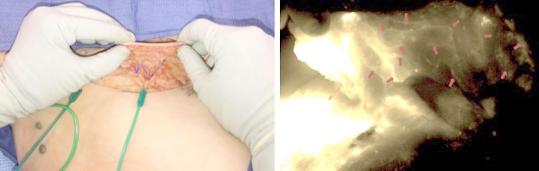
Fig. 1
(Left) Thin SCIP-based VLVT flap elevated with a 3–5 mm thickness, including subdermal lymphatic vessels. The flap’s nutrient vessels, the superficial branch perforator of the superficial circumflex iliac artery (A) and the superficial circumflex iliac vein (V), are visible. (Right) Dense lymphatic network seen in the VLVT flap (red arrows). The image was taken from the adiposal side of the flap. The bright areas on the left side represented the ICG injection site. No lymph nodes were seen in the flap. Adapted from Chen et al. [6] under CC BY 4.0 license. SCIP, superficial circumflex iliac artery perforator; VLVT, vascularized lymph vessel transplantation; ICG, indocyanine green
Fig. 2
Comparison of VLNT and VLVT flap inset. (Left) The bulk associated with transferred lymph node tissue disrupts the anatomic contour. (Right) The much thinner lymphatic vessel–containing flap avoids contour deformity. Adapted from Chen et al. [6] under CC BY 4.0 license. VLNT, vascularized lymph node transplantation; VLVT, vascularized lymph vessel transplantation
Technical proficiency in supermicrosurgery and continuous refinements in LVA technique have significantly broadened its indications. Initially reserved for early-stage disease, LVA has evolved, through ongoing research and procedural advances, into an effective option for treating fluid-predominant lymphedema [13, 28, 29]. This progress has driven a complete transition from VLVT to LVA as the preferred procedure for fluid-predominant cases (Fig. 3).
Fig. 3
(Left) A venule (V) and a lymphatic vessel (LV) dyed with isosulfan blue prepared for anastomosis. A short segment of 6 − 0 Prolene suture is placed inside the venule and the lymphatic vessel to act as a stent to prevent lumen collapse during the anastomosis. A yellow triangular background with 1-mm grid markings provides visual reference for scale. (Right) Washout of isosulfan blue dye into the venule is observed after completion of the anastomosis
Further advancements in lymphatic reconstruction led to the development of LNVA, which enables rapid and robust clinical improvement by diverting lymphatic fluid from lymph nodes directly into adjacent veins. Compared to LVA, LNVA is technically more straightforward and has gained popularity for its efficiency and consistent outcomes. Because LNVA drains an entire lymphosome containing multiple lymphatic vessels, it often produces a quicker and more pronounced response than LVA alone. In this sense, a single well-performed LNVA can theoretically achieve the effect of multiple LVAs (Fig. 4).
Fig. 4
Two lymph node-to-vein anastomoses in the right groin (white arrow: node-to-end; black arrow: node-to-side). SIEV, superficial inferior epigastric vein
The utility of LVA and LNVA extends beyond treating established, symptomatic lymphedema. These techniques can also proactively address subclinical, asymptomatic lymphedema.
LVA/LNVA for Subclinical LymphedemaFollowing axillary lymph node dissection or radiation therapy, lymphatic injuries lead to subclinical lymphedema (International Society of Lymphology [ISL] Stage 0 or Campisi Stage Ia) [30, 31]. The LYMPHA procedure [32] was proposed to provide immediate axillary lymphatic drainage post-dissection but demonstrated limited long-term efficacy [33, 34]. To address these limitations, delayed, distally-based LVA (DD-LVA) [35, 36] and LNVA (DD-LNVA) were introduced. Performing these anastomoses after cancer treatment and at a distal site facilitates the selection of small, low-pressure venules, promoting favorable pressure gradients and sustained anterograde lymphovenous flow. In addition, the superficial location of the distal lymphatics enables more straightforward supermicrosurgical manipulation while avoiding the radiation-related injury commonly seen in the axilla. When an epitrochlear lymph node is present, identified by CT or ultrasound, DD-LNVA offers more comprehensive decompression than DD-LVA alone [37].
LiposuctionFor solid-predominant lymphedema, liposuction remains the preferred method, effectively debulking pathologic fibrosis and adipose deposition [38,39,40] (Fig. 5). It significantly improves lymphatic function and patient quality of life [41, 42]. Following debulking with liposuction, LVA or LNVA can be performed using the “hybrid sequence” approach, leveraging the observed obligatory post-liposuction lymphatic regeneration. This sequence enables surgeons to operate on lymphatics that have been pre-conditioned and improved through lymphatic regeneration, yielding superior functional and clinical outcomes.
Fig. 5
(Left) Preoperative appearance of BCRL in the left upper limb, with significant lipodystrophy deposition. (Right) Postoperative appearance of the left upper limb after liposuction, with 3,100 cc of lipoaspirate removed. BCRL, breast cancer-related lymphedema
Bringing It all TogetherTo guide clinicians in selecting the most appropriate surgical strategy, Table 1 summarizes the core procedures historically and currently used at our center for managing BCRL. This overview highlights key aspects of each technique, including indications, technical considerations, benefits, risks, and emerging trends — serving as a practical framework for tailoring treatment plans to individual patient needs.
Today’s approach to BCRL represents a clear paradigm shift: what was once viewed as an intractable complication is now increasingly manageable with reliable, reproducible outcomes. Advances in diagnostic imaging, patient-centered care, and refined microsurgical methods have expanded therapeutic possibilities, enabling meaningful improvements in function and quality of life. These developments, driven by rigorous research and collaborative clinical practice, continue to shape the future of BCRL care — pushing boundaries and setting new standards for patient outcomes.
Table 1 Comparative overview of VLNT, VLVT, LVA, LNVA, and liposuction techniques for surgical management of breast cancer-related lymphedema. Abbreviations: VLNT, vascularized lymph node transplantation; VLVT, vascularized lymph vessel transplantation; LVA, lymphaticovenular anastomosis; LNVA, lymph node-to-vein anastomosis; ISL, International Society of Lymphology
Comments (0)