Glioblastoma with Sudden-Onset General Convulsions at 32 Weeks of Gestation
Background
Pregnancies complicated by brain tumors are rare and challenging. Contrast-enhanced magnetic resonance imaging (MRI), vital for evaluating malignancy, poses fetal risks. This case discusses diagnosing glioblastoma during pregnancy using traditional MRI.
Case Presentation
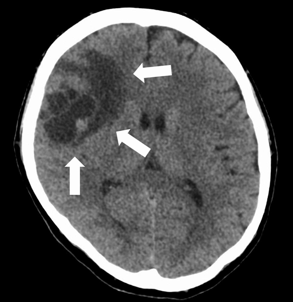
FA 42-year-old pregnant woman with a history of migraines experienced generalized convulsions at 32 weeks of gestation. Upon hospital arrival, her symptoms and normal blood pressure reduced the likelihood of eclampsia. A second convulsion occurred, which was treated with anticonvulsants. Transabdominal ultrasound showed no fetal bradycardia. Imaging studies by traditional MRI, to avoid fetal exposure to contrast agents, revealed a lesion in the right frontal lobe that showed signs of a cystic tumor with extensive edema and diffusion restriction, suggesting malignancy. Considering the occurrence of repeated convulsions and the fetus’s maturity, an emergency cesarean section was performed without any complications. Five days after surgery, contrast-enhanced MRI confirmed tumor malignancy. She underwent craniotomy and tumor resection, with histopathology supporting the diagnosis of a grade 4 glioblastoma. She subsequently underwent radiation and chemotherapy.
Discussion
Brain tumors during pregnancy are extremely rare, complicating standard management practices. Key considerations include tumor malignancy, maternal urgency, and gestational age. While contrast-enhanced MRI aids malignancy detection, it poses risks to the fetus. Traditional MRI can still indicate malignancy through features like signal heterogeneity, extensive edema, and diffusion restriction. Urgent maternal treatment is prioritized for malignant tumors, while benign tumors may allow pregnancy continuation. In this case, it was possible to obtain sufficient evidence with traditional MRI to make difficult choices of clinical management.


Comments (0)