Mpp6 gene-mutated mice
All animal experiments were performed in accordance with the guidelines of the Animal Care and Use Committee of Shinshu University. Mpp6 gene-mutated mice were generated using CRISPR-Cas9 genome editing, targeting the amino acid sequence just before the PDZ domain of the MPP6 protein, as previously reported (Saitoh et al. 2019).
Preparation of cerebral lysates and synaptic plasma membrane fractions, and western blot analysis
The cerebrum of 3- to 4-month-old Mpp6 +/+ or Mpp6 −/− mice (three mice per group) were lysed in TENT buffer [20 mM Tris (hydroxymethyl) aminomethane (pH 7.4), 1 mM ethylenediaminetetraacetic acid (EDTA), 50 mM NaCl, 1% Triton X-100] containing a protease inhibitor cocktail (Sigma, St. Louis, MI, USA). The lysates were centrifuged at 15,000 × g at 4 °C for 15 min, and the protein concentration was adjusted using a protein assay kit (ThermoFisher, Rockford, IL, USA). Synaptic plasma membrane fractions were prepared from mice and Wistar rats (male, 6 weeks old) as described previously (Suzuki et al. 2018).
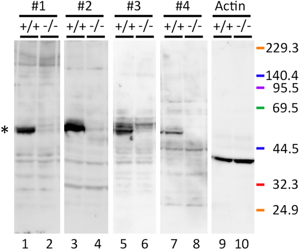
The samples were mixed with Laemmli sample buffer containing β-mercaptoethanol (β-ME) and subjected to sodium dodecyl sulfate–polyacrylamide gel electrophoresis (SDS-PAGE) on a 5–20% gradient gel, and transferred onto membranes. The membranes were probed with the following primary antibodies (Abs): rabbit polyclonal anti-MPP6 Abs targeting different regions [N-terminal region (MPP6 Ab no. 1: GeneTex; no. GTX108010), second L27 domain (MPP6 Ab no. 2: ATLAS (Stockholm, Sweden); no. HPA019085), central region (MPP6 Ab no. 3: GeneTex; no. GTX117969), C-terminal region (MPP6 Ab no. 4: originally produced by Saitoh et al. (2019)], rabbit polyclonal anti-MPP1 Ab (originally produced by Zhang et al. (2005), rabbit polyclonal anti-MPP2 Ab [GeneTex; no. GTX103908 (Yamada et al. 2022)], rabbit polyclonal anti-Lin7 Ab (GeneTex; no. GTX117114), mouse monoclonal anti-CASK Ab (NeuroMab, Davis, CA, USA; no. K56A/50), mouse monoclonal anti-actin Ab (Wako, Osaka, Japan; clone no. 2F3). After incubation with horseradish peroxidase (HRP)-conjugated anti-rabbit or anti-mouse immunoglobulin G (IgG) secondary Ab (ThermoFisher), the membranes were treated with a chemiluminescent detection solution (ThermoFisher) and visualized using a detection system (LAS4000: FUJIFILM, Tokyo, Japan).
Band intensities were quantified using Photoshop software (Adobe, San Jose, CA, USA), and the resulting values were analyzed using the following steps. First, actin protein levels in Mpp6 +/+ and Mpp6 −/− mice (three mice per group) were compared to confirm that there was no significant difference between the groups. Next, the levels of MPP1, MPP2, Lin7, and CASK proteins were normalized to actin protein levels in the three Mpp6 +/+ and three Mpp6 −/− mice. Finally, the relative expression levels of MPP1, MPP2, Lin7, and CASK proteins in Mpp6 −/− samples were determined by setting the corresponding Mpp6 +/+ sample values to 1.0. Statistical analysis was performed using the Mann–Whitney U test.
Immunoprecipitation study
Immunoprecipitation analyses were performed to examine the interactions between MPP6 and associated proteins in the mouse cerebrum. Cerebral lysates were prepared using TENT buffer containing a protease inhibitor cocktail, followed by centrifugation at 12,000 × g at 4 °C for 10 min. The lysates were pretreated with protein G-sepharose (GE Healthcare, Piscataway, NJ, USA) at 4 °C for 1 h to remove nonspecific proteins, including intrinsic mouse IgGs. The samples were then incubated with rabbit polyclonal anti-MPP6 Ab no. 2 at 4 °C for 2 h. Immunoprecipitated proteins were collected using protein G-sepharose at 4 °C for 1 h, eluted by boiling in Laemmli sample buffer containing β-ME, and subjected to SDS-PAGE on a 5–20% gradient gel and western blot analyses using Abs against MPP1, MPP2, or CASK, followed by a HRP-conjugated anti-rabbit or mouse IgG Ab. Blots were visualized using a chemiluminescent detection system.
Light microscopic immunohistochemistry
Anesthetized 3- to 4-month-old C57BL/6J mice were perfused via the heart with 2% paraformaldehyde in 0.1 M phosphate buffer (PB, pH 7.4). The cerebrum was dissected and immersed in the same fixative at 4 °C for 2 h, embedded in 30% sucrose, and sectioned into floating sections (20 μm) using a cryostat. Coronal sections corresponding to Allen Brain Atlas images 55–70 were obtained and subjected to antigen retrieval by microwave heating in ImmunoSaver™ (FUJIFILM Wako, Osaka, Japan) (Namimatsu et al. 2005). Sections were incubated with rabbit anti-MPP6 Ab no. 2, followed by biotinylated anti-rabbit IgG Ab (Vector Lab, Burlingame, CA, USA) at room temperature for 2 h. After treatment with a HRP-conjugated avidin–biotin complex (ThermoFisher) for 30 min, sections were visualized using metal-enhanced diaminobenzidine (ThermoFisher), sections were treated with 0.04% OsO4 for 60 s, and mounted with Vectashield™. They were observed under a light microscope (BX51: Olympus, Tokyo, Japan) using objective lenses of 4, 10, 20, and 40×. Images were captured with a digital camera (FX630: Flovel, Tokyo, Japan) at a resolution of 300 dpi.
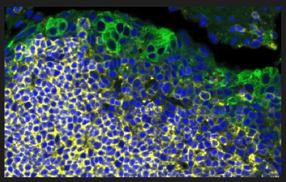
For double-fluorescence immunostaining, some sections were incubated overnight at 4 °C with rabbit polyclonal anti-MPP6 Ab no. 2 and mouse monoclonal anti-GluN1 (BioLegend, San Diego, CA, USA; no. MMS-5145). The sections were then treated with Alexa Fluor 594-conjugated anti-rabbit IgG (Invitrogen) and Alexa Fluor 488-conjugated anti-mouse IgG (Cell Signaling Technology, Danvers, MA, USA) at room temperature for 2 h. After mounting with Vectashield™, the cerebral cortex regions were examined using a confocal laser-scanning microscope (LSM880; Zeiss, Oberkochen, Germany) with a 40× objective lens. All light microscopic images were arranged using Photoshop (ver. 26.5) at a resolution of 300 dpi.
Post-embedding immunoelectron microscopy
Anesthetized 3- to 4-month-old C57BL/6 J mice were perfused via the heart with 2% paraformaldehyde and 0.25% glutaraldehyde in 0.1 M PB and post-fixed at 4 °C for 1 d. Cerebral cortex samples (Allen Brain Atlas images 60–70) from the right side above the corpus callosum were dissected, dehydrated in graded ethanol, and embedded in LR-Gold resin (Polysciences Inc., Warrington, PA, USA) under ultraviolet irradiation at −20 °C for 2 d. Ultrathin sections from cortical layers II/III–V were mounted on Formvar-coated grids, subjected to antigen retrieval by heating in ImmunoSaver™ at 95 °C for 10 min, and incubated with rabbit anti-MPP6 Ab no. 2, followed by 18 nm gold-conjugated anti-rabbit IgG Ab (Jackson ImmunoResearch, West Grove, PA, USA). Sections were observed using a transmission electron microscope (JEM-1400: JEOL, Tokyo, Japan).
Electron microscopy for ultrastructural analysis
Perfusion-fixed 4-month-old male Mpp6 +/+ and Mpp6 −/− mice (three per group) were analyzed. The cerebral cortex (Allen Brain Atlas images 60–70) was dissected, post-fixed in 1% OsO4 in 0.1 M PB for 2 h, dehydrated, and embedded in epoxy resin. Ultrathin sections were obtained from cortical layers II/III–V, stained with uranyl acetate and lead citrate, and examined using a transmission electron microscope. The width of synaptic clefts and thickness of the postsynaptic density (PSD) were measured in type I synapses from Mpp6 +/+ (n = 280) and Mpp6 −/− (n = 375) mice. All electron microscopic images were analyzed using Photoshop (ver. 26.5) at a resolution of 300 dpi. Statistical analyses of synaptic cleft width and PSD thickness in Mpp6 +/+ and Mpp6 −/− mice (three per group) were performed using the Mann–Whitney U test.
Elevated plus-maze test
Mpp6 +/+ and Mpp6 −/− mice of different age groups were tested (2–3 months: n = 6 and 7, respectively; 4–7 months: n = 3 and 4, respectively; 18 months: n = 3 per group). The elevated plus-maze was constructed using plastic rails (Pla-rail™, Takara-Tomii, Tokyo, Japan) and elevated 40 cm above the floor. Closed arms were enclosed using Truss bridge structures [Pla-rail™; 40 cm (length) × 6 cm (width) × 7 cm (height)]. Mice were placed at the center of the maze, and time spent in the open and closed arms was recorded for 10 min using video tracking. Statistical analysis of the average time spent in different areas (closed arm, center area, and open arm) by Mpp6 +/+ and Mpp6 −/− mice at different developmental stages (2–3 months, 4–7 months, and 18 months) was performed using the Mann–Whitney U test.
Locomotor activity (movement) test
To measure locomotor activity, Nano-tag™ devices (Kissei Comtech, Matsumoto, Japan) were implanted subcutaneously in 8-month-old Mpp6 +/+ and Mpp6 −/− mice (three per group) under anesthesia. After a 2-week acclimation period, movement was recorded for 7 d. The light cycle was set from 9:00 to 21:00, and the dark cycle from 21:00 to 9:00. Statistical analysis of the average locomotor activity counts per hour in Mpp6 +/+ and Mpp6 −/− mice (three per group) was performed using the Mann–Whitney U test.


Comments (0)