Animal information
Male Wistar rats, each weighing approximately 300 g, were obtained from the Animal House of the Mossakowski Medical Research Institute, Polish Academy of Sciences. Piglets of the Złotnicka White pig, weighing approximately 7–8 kg, were acquired from the Swadzim Agricultural Experimental Farm at the Poznań University of Life Sciences. All experimental procedures were conducted in accordance with Polish law and the guidelines of the Local Ethical Commission for Animal Research.
The MOTS-c peptide used in the experiments was purchased from Novazym (Poznań, Poland). For experiments involving rat islets, the rat-specific MOTS-c amino acid sequence—MKRKEMGYIFFSQRTLRNPL—was used. For experiments involving pig islets, the human MOTS-c sequence—MRWQEMGYIFYPRKLR—was used, as the porcine MOTS-c sequence has not yet been identified. Given that pigs are physiologically closer to humans than rats, especially in pancreatic structure, we chose to use the human MOTS-c peptide in our pig-based experiments.
Isolation of rat pancreatic islets
Male Wistar rats were decapitated and exsanguinated, after which the dissection procedures were carried out immediately. The abdominal cavity was opened, and clamps were placed on the duodenum near the pancreatic duct estuary. The pancreatic duct was then severed, and a cannula was inserted. A total of 13 ml of Hanks’ balanced salt solution (HBSS, 0.137 M NaCl, 5.37 mM KCl, 4.17 mM NaHCO3, 1.26 mM CaCl2, 0.84 mM MgSO4, 0.44 mM KH2PO4, and 0.34 mM Na2HPO4) containing 13 Wunsch units of Liberase TL (cat. no. 05401020001, Roche, Basel, Switzerland) was injected into the pancreas. The pancreas was excised, transferred to a Falcon tube, and incubated in a water bath at 37 °C for 11 min. Following incubation, the tube was shaken vigorously for 30 s. The enzymatic digestion was stopped by adding HBSS supplemented with 10% fetal bovine serum (FBS). The islets were washed and manually isolated under a binocular microscope (Delta Optical, Poznań, Poland), then transferred into Krebs Ringer buffer (KRB, 115 mM NaCl, 24 mM NaHCO3, 5 mM KCl, 1 mM MgCl2, 1 mM CaCl2, and 0.5% bovine serum albumin, BSA) containing 6 mM glucose. They were incubated at 37 °C with 5% CO2 for 1.5 h to allow regeneration. Approximately 50 islets were collected for RNA or protein extraction. For RNA isolation, islets were frozen in 500 µl of TRIzol (cat. no. 15596026, Thermo Fisher Scientific, Waltham, MA, USA); for protein extraction, they were frozen in 200 µl of radioimmunoprecipitation assay buffer (RIPA, cat. no. 20-188, Merck Millipore, Burlington, MA, USA). All samples were stored at − 80 °C until further processing.
Isolation of pig pancreatic islets
Pancreatic islets were isolated from the Złotnicka White piglets via enzymatic digestion of the pancreas. The animals were killed by an overdose of anesthetics and sedatives administration (medetomidine 200 µg/kg of body weight and ketamine 40 mg/kg of body weight) and then exsanguinated by intracardiac puncture. The pancreas was excised and placed in HBSS. Approximately 2 g of pancreatic tissue was transferred into a Falcon tube containing 10 ml of HBSS and finely chopped with scissors. Collagenase P (cat. no. 11213873001, Roche, Basel, Switzerland) was then added to the solution. The tube was incubated in a water bath at 37 °C for 12 min, followed by vigorous shaking outside the bath for 1 min. Enzymatic digestion was stopped by adding 90 ml of HBSS supplemented with 10% FBS. The islets were washed and manually isolated under a binocular microscope (Delta Optical, Poznań, Poland), then transferred to KRB containing 6 mM glucose. They were incubated at 37 °C with 5% CO2 for 1.5 h to allow for regeneration. Approximately 50 islets were collected for RNA or protein isolation. For RNA extraction, islets were frozen in 500 µl of TRIzol (cat. no. 15596026, Thermo Fisher Scientific, Waltham, MA, USA); for protein extraction, they were frozen in 200 µl of RIPA (cat. no. 20-188, Merck Millipore, Burlington, MA, USA). Samples were stored at − 80 °C until further analysis.
Protein isolation
Pancreatic islets were incubated for 24 h in a 12-well plate. Following incubation, the islets were collected using RIPA buffer supplemented with cOmplete protease inhibitor tablets (cat. no. 04693116001, Roche, Basel, Switzerland) for protein isolation. The lysate was mixed using a vortex and centrifuged (two times). The tubes were then placed on a thermomixer and shaken at 900 rpm at 4 °C for 10 min. The samples were then centrifuged for 10 min at 13,000 × g. The resulting supernatant was carefully transferred to new tubes. Protein concentrations were measured using the Pierce BCA Protein Assay (Thermo Fisher Scientific, Waltham, MA, USA), following the manufacturer’s protocol.
Western blot
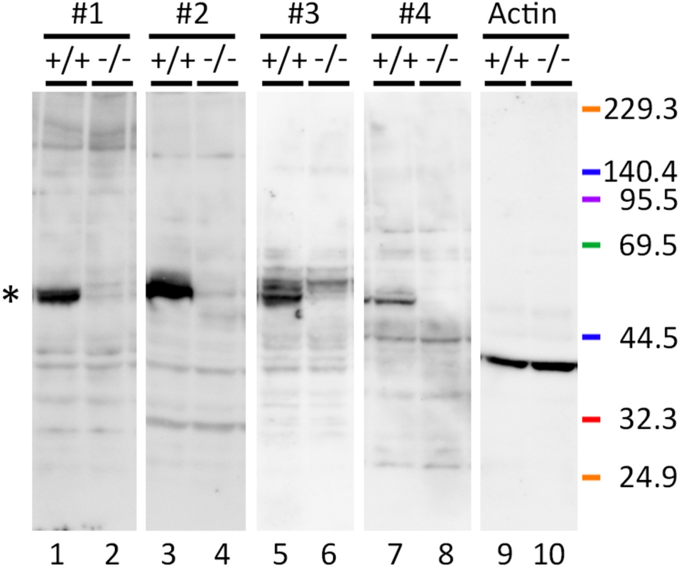
The Western blot procedure followed the protocol described in Bień et al. (2024). Briefly, equal amounts of protein (15 µg) were separated by 12% gel electrophoresis (SDS PAGE) and transferred onto a 0.2-µm PVDF membrane. The membrane was blocked using 3% BSA and incubated overnight with the primary antibody 1:1000. After the membrane was washed with tris-buffered saline with Tween-20 (TBST), it was incubated with a secondary antibody 1:5000 for 1.5 h. Secondary antibodies were conjugated with horseradish peroxidase and the signal detection method was chemiluminescence. The signal corresponding to the target protein was measured. Subsequently, the membrane was incubated overnight with anti-β-actin or antiGAPDH antibody 1:5000, washed with TBST, and incubated again with a secondary antibody 1:5000. The signal for the reference protein was visualized using Super Signal™ West Pico PLUS (Thermo Fisher Scientific, Waltham, MA, USA) and detected using the ChemiDoc MP Imaging system (Bio-Rad, Hercules, CA, USA).
The antibodies and blocking peptide used for Western blotting were as follows: antiMOTS-c, rabbit polyclonal (cat. no. MBS542112, MyBioSource, San Diego, CA, USA); blocking peptide (cat. no. MBS543991, MyBioSource, San Diego, CA, USA); anti-β-actin (cat. no. A1978, Sigma-Aldrich, St. Louis, MO, USA); antiGAPDH (cat. no. G8795, Sigma-Aldrich, St. Louis, MO, USA). The secondary antibodies used were as follows: antirabbit IgG (cat. no. 7074P2, Cell Signaling, Danvers, MA, USA) and antimouse IgG (cat. no. A2304, Sigma-Aldrich, St. Louis, MO, USA).
Immunofluorescence
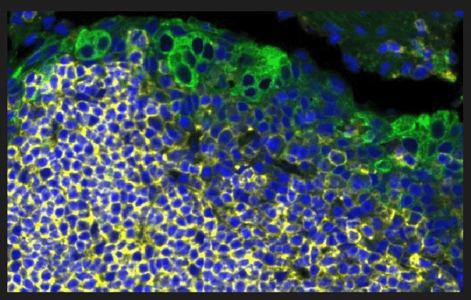
Rat and pig pancreases were dissected out and placed in Bouin’s reagent (150 ml picric acid, 50 ml formaldehyde, 10 ml acetic acid; Sigma-Aldrich, St. Louis, MO, USA) immediately. Subsequently, fixed pancreases were embedded in paraffin, and sliced in 5-µm sections using a microtome. Paraffin was removed by heating the slides at 60 °C for 45 min. The sections were then rehydrated through graded alcohol solutions with decreasing concentrations (100%, 85%, 70%, 60%, 50%, water). Antigen retrieval was performed by boiling the slides in citrate buffer (pH 6, 10 mM disodium citrate and 0.05% Tween-20) three times for 5 min each, then cooling at room temperature for 20 min. Afterward, the samples were rinsed with water for 5 min. The tissue sections on the slides were encircled using a Dako Pen to contain the reagents during incubation.
To reduce autofluorescence, the samples were incubated in 15 mM glycine solution for 5 min, washed in phosphate-buffered saline (PBS), incubated in 0.2% gelatin solution, and washed again with PBS. Primary antibodies were then applied to stain for MOTS-c 1:200 (with and without 100 µl of blocking peptide in a concentration 2.5 mg/ml), insulin 1:400, and glucagon 1:400 in PBS buffer with 0.2% gelatin. Sections were incubated with primary antibodies overnight. For MOTS-c staining with blocking peptide, the antibodies were preincubated with the peptide for 48 h before application. Following primary antibody incubation, all samples were washed and incubated for 1 h in PBS containing 0.2% BSA. The secondary antibody was added for 15 min also in a concentration of 1:400 in PBS buffer with 0.2% gelatin. Finally, cell nuclei were stained with 4′,6-diamidino-2-phenylindole (DAPI) for 1 min. Images were captured using a Leica DMI8 fluorescence microscope (camera Leica K5-14403549, 16 bits, 2048 pixels, software system LAS X, objective HC PL FLUOTAR L 20×/0.40 DRY, aperture 0.4).
The antibodies and blocking peptide used for immunofluorescence were as follows: antiMOTS-c, rabbit polyclonal (cat. no. MBS542112, MyBioSource, San Diego, CA, USA); blocking peptide (cat. no. MBS543991, MyBioSource, San Diego, CA, USA); anti-insulin, polyclonal guinea pig (cat. no. A0564, Agilent, Santa Clara, CA, USA); and antiglucagon, polyclonal guinea pig (cat. no. 4031-01F, Merck Millipore, Burlington, MA, USA). The secondary antibodies used were Alexa Fluor 488 goat antiguinea pig IgG (cat. no. A11073, Life Technologies, Carlsbad, CA, USA) and Cy3 Goat antirabbit IgG (cat. no. A10520, Life Technologies, Carlsbad, CA, USA).
MOTS-c secretion and cell culture medium with free fatty acids
Pancreatic islets were placed in a 48-well plate, with five islets per well, and incubated for 1.5 h in Krebs buffer. The control group received buffer without any additives, while experimental groups received Krebs buffer supplemented with: (a) 200 µM of oleic, stearic, or palmitic acid; (b) insulin at concentrations of 1, 10, or 100 nM; (c) glucagon at concentrations of 1, 10, or 100 nM. Additionally, rat and pig pancreatic islets were incubated for 1.5 h in experimental media containing glucose at concentrations of 2, 6, or 16 mM. MOTS-c levels in the incubation medium were measured using rat and pig MOTS-c ELISA kits (SunRed, Shanghai, China), following the manufacturer’s instructions.
Before islets were incubated in the medium, warm, free fatty acids were added to the medium, resulting in a final concentration of 200 µM of each free fatty acid. Then, the experimental medium was incubated for 1.5 h at 37 °C to couple free fatty acids to BSA in the medium.
Secretion of hormones
Rat and pig pancreatic islets were incubated for 1.5 h in a 48-well plate with varying concentrations of MOTS-c (1 nM, 10 nM, and 100 nM or alternatively 10 nM and 100 nM in Krebs Ringer buffer), along with a control group that received no MOTS-c. After incubation, hormone concentrations were measured using ELISA kits specific to each species. Insulin levels were assessed using the Rat Insulin (INS) ELISA Kit and the Pig Insulin (INS) ELISA Kit (SunRed, Shanghai, China), while glucagon levels were determined using the Rat Glucagon (GC) ELISA Kit and the Pig Glucagon (GC) ELISA Kit (all from SunRed, Shanghai, China). All measurements were conducted according to the protocols provided by the manufacturer.
RNA extraction, reverse transcription, and PCR
Pancreatic islets were incubated for 24 h in 12-well plates. Following incubation, total RNA was extracted using TRIzol according to the manufacturer’s instructions and was measured by Implen NP80 NanoPhotometer (Implen, Munich, Germany). Reverse transcription was then performed using the High Output cDNA Reverse Transcription Kit (Applied Biosystems, Waltham, MA, USA), following the protocol provided in the kit manual.
Quantitative PCR was carried out using the HOT FIREPol EvaGreen qPCR Mix Plus (Solis BioDyne OÜ, Tartu, Estonia) on the QuantStudio™ 12K Flex System (Thermo Fisher Scientific, Waltham, MA, USA). The primer sequences (5ʹ–3ʹ) were as follows. Rat insulin F: CCAGTTGGTAGAGGGAGCAG, R: AGACCATCAGCAAGCAAGCGGTC; insulin receptor F: CAGAAAAACCTCTTCAGGCAAT, R: TTCAAGGGATCTTCGCTTTC; glucagon F: AAGATGGTTGTGAATGGTGAAA, R: TGAGATGAACACGATTCTCGAT; glucagon receptor F: TTCTGTTGCAGACCAGCTCA, R: GTGACCAGTGCCACCACA; GAPDH F: CTGCACCACCAACTGCTTAG, R: TGATGGCATGGACTGTGG; pig insulin F: GTGGCATCGTGGAGCAGT, R: CGGCCTAGTTGCAGTAGTTCTC; insulin receptor F: AACGCCAGGGACATCGTCAA, R: CTTTGGACACCACCCCCAGG; glucagon F: CATCAGCCACTGCACAAAAT, R: AGGGCACGTTTACCAGTGAC; glucagon receptor F: GTCACGAAGGCAAACACCAC, R: CTGCCCTGGTACCACAAAGT; TATA-box binding protein (TBP) F: TTGAGAACATCTACCCTATCC, R: CGTCCACAACACCACCATT.
The PCR reaction was carried out in a total volume of 10 µl, consisting of 5 µl of reagent mix, 3 µl of cDNA, and 2 µl of primer mix (final primer concentration of 2.5 µM). Cycling conditions included an initial denaturation at 95 °C for 10 min, followed by 40 cycles of 15 s at 95 °C (denaturation), 1 min at 61 °C (annealing), and 20 s at 72 °C (extension). Melt curve analysis was performed with the following settings: 95 °C for 15 s, 60 °C for 60 s, and 95 °C for 15 s.
MTT
Pancreatic islets were incubated with different concentrations of MOTS-c for 24 h. Following incubation, a 0.05% MTT solution (Merck Millipore, Burlington, MA, USA) was added to the islets. The plate was then incubated for 20 min at 37 °C. Afterward, the medium was removed, and the islets were dissolved in 100 µl of dimethylsulfoxide. Absorbance was measured at a wavelength of 570 nm, with background correction performed at 650 nm, using the Synergy 2 microplate reader (Agilent, Santa Clara, CA, USA).
Apoptosis
Pancreatic islets were incubated with MOTS-c for 24 h in 96-well plates. The level of apoptosis was assessed using the Cell Death Detection ELISA Plus kit (Roche, Basel, Switzerland), following the manufacturer’s protocol. Absorbance was measured at a wavelength of 405 nm, with background correction performed at 490 nm, using the Synergy 2 microplate reader (Agilent, Santa Clara, CA, USA).
Statistical analysis
All analyses were carried out using GraphPad Prism 6.0 software (GraphPad Software, San Diego, CA, USA). The results are presented as the arithmetic mean ± SEM. The significance of differences was determined using one-way analysis of variance (ANOVA) with a Dunnett post hoc test, comparing the results with the control group. Additionally, a Tukey post hoc test was employed when comparing groups with each other (experiments with glucose). Statistical significance is denoted by * for p < 0.05 and ** for p < 0.01.


Comments (0)