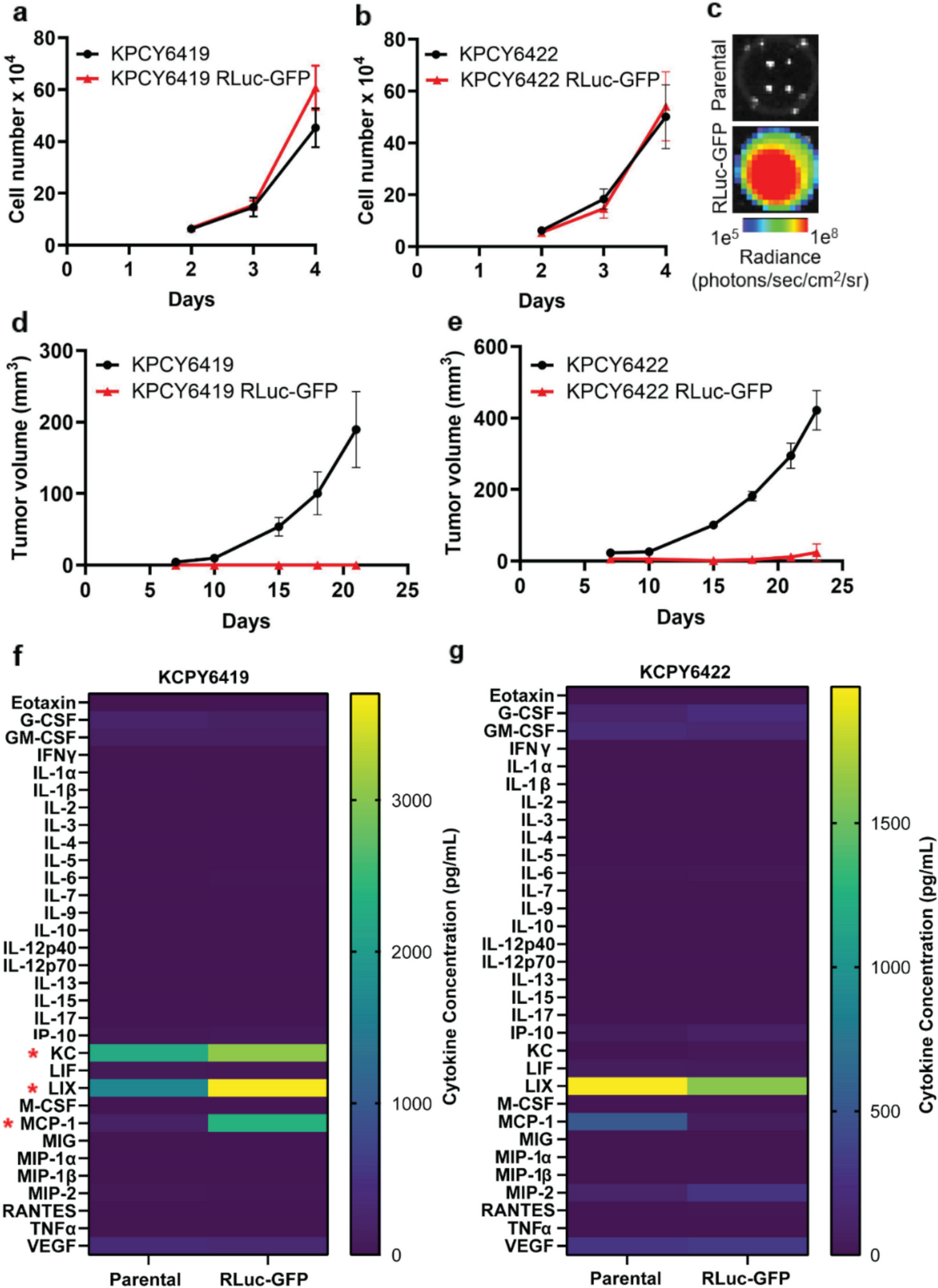
Our studies characterized the immunogenicity of RLuc-GFP and CBG-GFP reporters expressed in cancer cells. Ensuring that the incorporation of imaging reporters do not alter the tumor-immune microenvironment is essential when using immunocompetent murine models, especially for studying tumor-immune interactions. This work presents a strategy to assess whether the stable incorporation of an imaging reporter in tumor cells triggers an immune response. Our data demonstrate that while comparing the in vitro proliferation of parental and reporter-expressing cells is useful for confirming stable reporter incorporation, it is insufficient to predict in vivo tumor growth. Although no differences in proliferation were observed between parental cells and those expressing RLuc-GFP or CBG-GFP in vitro (Fig. 1a & b, Fig. 2), only cell lines expressing CBG-GFP successfully established tumors in vivo in immunocompetent mice (Fig. 3). The incorporation of RLuc-GFP in tumor cells resulted in no in vivo tumor growth, most likely a result of the immunogenicity of RLuc which elicited an enhanced activation and cytotoxicity of T cells (Fig. 5e & f). Although both PDAC RLuc-GFP cell lines failed to develop tumors in vivo (Fig. 1d & e), increased levels of KC, LIX and MCP-1 were observed only in KPCY6419 RLuc-GFP cells compared to parental cells (Fig. 1f). In contrast, no differences in cytokine expression were detected between KPCY6422 parental and RLuc-GFP cells (Fig. 1g). This variability in secreted cytokines suggest that cytokine expression did not contribute to the lack of in vivo tumor growth. Similarly, incorporation of the CBG-GFP reporter altered the in vitro secreted cytokine expression in several cell lines; a decrease in KC and LIX was observed in KPCY6419 CBG-GFP cells compared to parental cells (Fig. 4b). While an increase in KC was observed in KPCY6422 CBG-GFP cells compared to parental cells (Fig. 4d). An increase in IP-10, MCP-1, and RANTES levels in CBG-GFP cells compared to parental cells (Fig. 4f) while B16F10 CBG-GFP cells had an increase in IP-10 and RANTES (Fig. S4d). KC and LIX are cytokines known to recruit neutrophils and monocytes, with both cytokines binding the CXCR2 receptor [26, 27]. IP-10 and MCP-1 are involved in attracting various immune cells such as T cells, natural killer cells, and dendritic cells to sites of inflammation while RANTES is a cytokine that is involved in the activation and proliferation of T cells [28,29,30]. However, these changes in secreted cytokines did not correspond to parallel changes in immune cell recruitment to the tumor microenvironment (Fig. 5). Furthermore, no consistent pattern of increased or decreased cytokine levels was observed following the incorporation of the reporter. Similar to RLuc-GFP reporter cells, the incorporation of the CBG-GFP reporter resulted in alterations to a few cytokines; however, CBG-GFP expressing cells demonstrated comparable in vivo tumor growth rates and minimal differences in tumor immune cell composition. These findings suggest that the altered cytokine secretion does not significantly impact in vivo tumor growth or elicit an immune response. This lack of correlation may stem from the low cytokine concentrations observed in vitro, measured in the pg/mL range, or from the host immune system's ability to neutralize and regulate these cytokines in vivo, thereby mitigating their effects. These variations in cytokine profiles suggest cell line-specific responses to CBG-GFP expression, yet overall indicate that CBG-GFP does not broadly alter the immune landscape. Thus, in vitro proliferation and cytokine analysis alone are insufficient to predict in vivo tumor growth and immunogenicity.
How the tumor immune cell composition was affected by the presence of the RLuc-GFP reporter could not be evaluated given the lack of established RLuc-GFP tumors in vivo. Several studies have shown that firefly luciferase can provoke an immune response against the reporter protein when used in vivo in an immunocompetent mouse [15, 18, 31]. This response may result from the presentation of foreign firefly luciferase antigens to major histocompatibility molecules, which can activate cytotoxic and memory T cell responses. This could enable T cells to eliminate luciferase-expressing tumor cells before they can develop or lead to inconsistent tumor growth patterns [19, 31, 32]. Moreover, a recent study demonstrated that KPC-firefly-luciferase-expressing cells elicited a potent anti-tumor immune response in a PDAC mouse model [18]. This response was characterized by an increase in CD8+ T cell and natural killer cell infiltration, resulting in significantly smaller or even absent tumors in vivo [18]. While previous studies have suggested that GFP may be immunogenic [33, 34], our findings indicate that the immunogenicity observed in our study is primarily attributed to firefly luciferase rather than GFP. This is supported by the fact that CBG-GFP-expressing tumor cells, which also contain GFP, did not provoke a significant immune response or lead to tumor rejection.
Our studies suggest that click beetle green luciferase has minimal immunogenicity, as CBG-GFP-expressing tumor cells exhibited similar in vivo tumor growth rates to parental tumor cells. This was consistent across several PDAC and melanoma cell lines in an immunocompetent C57BL/6 mouse model. Minimal differences in tumor immune cell composition were observed with the incorporation of the CBG-GFP reporter in the tumor cells studied (Fig. 5), with the exception of KPCY6419 and B16F10. While a significant increase in intratumoral CD4+ T cells was observed in KPCY6419 CBG-GFP tumors compared to parental tumors (Fig. 5a), the overall percentage of CD4+ T cells remained low, and no other significant changes in immune composition were noted. A significant increase in monocytes was observed in B16 F10 CBG-GFP tumors compared to parental tumors (Fig. S4e). However, no other significant changes in immune cells were observed. Given the absence of differences in in vivo tumor growth, the single alteration in immune cell subsets resulting from the incorporation of CBG-GFP into KPCY6419 and B16F10 cells is unlikely to significantly impact future studies evaluating tumor-immune interactions during tumor development. Collectively, these findings highlight the minimal immunogenic effects of CBG-GFP expression in tumor cells demonstrating its suitability as an optimal reporter for preclinical tumor immunology studies.
While this study provides valuable insights into the use of bioluminescence reporters, several limitations must be acknowledged. One significant limitation is that the immune composition of tumors was only evaluated at tumor endpoint. Evaluating immune composition only at the endpoint may miss early immune responses critical for understanding tumor-host interactions and influencing tumor progression [35]. Although no differences in tumor growth were observed, the potential impact of CBG-GFP incorporation on early immune events cannot be excluded. Future studies should incorporate temporal analysis of immune responses following tumor inoculation to more comprehensively understand immune dynamics. Additionally, loss of reporter plasmids represents another limitation, as observed in Pan02 PDAC cells labeled with the CBG-GFP reporter (Fig. S6). This issue resulted in the formation of palpable tumors with similar in vivo tumor growth rates but lacked a bioluminescence signal, even after three in vivo selection passages. This highlights the cell line-specific variability in response to CBG-GFP expression and emphasizes the need for developing more stable integration methods to ensure consistent expression throughout in vivo studies. While CBG-GFP showed minimal immunogenicity in C57BL/6 mice, its immunogenic profile in other mouse models remains uncertain. Variations in tumor microenvironments and host genetic backgrounds may significantly shape the immune response to reporter genes. While our data demonstrate the applicability of CBG-GFP in five cancer cell lines, we were unable to establish a stable Pan02 CBG-GFP cell line. Additionally, lentiviral infection results in the random integration of reporter genes, which can disrupt cellular pathways and alter cell behavior, potentially increasing immunogenicity depending on the cell line and mouse model. These studies utilized polyclonal reporter cell populations; isolating monoclones may reveal differences in immunogenicity. Future studies will assess the immunogenicity of these monoclones. Therefore, it is essential to validate the stability and lack of immunogenicity of the CBG-GFP reporter when incorporating it into new cancer models. Lastly, our study did not evaluate the immunogenicity of GFP. However, GFP has been widely used in cancer models since the 1990 s and is well-established for tracking tumor cells in vivo [36, 37]. Our focus was on the impact of bioluminescence reporters within dual bioluminescence-fluorescence systems. These dual reporters offer several advantages; GFP facilitates the efficient isolation of labeled tumor cells, a process that is challenging when relying solely on bioluminescence reporters and eliminates the need for antibiotic selection genes. Furthermore, dual reporters synergistically combine the strengths of both imaging modalities. GFP allows for high-resolution cellular microscopy, though its effectiveness is constrained by limited tissue penetration and potential phototoxicity. In contrast, bioluminescence imaging provides quantitative capabilities, enhanced sensitivity, minimal background noise, and the ability to perform real-time, non-invasive, longitudinal whole-body imaging. Together, these features make dual reporters highly versatile tools for studying tumor immunology.

Comments (0)