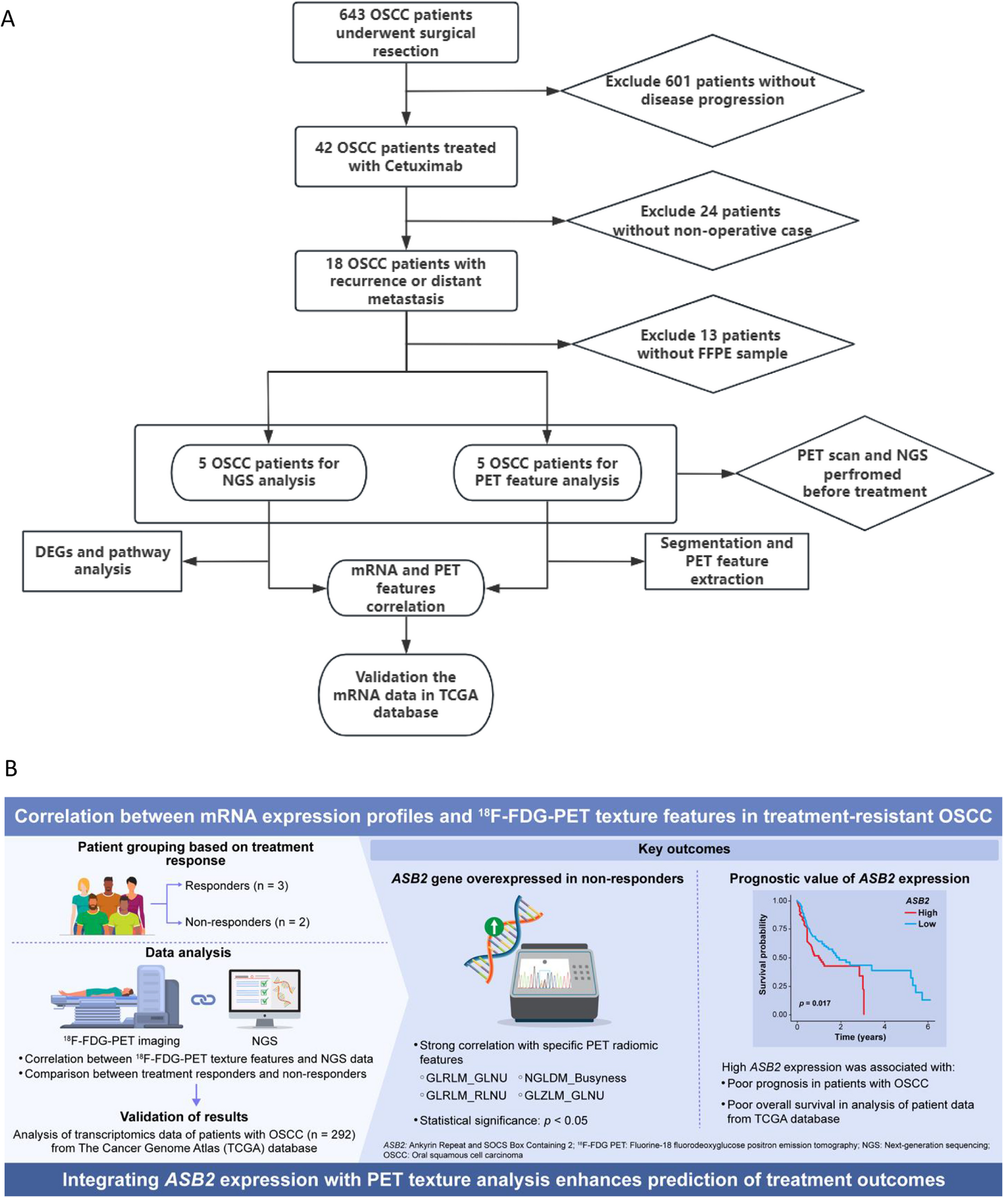
We investigated the relationship between mRNA expression using NGS and 18F-FDG PET texture analysis in treatment-resistant OSCC patients undergoing molecularly targeted therapy. Although MTV has been established as a prognostic factor in previous studies, our analysis may provide supplementary insights to conventional metrics. Specifically, our findings suggest that advanced radiomic features such as GLRLM_GLNU, GLRLM_RLNU, and GLZLM_GLNU may serve useful imaging biomarkers for assessing treatment efficacy. Additionally, the correlation between PET and NGS data with high ASB2 expression indicates the potential to predict poor prognosis. Integrating mRNA data with 18F-FDG PET texture features may enhance predictions of OSCC treatment efficacy. Combining these features with extensive databases could enable the creation of subgroups that account for genes expressed related to poor prognostic factors and individual patient risk, proving beneficial for non-invasive applications.
Currently, 18F-FDG PET imaging is primarily used for detecting metastases in lymph nodes and distant organs before initial treatment and for assessing regions of interest using SUVmax in OSCC management. Besides morphological imaging modalities such as CT and MRI, integrating functional metabolic changes that reflect tumor glucose metabolism and comprehensive tumor gene analysis through 18F-FDG PET may prove valuable for monitoring treatment efficacy. This method is a useful tool for assessing treatment effectiveness. Our study demonstrated the utility of 18F-FDG PET texture analysis in correlating features with mRNA expression and confirmed the effectiveness of radiogenomics in treatment-resistant OSCC. The novelty of our study lies in its focus on combining PET texture features with mRNA fusion data to investigate radiogenomic associations in oral squamous cell carcinoma that has progressed despite standard treatment. By integrating these data, we identified potentially useful genes and validated their prognostic significance using external datasets from TCGA. This distinct approach highlights the potential of radiogenomics as a valuable method for understanding tumor biology and evaluating prognosis in challenging clinical scenarios. Additionally, ASB2, a gene strongly correlated with radiomic features, was associated with poor prognosis, corroborated by a large dataset from TCGA.
Jiaving et al. (2021) conducted a survival curve analysis of OSCC patients from The Cancer Genome Atlas (TCGA), identifying six candidate genes (CXCL10, OAS2, IFIT1, CCL5, LRRK2, and PLAUR) associated with patient survival [19, 20]. In this study, NGS analysis of cases treated with molecular-targeted drugs led to the identification of new genes through enrichment analysis. We found that the non-response group exhibited enhanced cancer proliferation and metabolic pathway activity, whereas the response group demonstrated greater immune activation. The diagnostic and prognostic value of mRNA in OSCC, along with its correlation with the tumor's clinicopathological profile, has been documented. Future research should explore these genes’ role in survival and treatment response prediction in OSCC patients. Aberrantly expressed mRNAs influence tumorigenesis, and the PI3 K/Akt signaling pathways and target genes for the repression of the p53 signaling pathway have been identified. Further investigation is necessary to understand therapeutic sensitivity and these signaling pathways.
Radiogenomic features based on SUVmax in FDG PET and the epithelial-mesenchymal transition (EMT) phenotype based on mRNA expression have been studied in non-small cell lung cancer (NSCLC). This study explored the association between radiogenomic features based on SUVmax in 18F-FDG PET and EMT based on mRNA expression, confirming that higher EMT protein expression was linked to significantly different cell migration, glucose uptake, and hexokinase activity. Additionally, NSCLC cells exhibited increased resistance to chemotherapy in vitro [21]. In our study, ASB2-positive cases were categorized within the tumor cytoplasm of non-responder group cases, though some reports suggest that ASB2 knockdown inhibits the migration of human NK cells [22]. The efficacy of the tumor microenvironment in immunotherapy, particularly concerning NK cells, warrants further investigation. In colorectal cancer, ASB2α has been reported to induce a tumor-promoting immune pathway via Th2 cells and to facilitate tumor progression through the evasion of immune surveillance [23]. In oral squamous cell carcinoma, Th2-related cytokines, such as IL- 4 and IL- 10, have also been reported to show elevated serum levels compared to healthy individuals [24], suggesting that the expression of these cytokines, involved in cancer immune responses, may contribute to their potential role as prognostic biomarkers reflected in PET imaging findings in OSCC [25].
This study conducted mRNA expression analysis and 18F-FDG PET texture analysis treatment-resistant OSCC patients, but the sample size was limited to only five patients. This small sample size is a significant limitation, reducing statistical power and limiting the generalizability of the findings. However, we supplemented our analysis with data from 292 OSCC patients from the TCGA database, which helped validate the associations between ASB2 expression and 18F-FDG PET texture features observed in our study. The use of TCGA data adds reliability to our conclusions, despite the small initial sample size. However, future studies should consider public datasets that include both PET imaging and NGS data, as the TCGA-OSCC dataset does not contain PET-CT imaging data.
As an approach to the clinical implementation of radiogenomics, a functional metabolic imaging database based on TMB levels derived from cancer-related genes following standard treatment enables non-invasive and dynamic monitoring. Integrating radiogenomic insights into clinical workflows may facilitate personalized treatment planning by identifying patients most likely to benefit from targeted therapies or immunotherapies and detecting early treatment resistance, thereby enabling timely therapeutic modifications. Furthermore, non-invasive metabolic imaging may enhance patient follow-up by providing real-time assessments of tumor evolution. This process, in turn, supports the development of strategic treatment plans that optimize the timing of drug therapy, treatment costs, and toxicity, potentially contributing to improved survival rates. These applications have the potential to improve treatment efficacy, optimize resource utilization, and enhance clinical outcomes.
Nevertheless, clinical and treatment-related differences between the patient data from our study and the TCGA cohort could influence the results. The conclusions drawn from our five cases need further validation in larger independent cohorts to ensure robustness and broader applicability.
Future research should focus on collecting data both before and after initial treatment and analyzing them longitudinally to better understand the mechanisms underlying treatment resistance and to improve the accuracy of treatment efficacy predictions. By predicting tumor growth rate based on gene mutation levels and stratifying TMB-related therapeutic resistance mechanisms, the improved matching accuracy of functional metabolic imaging will enable healthcare providers to anticipate therapeutic resistance before initiating drug therapy and determine the optimal timing for therapy initiation. Additionally, expanding the study to include a larger patient population will strengthen the findings and ensure their applicability across a broader range of clinical settings.

Comments (0)