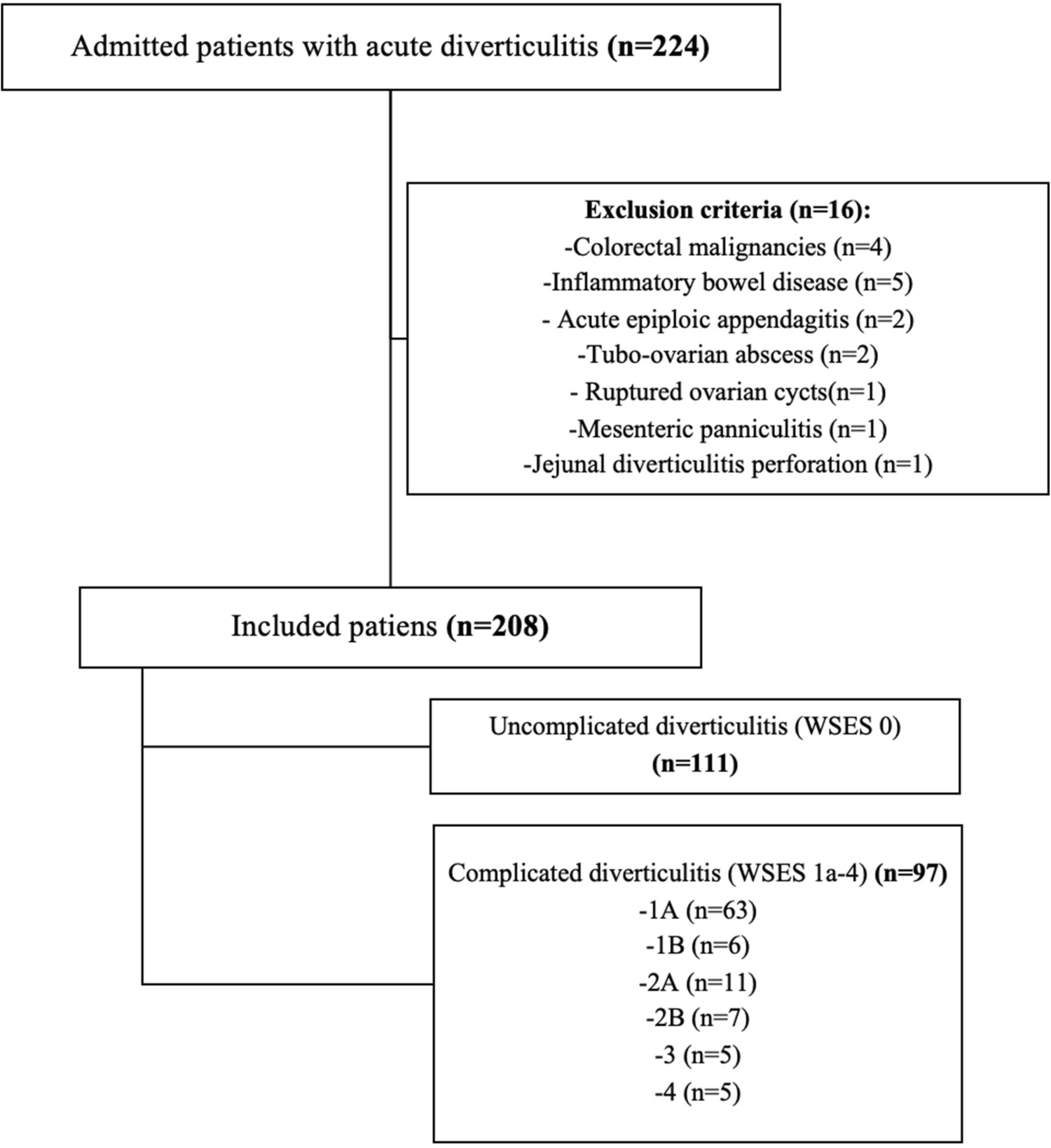
The study included 208 patients, of which 109 were female patients (52.4%) and 99 were male (47.6%), with a mean age of 57.2 (± 15.2) years. Uncomplicated group comprised 53.4% (n = 111) while complicated group comprised 46.6% (n = 97) (Table 1).
Table 1 Comparison of Demographics, Laboratory Findings, Clinical Course, Treatment, and Follow-up Parameters of Complicated and Uncomplicated DiverticulitisWhen clinical and laboratory findings were compared, CRP, WBC count, neutrophil count, neutrophil percentage, procalcitonin, and direct bilirubin were found to be significantly higher in the complicated diverticulitis group compared to the uncomplicated group (p < 0.001, p = 0.028, p = 0.003, p = 0.001, p = 0.005, p = 0.02, respectively) (Table 1). There were no significant differences in vital signs between the groups.
The overall mean follow-up period was 30.7 ± 14.8 (6–61) months, with the complicated group at 33.2 ± 15.9 months, significantly longer than the uncomplicated group at 28.5 ± 13.6 months (p = 0.026). Additionally, 32.7% of patients had a previous history of multiple episodes, while 67.3% had a single episode. There were no statistically significant difference in number of episodes between uncomplicated and complicated groups. Among the cohort, 154 individuals (74%) exhibited involvement of the sigmoid colon, whereas 23 patients (11.1%) were affected in the cecum, 19 patients (9.13%) in the descending colon, 9 patients (4.33%) in the ascending colon, and 3 patients (1.44%) in the transverse colon (Table 1). Sigmoid colon involvement was significantly more frequent in the complicated group (p = 0.005). Of the 208 patients, 177 (85.1%) received conservative treatment, 7 (3.37%) had percutaneous abscess drainage, 8 (3.85%) underwent elective surgery, and 16 (7.69%) required emergency surgery. There were no differences in operative indications between complicated and uncomplicated diverticulitis.
The mean BMI of patients was determined to be 29.1 ± 5 kg/m2 (Table 2). The mean BMI of the complicated diverticulitis group (28.3 ± 5.05) was statistically significantly lower compared to the uncomplicated group (29.8 ± 4.88) (p = 0.037). Similarly, the VFA of the complicated group (179 ± 82.8 cm2) was found to be statistically significantly lower compared to the VFA of the uncomplicated group (203 ± 86.3 cm2) (p = 0.046). The mean SFA of the patients was 234 (± 96.4) cm2. The V/S ratio was calculated as 0.89 (± 0.44). There was no statistically significant difference between the complicated and uncomplicated groups in terms of SFA and V/S.
Table 2 Statistical Analysis of Body Mass Index, Visceral Fat Area, and Subcutaneous Fat Area of Complicated and Uncomplicated Groups Based on World Society of Emergency Surgery ClassificationWhen comparing patients undergoing conservative treatment with those undergoing interventional treatment, the BMI in the interventional treatment group (25.9 kg/m2) was found to be lower compared to the conservative treatment group (29 kg/m2) (p = 0.007) (Table 3). Similarly, a significant difference was found in terms of VFA between the interventional treatment group (156 cm2) and the conservative treatment group (193 cm2) (p = 0.025). There was no significant difference observed in terms of SFA and V/S ratio regarding treatment modalities.
Table 3 Comparison of conservative treatment and interventional treatment groups concerning body mass index (BMI), visceral fat area (VFA), subcutaneous fat area (SFA), and visceral-to-subcutaneous fat ratio (V/S)The median BMI value (23.9) and VFA (142 cm2) of the 16 patients who underwent emergency surgery was 23.9, and it was were found to be statistically significantly lower compared to patients who did not undergo emergency surgery (29 kg/m2, 193 cm2, respectively) (p = 0.002, p = 0.034, respevtiveley) (Table 4). There was no significant difference between patients who underwent emergency surgery and those who did not in terms of SFA and V/S.
Table 4 Comparison of patients who underwent emergency surgery with other patients and Comparison of patients who underwent elective surgery with other patients concerning BMI, VFA, SFA, and V/S andThe median BMI value (25.2 kg/m2) and and VFA (112 cm2)of the 8 patients who underwent elective surgery were significantly lower compared to who did not undergo elective surgery (28.9 kg/m2, 191 cm2, respectively) (p = 0.031, p = 0.043, respectively) (Table 4). There was no significant difference between patients who underwent elective surgery and those who did not in terms of SFA and V/S.
A more comprehensive analysis was also conducted where the complicated group (radiological and/or clinical) are comprised of those categorized as WSES Stage 1 A, 1B, 2 A, 2B, 3, and 4 at the time of admission, as well as cases presenting with obstruction or fistula, abscess formation during follow-up, emergency or elective surgeries, percutaneous abscess drainage, and postoperative complications (n = 94) (Table 5). Patients who did not have any of these complications were classified into the non-complicated group (n = 114). The median BMI value of patients in the complicated group (27.3 kg/m2) was significantly lower than the non-complicated group (29.5 kg/m2) (p = 0.006). Similarly, there was a significant difference in VFA examination between the two groups (179 cm2, 201 cm2 respectively, p = 0.033) There was no significant difference in SFA and V/S between the two groups.
Table 5 Comparison of patients exhibiting radiological and/or clinical complications at presentation, and those who developed complications during follow-up, with Non-complicated patients regarding BMI, VFA, SFA, and V/SUnivariate Cox proportional hazards regression analysis identified several clinical, laboratory, radiologic and demographic factors significantly associated with the risk of complicated diverticulitis, including VFA (HR: 1.005, p = 0.0009), SFA (HR: 0.998, p = 0.0061), V/S ratio (HR: 3.40, p = 0.0002), CRP (HR: 1.021, p < 0.0001), WBC count, neutrophil metrics, procalcitonin, bilirubin levels, male gender, age ≥ 60, multiple episodes of diverticulitis, and sigmoid colon localization (Table 6).
Table 6 Univariate and Multivariate Analysis of Predictors of Uncomplicated versus Complicated DiverticulitisIn the multivariable Cox regression model, independent predictors of complicated diverticulitis included:
V/S Ratio (HR: 2.64, 95% CI: 1.44–4.84, p = 0.0021), CRP (HR: 1.014, 95% CI: 1.004–1.023, p = 0.0089), Total Bilirubin (HR: 1.24, 95% CI: 1.01–1.53, p = 0.0382), Multiple Episodes (HR: 1.42, 95% CI: 1.07–1.88, p = 0.0145), Male Gender (HR: 1.30, 95% CI: 1.06–1.60, p = 0.0129) and Sigmoid Colon Localization (HR: 0.84, 95% CI: 0.71–0.99, p = 0.0451) (Table 6). VFA lost significance in multivariate analysis (p = 0.0847), likely due to collinearity with the V/S ratio, while variables such as age and procalcitonin also became insignificant. These results indicate that fat distribution, inflammatory markers, number of episodes, and demographic factors are critical independent risk factors for complicated diverticulitis.

Comments (0)