The aim of our study was to evaluate the outcome of femoral shaft fractures, which were treated with open reduction, cerclage wiring and intramedullary nailing.
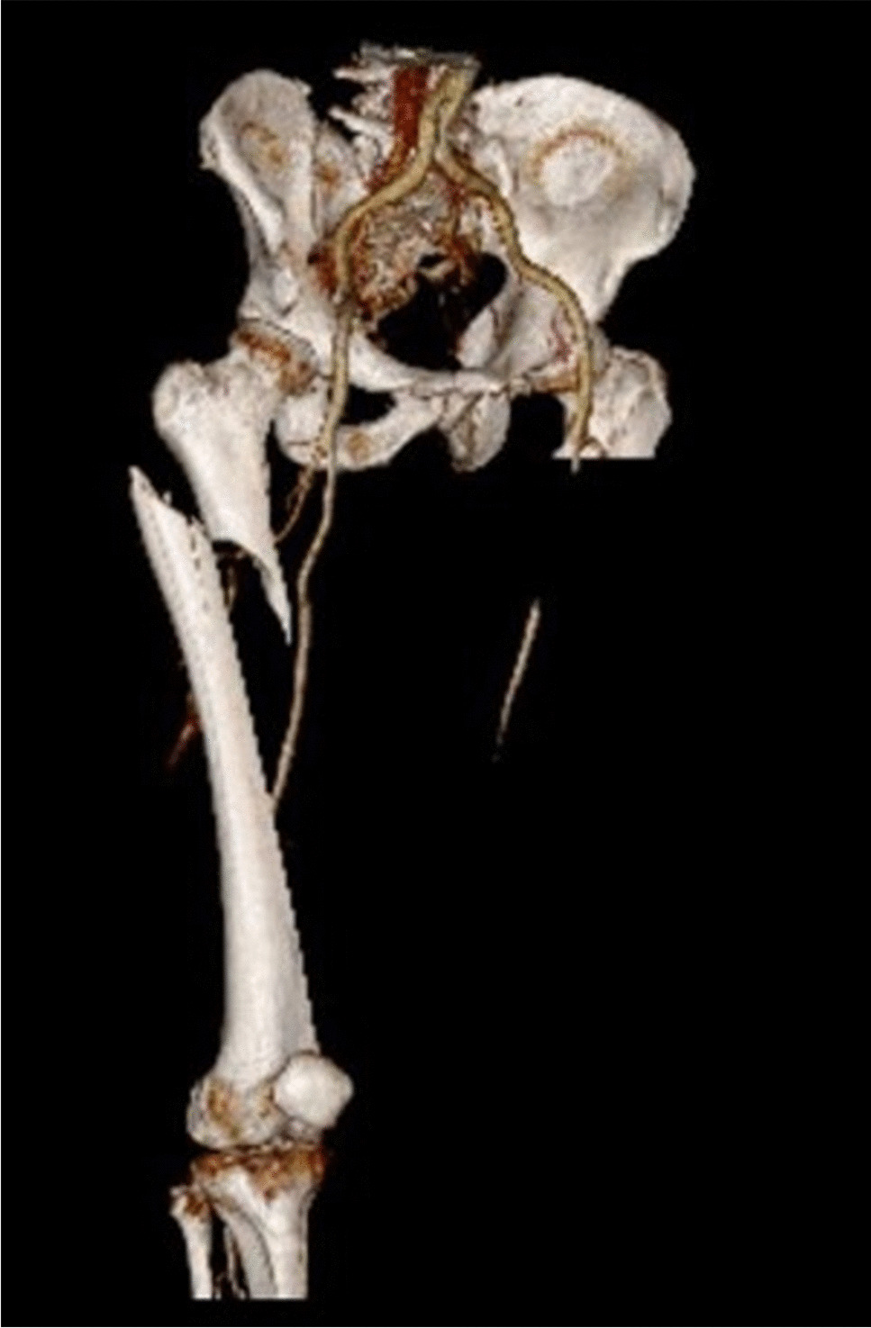
Our analyzed patient cohort was mainly male, middle-aged, mostly suffered from high velocity traumas and were frequently diagnosed with multifragmentary subtrochanteric fractures. In the provided figures, the pattern of injury as well as the intraoperative and postoperative X-Rays from a patient of the study cohort are exemplarily illustrated (Figs. 1–3).
One of our major results was, that these fractures were associated with union in 71%, delayed union in 10% (but eventually healed) and nonunion in 19% with the incidence of surgical site infection in 4% and the overall need for revision surgery in 19%.
Multiple studies exist about differences in outcome between open and closed reduction in intramedullary treatment of femoral shaft fractures [19,20,21]. Although there is common sense, that closed reduction is the preferred technique if an adequate reduction is possible, concrete details about the outcome after open reduction and internal fixation are not well described due to the small sub-cohort in most of the studies [22,23,24].
Union/nonunion
Salman et al. performed a systematic review and meta-analysis of 12 studies regarding outcome to compare open to closed reduction methods in intramedullary nailing treatment in femoral shaft fractures [22]. They found a significant better union rate following closed vs. open reduction, with an odds ratio of 2.06 (95% CI, 1.23–3.44). Our incidence of nonunion (19%) is in accordance with those findings, as well as with the results of a retrospective study by Kisan et al., who described a nonunion rate of 14.3% after open vs. 13% after closed reduction [22, 24].
In addition, we found a delayed union rate of 10%, while the review of Kortykowski et al. demonstrated in their review that open reduction was associated with a significantly longer time to union, which was reported by ten studies [25]. In the closed reduction group a time to union was 22 weeks, compared to 24 weeks in the open reduction group [25].
Overall, we noted a union rate of 81% without revision surgery, which is a good result considering that the technique of open reduction should be preferably applied in complex fractures when the attempt of closed reduction failed.
Infections
Korytkowski et al. performed a meta-analysis of comparative studies and described a lower overall infection rate in the closed reduction cohort ((OR = 0.581; 95% CI, 0.344- 0.981; p = 0.042) [25]. Similarly, the meta-analysis of Salman et al. revealed that the incidence of infection following open reduction was significantly higher with rates up to 24% compared to the closed reduction group in the analysed studies [22]. Pooled analysis showed a doubled risk of infection (OR = 1.94; 95% CI, 1.16–3.25, p < 0.05) [22]. We found a low number of surgical site infections with a rate of 4%. This may be due to the high medical standard and strict use of perioperative antibiotic prophylaxis, nevertheless, we cannot offer conclusive explanation for this low number.
Revision surgeries
Altogether, we registered a surgical revision rate of 19%. Salman et al. reported similar findings, but no significant difference in revision rates between both groups (OR = 0.85; 95% CI, 0.53–1.35) [22]. Our rate is based on the avascular femoral head necrosis as well as the nonunions.
Malunion
We did not observe any malunion. We attribute this to improved intraoperative control for restoring length, axis and rotation with open reduction techniques.
According to the literature malalignment was described to be significantly less with an OR of 0.32 in the open-reduction group in the literature, which shows the strength of this method to achieve a better anatomical reduction compared to closed reduction [22]. Malrotation is one of the most frequent malalignments following intramedullary nailing [26]. Even though not always clinically symptomatic, these malrotations can lead to an increased incidence of osteoarthritis of the hip, knee and ankle [27]. Karaman et al. showed that patients treated with closed reduction and intramedullary nailing with a malrotation of more than 10 degrees, had significantly lower functional scores compared to the cohort without rotational malalignment [28]. Malrotation preventing the achievement of the neutral rotational position bears the highest risk for clinical complaints [29]. This underlines the benefit of open-reduction techniques for precise and anatomical reconstruction in femoral shaft fractures.
In the past, there had been adverse discussions about the use of cerclage wiring which was believed to cause impairment of periostal perfusion and regional bone vascularity. This assumption had been refuted by studies describing no relevant effects of the cerclage wires on periosteal perfusion [30, 31].
In our study we used in 36% of cases two cerclages but had applied up to four cerclages per femur in selected cases of 13% which underscores the strategy of minimizing additional soft tissue trauma by confinement of the number of cerclages.
Regarding the limitations of our study, it is of paramount importance to acknowledge the scope of our dataset derived from a single center. As we focused in this study mainly on the description of the outcome of the patients treated with open reduction and cerclage wiring, our study lacks a CRIF cohort as a control group. As such, we were not able to compare directly a CRIF cohort with the ORIF group in terms of outcome. This constraint inherently limits to draw conclusions/the generalizability of our findings across a broader spectrum of clinical settings and populations. Associated injuries might necessitate different surgical approaches, rehabilitation protocols, and post-operative care, all of which could influence the outcomes observed with cerclage wiring.
Additionally, the diversity of cerclage materials and designs used in clinical practice further complicates the extrapolation of our findings. Different materials may have varying effects on bone vascularity and bone healing outcomes, and the design of the cerclage system (e.g., the number of wires used, their configuration) could also play a critical role.
Since we only included patients with cerclage wiring left in situ, the question if the reduction aid should be removed after implantation of the intramedullary nail or not cannot be addressed. This topic is still a matter of ongoing debate and will need further investigations.


Comments (0)