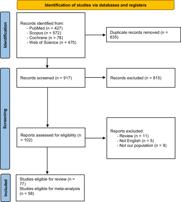
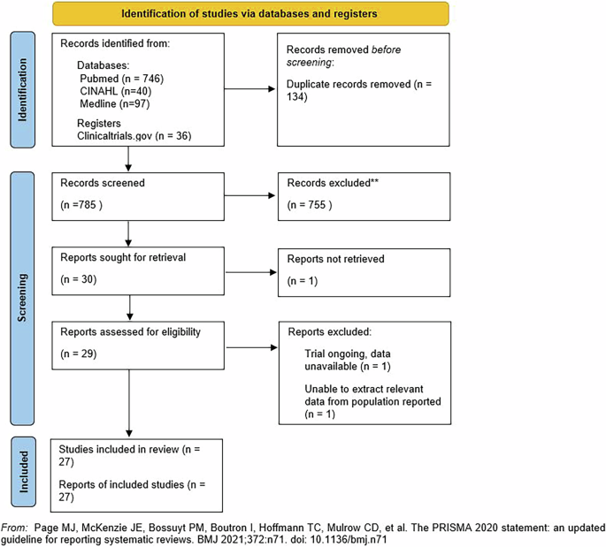
This systematic review and meta-analysis aimed to rigorously search the literature, providing an analysis of the current understanding of the role of OCTA in SCR diagnosis and monitoring. A total of 31 studies met our inclusion criteria [1, 6, 8,9,10,11,12, 16,17,18,19,20,21,22,23,24,25,26,27,28,29,30,31,32,33,34,35,36], with 17 (57%) of these studies being published since a previous systematic review was conducted by Guerra et al. in 2019 [13]. Significantly, the growing evidence base exploring OCTA as a tool for diagnosis and staging of SCR enabled us to conduct the first quantitative synthesis of published data, identifying several statistically significant markers of macular ischaemia in patients with SCR. A discussion of the review’s narrative and quantitative findings contextualised within the existing literature is detailed below.
The meta-analysis results show that OCTA is a useful tool for isolating quantitative markers of SCR. Across the studies in this review, FAZ size and vessel density were the most widely reported OCTA metrics. Mean vessel density was significantly lower in patients with SCR than in healthy controls [11, 17, 28, 29, 34, 37]. The FAZ area was larger, indicating that both could represent useful markers of sub-clinical macular ischaemia in patients with SCR [8, 11, 17, 21, 29, 34, 37]. Notably, several studies achieved greater sensitivity and specificity for SCR detection by combining several OCTA parameters to generate a single metric that indicates macular ischaemia. For example, Alam et al. conducted computer-aided SCR classification using six quantitative OCTA parameters, highlighting the potential for OCTA to produce biomarkers that facilitate objective and automated SCR classification [10]. Developments in this area could be instrumental in setting up cost-effective screening programmes for SCR utilising OCTA as a rapid, non-invasive primary screening tool.
Additionally, less commonly reported quantitative indicators of retinovascular remodelling, such as vessel tortuosity [23, 35], space between vessels [32] and fractal dimension [21], were found to be of value in detecting macular ischaemia in patients with SCR. However, analysis of less widely utilised OCTA measures requires specialist expertise in image processing and analysis, and therefore, despite the widespread use of OCTA, its potential to shed light on the vascular remodelling taking place in SCR remains relatively untapped. However, built-in software that automatically provides quantitative metrics is on the horizon and would quickly advance the clinical utility of quantitative OCTA metrics [39].
As anticipated, studies in this review showed a positive correlation between the retinal thinning on SD-OCT ETDRS retinal thickness maps and OCTA assessments of vessel density [1, 10, 17, 30, 33]. Hence, as demonstrated by Grego et al., OCTA has the potential to help characterise the distinct process of vascular remodelling that underlies the characteristic temporal retinal thinning widely documented in SCR [38]. Furthermore, OCTA has been demonstrated to work synergistically with additional imaging techniques, such as adaptive optics scanning light ophthalmoscopy, to improve SCR prognostication and predict response to systemic treatment. Therefore, the potential to characterise the pathophysiology of SCR and identify non-invasive biomarkers of SCR progression utilising a combination of OCTA metrics and multimodal imaging is promising. [30].
Notably, due to their small sample size, some studies in this review opted to report the relative frequencies of qualitative findings, such as the presence of flow voids or FAZ enlargement, rather than conducting a quantitative analysis. Generalisations of qualitative data analysis across studies cannot be made due to inter-study variations in image analysis and the inherent subjectivity of qualitative image grading. However, it is reassuring that where qualitative analysis was performed, results aligned with quantitative findings. FAZ size and flow voids in the DCP and SCP were assessed by most graders to be increased in patients with SCR, suggesting clinicians with limited expertise in OCTA data analysis may be able to utilise OCTA clinically to aid diagnosis and clinical decision-making (Table 2).
Importantly, several studies contrasted the role of OCTA with that of the current gold standard, FFA [5]. OCTA has garnered attention due to its non-invasive nature and ease of use [26], and studies in this review highlight the increased sensitivity of OCTA in detecting macular microvascular abnormalities in SCR compared to FFA [9, 26]. Minvielle et al. demonstrated that OCTA identified parafoveal vascular changes in all patients with SCD, whereas FFA detected abnormalities in only half [9]. Similarly, the case series by Sambhav et al. found that all patients exhibited normal FFA findings despite evidence of macular changes on OCTA [26]. This discrepancy is likely due to OCTA’s ability to provide depth-resolved imaging of the retinal capillary plexuses, particularly the DCP, which appears disproportionately affected in SCR [9, 26].
Furthermore, Sanfilippo et al. demonstrated that ultra-widefield OCTA can reveal DCP flow deficits that are less apparent on conventional FFA, suggesting its potential for comprehensive assessment of microvascular compromise extending into the retinal far periphery, an area of premium importance in SCR [18]. Nonetheless, while ultra-widefield OCTA may help bridge the gap between OCTA and FFA [18], FFA remains the gold standard due to its ability to reliably detect hallmark SCR features, including peripheral ischaemia, vascular leakage, arteriovenous anastomoses, and sea-fan neovascularization [5]. Further research is needed to establish the clinical utility of ultra-widefield OCTA and determine its optimal integration alongside FFA for the diagnosis and monitoring of SCR.
Concerning participant characteristics, it is significant that despite many of the included studies documenting participant data such as Goldberg stage, genotype, serology, and visual acuity, only a few studies had sufficient power to establish correlations between these metrics and OCTA changes. Consequently, our ability to establish associations between patient characteristics and OCTA imaging is limited. The absence of analytical statistics examining the relationship between the Goldberg stage—a grading system emphasizing peripheral retinal changes—and OCTA scans centred on the fovea is a significant limitation. This gap hinders our understanding of whether peripheral and central disease processes are distinct entities or represent different stages of a unified pathophysiological mechanism. Moreover, there is a pressing need for a more comprehensive understanding of the risk factors predicting adverse outcomes in SCR, given the non-linear evolution of its disease course and striking phenotypic diversity exhibited by each genotype [40]. For example, some patients with SCR progress rapidly, developing sight threatening complications, while others appear to be safeguarded by the phenomenon of autoinfarction, wherein bleeding vessels spontaneously occlude [41]. Moreover, investigating which imaging OCTA biomarkers are associated with adverse clinical outcomes (such as reduced visual acuity) and how these relate to existing feature-based grading systems is critical for determining the clinical utility of OCTA.
Patient genotype was measured by 27 out of 30 studies (90%). The HbSC genotype is commonly thought to possess the highest risk of proliferative SCR development [39], while HbSS is associated with non-proliferative SCR. However, in this review, only one cross-sectional study conducted by Han et al., with a relatively small sample size of ten eyes, corroborated this finding [19]. Instead, most studies showed no relationship between genotype and SCR. Surprisingly, the cross-sectional analysis conducted by Fares et al., which utilised a dataset of 151 eyes, reported that HbSS genotype, rather than HbSC genotype, was associated with an increased risk of sickle cell maculopathy [8]. It has been posited that genetic differences in endothelial function in patients with sickle cell disease may contribute to its phenotypic diversity. Endothelial cells that release VEGF in response to inflammation, particularly when unopposed by growth-inhibiting factors like Thrombospondin-A, are more likely to develop neovascular SCR. However, accounting for diverse endothelial responses and, therefore, phenotypic variability risk stratifying genotypes presents a challenge and may explain the inconsistent findings in this review [42]. Furthermore, it is important to highlight that out of all participants included in the studies synthesised within this review, only 19.89% of participants were of the HbSC genotype. While this reflects the population prevalence of HbSS and HbSC genotypes, future research should focus on sampling those with HbSC genotypes, such that sample sizes of HbSC patients suitable for analytical statistics can be generated.
Additionally, serological markers for disease activity could prove pivotal in enhancing our understanding of which patients with sickle cell disease are most likely to develop active SCR. Two cross-sectional studies linked lower levels of foetal Hb, high reticulocyte count and prothrombin ratio to evidence of macular ischaemia on OCTA, indicating that patients with hypercoagulability may be less at risk of retinal complications [8, 38]. While this finding aligns with the hypothesis that hypercoagulability may promote autoinfarction and be protective in the retina, more research is required to prove the link between serological markers and SCR progression in patients with sickle cell disease.
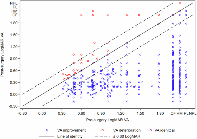
Understanding the relationship between OCTA metrics and clinical outcomes, such as visual acuity, is key to determining the clinical utility of OCTA imaging. While many studies recorded participant visual acuity, reporting a high mean visual acuity of 20/20 across study participants, only Han et al. correlated visual acuity data with OCTA findings [33]. Han et al. recorded the visual acuity of 46 patients with SCR and found a significantly positive correlation between the extent of parafoveal flow loss (assessed qualitatively) and patients’ logMar acuity [33]. This finding complements previous studies that have successfully correlated OCTA quantitative measures such as FAZ size and macular vascular density with visual acuity in diabetic retinopathy and vein occlusions [43]. However, based on this study alone, no conclusions can be drawn about which OCTA changes are likely to be subclinical and which could lead to a fall in acuity. Altogether, more evidence is needed to characterise the relationship between OCTA markers of vascular remodelling and visual outcomes.
The Goldberg staging system, produced in 1980, remains the most widely recognised system for grading the severity of SCR [3]. The feature-based stages use macroscopic retinal changes to predict which patients are most likely to develop clinically significant, sight-threatening complications. Only one study in this review that attempted to correlate OCTA imaging findings and Goldberg staging could prove a statistically significant relationship [31]. The lack of correlation may indicate two distinct disease processes, i.e. macular disease and peripheral disease in people with SCD. However, statistically significant correlations between markers of macular ischaemia identified on OCTA and evidence of temporal retinal thinning on SD OCT increasingly suggest macular and peripheral remodelling are interrelated [6]. Alternatively, the lack of correlation may reflect the limited sample sizes in many of the studies included in this review. A grading system for SCR, which incorporates microscopic indicators of vascular remodelling in the macula, could aid SCR staging and prognostication.
Limitations and future research
A key strength of this review was the quantitative analysis of several OCTA metrics across several studies evaluating SCR in patients with sickle cell disease. Nonetheless, the conclusions of our analysis remain dependent on the quality of the available evidence. All studies that met our inclusion criteria were observational rather than randomised controlled trials. Consequently, uncontrolled confounders limit the extent to which conclusions can be drawn about cause and effect. The heterogeneity in OCTA technology, imaging schedules, and reporting between studies also limited our findings and meant some evidence could not be included in our final analysis. For example, only studies with comparable imaging areas could be combined and it is possible that incorporating Heidelberg, Optovue and Topcan devices introduces bias. Moreover, the small sample size available for evaluation included in most studies made it difficult to draw statistically significant associations between participant characteristics and OCTA imaging results. Additionally, the studies included in this review sought to identify statistically significant differences between patients with SCR and healthy controls. However, it is well established that OCTA metrics, such as FAZ size, exhibit considerable variation within healthy populations due to confounding factors such as age and sex (https://www.sciencedirect.com/science/article/abs/pii/S0014483519305895). Consequently, these findings must be interpreted within the context of population-level data, emphasizing the need for further studies to establish normative OCTA parameters across diverse age groups, genders, and ethnicities.
Future research focusing on prospective, longitudinal analysis of OCTA data from a large cohort of patients with SCR would be of great value in consolidating our understanding of how OCTA could be used to improve SCR diagnosis and management. The movement toward developing national and international imaging banks similar to those available for diabetic eye disease would be of great value in linking imaging results with clinical outcomes such as visual acuity or sight-threatening complications. The aggregation of such large imaging datasets, combined with advancements in machine learning, could aid the development of automated SCR screening and detection [44]. Furthermore, an improved understanding of which patients are at risk of ocular and systemic disease progression based on OCTA imaging data could prove vital for guiding the management of both ocular and systemic treatments for patients with sickle cell disease.

Comments (0)