
Remember me
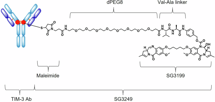


KK2845 consists of a fully human IgG1 antibody TIM-3 Ab, which targets human TIM-3, and SG3249 stochastically conjugated at cysteines of TIM-3 Ab via thiol-maleimide coupling. SG3249 is composed of a PEG-8 spacer, a valine-alanine linker and PBD dimer warhead SG3199 (Fig. 1). The valine-alanine linker is cleaved by cathepsin in the lysosome compartment to release the warhead [11].
Fig. 1: Structure of KK2845.
KK2845 is an ADC that consists of an anti-TIM-3 fully human IgG1 antibody, a valine-alanine linker, and a highly potent DNA cross-linking PBD dimer SG3199.
Binding of KK2845 to TIM-3 antigenThe binding affinity of TIM-3 Ab and KK2845 for human, cynomolgus monkey, mouse, rat, and rabbit TIM-3 was evaluated by a surface plasmon resonance (SPR) analysis. The binding parameters are summarized in Supplemental Table 1 and Supplemental Table 2. TIM-3 Ab and KK2845 clearly bound to human and cynomolgus monkey TIM-3. Meanwhile, no specific binding of TIM-3 Ab and KK2845 to mouse, rat, or rabbit TIM-3 were observed.
Internalization of KK2845To evaluate internalization, we used Kasumi-3 [16], an AML cell line expressing both TIM-3 and CD33 (supplemental Fig. 1). The incubation of Kasumi-3 with KK2845-Alexa488 for 30 minutes on ice showed restricted localization of KK2845-Alexa488 on the cell surface (Fig. 2). When Kasumi-3 was incubated with KK2845-Alexa488 at 37 °C for 2 or 24 hours, KK2845-Alexa488 was translocated into cells, and a part of the intracellular signal was co-localized with LysoTracker signal (Fig. 2). The internalization rate of KK2845 was almost comparable with that of CD33-ADC after 24 hours incubation in Kasumi-3 cells (Supplemental Fig. 2).
Fig. 2: Localization and trafficking of KK2845.
KK2845-Alexa488 was incubated with Kasumi-3 cells on ice (0 hour), or at 37 °C for 2 hours or 24 hours. The Alexa Fluor 488 signal is displayed in green. The LysoTracker signal is displayed in red. The DAPI signal is displayed in blue. Yellow colored signals indicated by arrows show colocalization of KK2845-Alexa488 and Lysotracker. Original magnification ×100 for all the imaging.
Cytotoxicity of KK2845 against AML cell lines in vitroThe cytotoxicity of KK2845 was assessed in AML cell lines Kasumi-3 and EoL-1/hTIM-3 [17]. The TIM-3 expression in EoL-1/hTIM-3 is shown in Supplemental Fig. 1. The cells were treated with KK2845 or negative control KM8047-SG3247 at concentrations from 0.01 to 10000 ng/mL (Fig. 3). The IC50 of KK2845 and KM8047-SG3247 against Kasumi-3 was 49.58 ng/mL and 1576.30 ng/mL, respectively (Supplemental Table 3). The IC50 of KK2845 and KM8047-SG3247 against EoL-1/hTIM-3 was 1.09 ng/mL and 495.30 ng/mL, respectively (Supplemental Table 3). Both KK2845 and KM8047-SG3249 showed weak cytotoxicity against TIM-3-negative EoL-1 cells, with IC50 values of 553.70 ng/mL and 640.80 ng/mL, respectively (Supplemental Table 3).
Fig. 3: Cytotoxicity of KK2845 against AML cells.
A-C Kasumi-3, EoL-1/hTIM-3, or EoL-1 cells were incubated with KK2845 (0.01-10000 ng/mL) or KM8047-SG3249 (0.01-10000 ng/mL) at 37 °C for 4 days. CCK-8 was then added to these cells, and the absorbance was measured. The mean ± SD cell viability (%) is shown (n = 3). The data presented are representative results from three independent tests.
Cytotoxicity of KK2845 against primary AML cells in vitroThe TIM-3 and CD33 expression levels in LSC fraction (CD45dimCD34+CD38-), leukemic progenitor fraction (CD45dimCD34+CD38+), and CD34- blast fraction (CD45dimCD34-) of 13 different AML patients is quantified and described in Supplemental Fig. 3.
Next, the cytotoxicity of KK2845 and CD33-ADC against three different lots of primary AML cells, AML818BM (Fig. 4A and B), AML817BM (Fig. 4C), and AML942 II BM (Fig. 4D), was tested. In AML818BM, CD34+ cells that contain LSCs and leukemic progenitors were the major fraction. In AML817BM, CD34- blast cells were the major fraction. In AML942 II BM, CD34+ leukemic progenitors were the major fraction. The numbers of TIM-3 and CD33 molecules on each fraction of these three lots of primary AML cells are described in Supplemental Table 4. Living CD34+ cells, CD34+CD38- cells where LSCs were concentrated, and CD34- cells were counted 4 days after treatment of KM8047-SG3249, KK2845, or CD33-ADC (Fig. 4). As a result, in AML818BM, both CD34+ cells and CD34+CD38- cells were killed by KK2845 or CD33-ADC in a dose-dependent manner from 1 or 10 ng/mL, respectively, in comparison to KM8047-SG3249 (Fig. 4A, B, and Supplemental Fig. 4). At 1 ng/mL, the percentage of living CD34+ cells and CD34+CD38- cells after treatment with KK2845 was lower than that with CD33-ADC treatment. At 10 and 30 ng/mL, the percentage of living CD34+ cells and CD34+CD38- cells after treatment with KK2845 was almost comparable to that with CD33-ADC. In AML817BM, living CD34- cells were killed by KK2845 and CD33-ADC in a dose-dependent manner from 1 ng/mL in comparison to KM8047-SG3249 (Fig. 4C). At all concentrations tested, the percentage of living CD34- cells after treatment with KK2845 was almost comparable to that with CD33-ADC. In AML942 II BM, CD34+ cells were killed by KK2845 and CD33-ADC in a dose-dependent manner from 3 ng/mL in comparison to KM8047-SG3249 (Fig. 4D). At all tested concentrations, the percentage of living CD34+ cells after treatment with KK2845 was almost comparable to that with CD33-ADC.
Fig. 4: Cytotoxicity of KK2845 against primary AML cells.
A-D The primary AML cells were incubated with KM8047-SG3249, KK2845, or CD33-ADC at 37 °C for 4 days. The payload of the CD33-ADC used in this study was SGD-1910. The living cells in the CD34+ fraction, CD34+CD38- fraction, or CD34- fraction were then counted by flow cytometry. Using the number of viable cells in the untreated group as 100%, the percentage of living cells in each treatment group was calculated. A, B Lot: AML818BM. The CD34- fraction was not evaluated due to a low number of cells. C Lot: AML817BM. The CD34+ fraction was not evaluated due to a low number of cells. D Lot: AML942 II BM. The CD34- fraction was not evaluated due to a low number of cells.
In conclusion, KK2845 had different sensitivities to distinct lots of primary AML cells, with the IC50 of approximately 1-20 ng/mL. The cytotoxicity was almost comparable between KK2845 and CD33-ADC.
ADCC of KK2845To evaluate ADCC activity, KK2845 was added to EoL-1/hTIM-3 cells co-cultured with human PBMCs derived from three different healthy donors. No cytotoxicity was observed when EoL-1/hTIM-3 were incubated with KK2845 alone without PBMCs (Supplemental Fig. 5), indicating that cytotoxicity observed in this assay system was dependent on effector cells but not on the payload of KK2845. Under this assay condition, the ADCC activity was observed when EoL-1/hTIM-3 cells were treated by 0.1 μg/mL or 10 μg/mL of KK2845 in the presence of PBMCs (Fig. 5).
Fig. 5: ADCC activity of KK2845.
A-C EoL-1/hTIM-3 cells were incubated with KK2845 (0.001-10 μg/mL) or KM8047-SG3249 (10 μg/mL) in the presence of human PBMCs at 37 °C for 4 hours. After incubation, the number of viable EoL-1/hTIM-3 cells and BD Trucount beads were counted by flow cytometry. The number of living EoL-1/hTIM-3 cells was normalized by the number of beads. ADCC activity was calculated as follows: ADCC activity (%) =100 - (Normalized cell number of each sample / Average (n = 4) of normalized cell number of sample A) ×100. The mean + SD is shown (n = 4). A PBMC donor 1, B PBMC donor 2, C PBMC donor 3.
Hematotoxicity of KK2845 in vitroTIM-3 is expressed only in a fraction of granulocyte/macrophage progenitor cells, but not on normal HSCs, whereas CD33 is broadly expressed on both normal HSCs and myeloid progenitor cells [12, 13]. To compare the hematotoxicity between KK2845 and CD33-ADC in vitro, we cultured human bone marrow-derived CD34+ cells in the presence of KM8047-SG3249, KK2845, or CD33-ADC at concentrations from 0.1 to 1000 ng/mL (Fig. 6). The viability of CD34+ cells was not affected by KK2845 treatment at concentrations of up to 1000 ng/mL, whereas it was inhibited by CD33-ADC treatment at concentrations of 100 and 1000 ng/mL (Fig. 6A).
Fig. 6: Cytotoxicity of KK2845 against human normal bone marrow cells.
A-B CD34+ bone marrow cells, or myeloid progenitor cells were incubated with PBS or increasing concentrations of KM8047-SG3249, KK2845, or CD33-ADC (0.1-1000 ng/mL) for 4 days. The payload of the CD33-ADC used in this study was SG3249. To assess cell viability, CellTiter-Glo® reagent was then added to these cells, and the luminescence was measured. Using cell luminescence, the percentage in each treatment group was calculated by setting the PBS treatment group (untreated control) as 100%. The mean ± SD of cell viability (%) is shown (n = 3). The data presented are representative results from two independent tests.
Then, we induced myeloid progenitor cells from CD34+ cells in vitro and monitored hematotoxicity as well. The viability of myeloid progenitor cells was not affected by KK2845 treatment at concentrations of up to 1000 ng/mL, whereas it was inhibited by CD33-ADC treatment at concentrations of ≥10 ng/mL (Fig. 6B).
Anti-tumor activity of KK2845 in vivoThe anti-tumor activity of KK2845 in vivo was examined in two subcutaneous xenograft models (Kasumi-3 and CMK11-5 [18]) and one disseminated xenograft model (EoL-1/hTIM-3). The antigen expression in these cell lines is shown in Supplemental Fig. 1. In the Kasumi-3 subcutaneous model, a single dose of KK2845 (0.25 mg/kg, 0.5 mg/kg, and 1 mg/kg) inhibited tumor growth (Fig. 7A). In the CMK11-5 subcutaneous model, a single dose of KK2845 (0.25 mg/kg and 0.5 mg/kg) also inhibited tumor growth (Fig. 7B).
Fig. 7: Anti-tumor activity of KK2845 in murine models of AML.
SCID mice were subcutaneously inoculated with Kasumi-3 (A) or CMK11-5 cells B. Vehicle, KK2845, or KM8047-SG3249 was intravenously administered once on day 1. Each plot represents the mean ± SE of tumor volume. (A: n = 7, B: n = 5). The difference in tumor volume on day 14 (A, B) between KK2845 and KM8047-SG3249 treated groups at each dose, was analyzed by Student’s t test or the Aspin-Welch test. *: p < 0.05. C A scheme for KK2845 efficacy evaluation using a disseminated model is presented. EoL-1/hTIM-3 cells were intravenously inoculated into NOD-SCID mice. After confirming EoL-1/TIM-3 engraftment in the bone marrow of satellite mice (n = 2) by detecting human CD45+ cells using flow cytometry, vehicle, 0.3 mg/kg of KM8047-SG3249, KK2845, or CD33-ADC was intravenously administered once. The payload of the CD33-ADC used in this study was SG3249. Five days after the administration of ADC, bone marrow cells were collected from the femur of each mouse. The residual cancer cells were analyzed by flow cytometry. D Each plot represents the individual value for the percentage of residual cancer cells. Horizontal lines represent the mean of each group (n = 10). E Overall survival curves in the disseminated EoL-1/hTIM-3 xenograft model. EoL-1/hTIM-3 cells were intravenously inoculated into NOD-SCID mice. After confirming EoL-1/TIM-3 engraftment in the bone marrow of satellite mice, vehicle, 0.3 mg/kg of KM8047-SG3249 or KK2845 was intravenously administered once. The survival time of the mice was monitored. The difference in overall survival between KK2845 and KM8047-SG3249 treated groups was evaluated by Log-Rank test (n = 8/group). *: p < 0.001. F The combination of KK2845, Venetoclax and Aza using subcutaneous xenograft model. SCID mice were inoculated with Kasumi-3 cells. Mice received 0.5 mg/kg KK2845 alone (single administration, i.v.), 100 mg/kg venetoclax (qd×7, p.o.) in combination with 5 mg/kg Aza (qd×7, i.v.), or 0.5 mg/kg KK2845 in combination with 100 mg/kg venetoclax and 5 mg/kg Aza from day 1. The time course of the mean tumor volume is shown. Each plot represents the mean ± SE of tumor volume (n = 5). The difference in tumor volume on day 21 between non-treatment and the triple-combination (KK2845, venetoclax and Aza), the dual-combination (venetoclax and Aza) and the triple-combination, or KK2845 monotherapy and the triple-combination were analyzed by the Aspin-Welch test. ‡: p < 0.05, difference between non-treatment and the triple-combination. #: p < 0.05, difference between the dual-combination and the triple-combination. *: p < 0.05, difference between KK2845 monotherapy and the triple-combination.
In the disseminated model (Fig. 7C), a single dose of KK2845 (0.3 mg/kg) completely eliminated EoL-1/TIM-3 cells engrafted in mouse bone marrow (Fig. 7D). Moreover, the anti-tumor activity was almost comparable between KK2845 and CD33-ADC at the same dose (Fig. 7D). Additionally, KK2845 significantly enhanced survival in this model, demonstrating potent anti-leukemia activity (Fig. 7E).
Next, we evaluated the anti-tumor activity of KK2845 in combination with venetoclax and Aza in Kasumi-3 subcutaneous xenograft model. As shown in Fig. 7F, the dual-combination of venetoclax and Aza did not show obvious anti-tumor activity. Meanwhile, KK2845 monotherapy clearly inhibited tumor growth. The triple-combination of KK2845, venetoclax and Aza also showed superior anti-tumor activity in comparison to KK2845 monotherapy.
KK2845 PK in cynomolgus monkeyKK2845 was administered to cynomolgus monkeys by intravenous infusion at doses of 0.2, 0.4, or 0.6 mg/kg, and the concentrations of the conjugated antibody and the total antibody in serum were evaluated. No animals were found dead or were euthanized due to moribundity in any group in the study period. No major differences were noted in the serum concentrations or pharmacokinetic parameters between the conjugated antibody and the total antibody (Supplemental Table 5 and 6), suggesting that free payloads were rarely released from ADC in the monkey blood stream.
Comments (0)