Cytotoxicity tests are a fundamental step in evaluating the biocompatibility of a material. This study was conducted to assess the toxicity of universal adhesives with different chemical compositions to human pulp cells.
Animal experiments and cell culture tests are commonly used to assess the cytotoxicity of dental materials. However, animal experiments are controversial, time-consuming, and costly methods [36]. Due to their advantages, such as low cost, controllability, and ease of application, cell culture tests have been increasingly used as an alternative to animal experiments [37]. In vitro cell culture testing is widely utilized in restorative dentistry to evaluate the biocompatibility of dental materials [38]. Various in vitro testing methods such as indirect and extract tests are used to evaluate the cytotoxic effects of biomaterials in cell culture studies [39, 40]. The most widely method used for these tests is the extract method. This method allows for the simulation of the clinical environment, since the adhesives are not in direct contact with the cells. Pagano et al. reported that the extract test method was more suitable for evaluating adhesives [41]. Therefore, the extract test method was chosen for this study.
The MTT test is an objective method for determining cell vitality and is applied to all cells that are metabolically active and, therefore, alive [42]. It is frequently cited in the literature as the chosen method to test the cytotoxicity of dental adhesives and other dental materials [43, 44]. In this study, the MTT test was used to detect cell vitality.
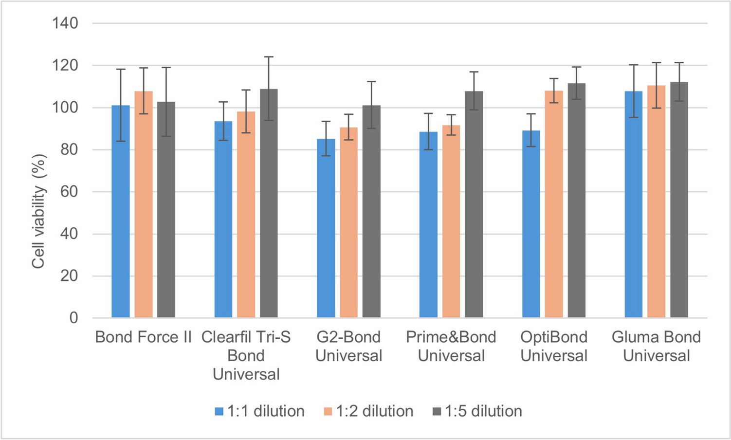
In addition to the composition of adhesives, pH is an important factor that can affect cytotoxicity. The pH values of universal adhesives are categorized into four groups: moderately strong (<1), strong (1,5), mild (>2), and ultra-mild (2,5) [45]. In this study, universal adhesives with a strong pH (GBU and G2BU, pH: 1.5), mild pH (OBU, pH: 2,3), and ultra-mild pH (BFII, pH: 2,8; C3SUB, pH: 2,5; PBU, pH: 2,5) were used. According to previous studies, the toxicity of adhesives has been reported to increase with higher acidic levels. However, it was not possible to evaluate the sole effect of pH on cytotoxicity in this study [46]. In addition, when the results of the study were examined, it was observed that as dilution increased, cytotoxic effects decreased, which may be related to the reduction in acidity. However, further studies are needed to investigate this topic in more detail.
It has been reported that parameters such as the type of curing device tip, light spectrum, and intensity used in the polymerization of dental adhesives affect their cytotoxic properties [47]. Polymerization with low-power light may prevent the complete conversion of monomers into polymers, leading to an increase in residual monomer content and a risk of leakage into surrounding tissues [48]. Therefore, the characteristics of the light-curing devices used during polymerization are of great importance. In a study conducted by Ergün et al., the cytotoxic effects of LED and halogen light sources on three different adhesive systems were examined [47]. Another study evaluated the cytotoxicity of an adhesive system on L929 mouse fibroblast cells using both LED light-curing devices and chemical polymerization methods [28]. In both studies in the literature, it was determined that the number of cells in the experimental group polymerized with an LED light device was higher, indicating lower cytotoxic effects [28]. Therefore, in this study, an LED light source with a power of 1200 MW/cm2 was applied to ensure proper polymerization and light intensity.
Bis-GMA, UDMA, TEGDMA and HEMA monomers in adhesives are associated with cytotoxicity [49, 50]. Among the monomers used in adhesives, Bis-GMA has relatively high cytotoxicity, but due to its high molecular weight it has a low capacity to penetrate dentine [51]. Bis-GMA has been documented to induce cytotoxicity in pulp cells by stimulating prostanoid production, potentially leading to inflammation or pulpal necrosis as a consequence of increased reactive oxygen species (ROS) generation [52]. On the other hand, HEMA has the ability to rapidly diffuse through dentin and can cause toxicity in the pulp tissue of cells [51]. It can also suppress the growth of many cell types and causes a delay in primary fibroblast cell cycle progression, especially by increasing the formation of reactive oxygen species (ROS) [53]. If ranking these monomers by toxicity, the most toxic is Bis-GMA, followed by UDMA, TEGDMA and HEMA, the least toxic [28]. The release of residual monomers, such as HEMA, Bis-GMA, and TEGDMA, which cause toxicity, is directly influenced by the degree of monomer conversion that occurs during adhesive polymerization [54]. It is known that current adhesive systems cannot hermetically seal deep dentin, and therefore, these residual monomers and other components of adhesive systems may penetrate through the dentinal tubules into the dental pulp chamber, potentially causing irreversible damage to the cellular tissue [55].
In addition to the cytotoxicity of traditional monomers found in dental adhesives, the cytotoxicity of acidic functional monomers is also important. Universal adhesives contain different acidic functional monomers, such as 10-methacryloyloxydecyl dihydrogenphosphate (10-MDP) and 4-methacryloyloxyethyl trimellianhydride (4-META). 10-MDP is the most commonly used acidic monomer in universal adhesives. It contains a dihydrogen phosphate group that causes tooth corrosion and a methacrylate group for cross-binding with other resin monomers [56]. 10-MDP promotes an inflammatory response and suppresses odontoblastic differentiation of human pulp cells [57]. 10-MDP also stops odontoblastic differentiation, but it is also thought to easily bind to calcium made by odontoblast-like cells and stop mineralization directly [58].
Although 4-META, another functional monomer used in adhesives, has been shown to have a high level of biocompatibility with dental pulp cells, studies investigating its cytotoxicity are limited [59, 60]. In addition, the interaction between monomers is also very important in terms of toxicity, as it has been shown that synergistic effects between monomers may develop when combinations of 4-META with TEGDMA, UDMA or especially Bis-GMA are tested [28].
The results of in vitro studies on adhesives provide useful information, although this information is not always interrelated. Many in vitro studies have reported different levels of cytotoxic effects of the adhesive system [40, 41, 61]. Testing nine adhesives, Tu and et al. [62] examined their cytotoxic effects on oral epithelial cells. Özen and et al. [63] noted that four different adhesives exhibited cytotoxicity to gingival fibroblast cells after 72 h. Demirel et al. [40] noted that the 1:1 adhesives extracts showed 60–90% cell vitality on L929 cells after 72 h. Yavuz and Surmelioglu reported that the cytotoxicity of adhesives varies depending on the exposure time [64].
The universal adhesives used in this study showed a cytotoxic effect after 48 h and 72 h, although they did not exhibit a 24-h cytotoxic effect. These results are similar to the results of studies discussed in the literature [40, 63]. The toxic effects of universal adhesives decreased as the dilution rate increased. Among the universal adhesives, those containing 4-META (GBU) affected pulpal cell viability less than those without 4-META. There were no statistically significant differences in composition between the universal adhesives containing 4-MET (G2BU), 10-MDP (C3SB, G2BU, PBU), 10-MDTP (G2BU) and GPDM (OBU) at all three times.
Inflammatory mediators are released by cells exposed to adhesive materials [14]. Increased mediators have characteristic functions, such as involvement in the inflammatory process, differentiation and activation. For example, IL-6 is a pleomorphic cytokine that plays a particular role in the development and progression of inflammation. It is usually found in low doses in healthy teeth, but high rates are associated with irreversible pulpitis [15]. In this study, the universal adhesives caused an increase in the release of TNF- α and IL-1 at the end of the first 24 h, but the release of TNF-α and IL-1 decreased in the control group after 72 h. The inflammatory response observed at the end of 24 h is thought to be caused by the monomers present in the adhesives, including Bis-GMA, TEGDMA, UDMA, HEMA, 10-MDP, 10-MDTP, 4-MET, and PENTA. In addition, among the tested adhesives, GBU induced the highest increase in TNF-α and IL-1 release at the end of the first 24 h. The low pH (1.5) and HEMA content of GBU can be considered potential factors contributing to the severity of the inflammatory response.
Monomers, such as TEGDMA, that leak from resin-based restorative materials have been shown to activate and induce the production of MMP-2, MMP-8 and MMP-9 in pulp cells [65]. In the studies of Lehmann N et al. and Van Meerbeck B et al., an increase in pulp MMP-2 and MMP-9 expression was found in self-etch adhesive application [66, 67]. Similarly, Orsini et al. observed an increase in MMP-2 expression after applying it directly onto human pulp cells, although using different adhesives than those used in this study [68]. In this study BFII containing TEG–DMA increased MMP-2 and MMP-8 levels, although statistically not significant, while PBU and OBU decreased MMP-8 levels. This could be due to the pH levels of PBU (2,5) and OBU (2,3).
Matrix metalloproteinases (MMPs), which are among the most important proteolytic enzymes of the extracellular matrix (ECM), have the ability to degrade all protein-based ECM components. MMP activity plays a role in various oral pathologies, including dental erosion, tooth decay, pulpitis, periapical periodontitis, and oral cancer invasion and metastasis [6,7,8]. In addition, MMP activity is involved in odontogenesis and tissue regeneration, and its dysregulation can lead to inadequate enamel mineralization or gingival overgrowth [9]. Therefore, fluctuations in MMP levels can disrupt the balance between degradation and repair processes. In particular, the direct application of adhesives to the pulpal wall in deep cavities may lead to pulpal pathologies and, consequently, periapical inflammation.
In this study, universal adhesives showed an increase in TNF- α and IL-1 levels in pulp cells after 24 h. However, at the end of 72 h this increase was not statistically significant. Universal adhesives also increased the amount of DMP-1 in pulp cells after 24 h, but did not make a statistically significant increase after 72 h. This increase in the DMP-1 level of pulpal cells at the end of 24 h is considered to be related to the increase in the amount of cytokines (TNF-a and IL-1) released from pulpal cells after application of universal adhesives.
Yoshioka et al. reported on the critical molecules involved in tertiary dentin formation using a comprehensive gene expression analysis on pulp cells [69]. According to the results, hybridization reveals that TIMP-1 is expressed by pulpal cells. It is also noted that TIMP-1 plays a role in the wound healing process in pulp tissue and could be a biological target for regenerative therapy [61].
In this study, universal adhesives did not produce a statistically significant increase in the amount of MMP-2 and MMP-8 in pulp cells. They did, however, cause a decrease of the tissue inhibitor TIMP-1 in the pulp cells. Although this decrease is not a statistically significant difference, it is estimated to be associated with an increase in MMP-2 and MMP-8.
The level of MMP, responsible for proteolytic activity, remained unchanged; however, the level of TIMP-1, which is responsible for tissue regeneration and the suppression of MMP’s proteolytic activity, decreased. This may hinder the proper healing of the dentin–pulp complex, negatively affecting reparative dentin formation and tissue regeneration. In addition, insufficient TIMP-1 levels may contribute to inflammation and excessive tissue remodeling, which could impair pulp tissue healing and long-term stability. Therefore, the decrease in TIMP-1 may indicate a disruption in the regulation of MMP activity, potentially leading to an impaired dentin-pulp healing process.
The limitations of this study are that it took place in an in vitro environment and was carried out on pulp cells for a short length of time. After the adhesives are applied to the tooth, they remain longer in the oral environment than the time tested in this study.
Within the limitations of this in vitro study, universal adhesives showed time- and dose-dependent cytotoxic effects on human pulp cells. Universal adhesives increased the amount of the DMP-1 protein in the pulp cells within the first 24 h, but did not affect it after 72 h. The level of the tissue inhibitor TIMP-1 in the pulp cells was reduced by the universal adhesives used, whereas the levels of MMP-2 and MMP-8 were unaffected.
Therefore, the application techniques of universal adhesives should be carefully reviewed to minimize cytotoxicity risks. In deep dentin cases with a thin dentin barrier, adhesives should be applied with caution, while their use should be avoided in areas with direct contact with the dental pulp. In addition, to prevent residual monomers and potential cytotoxic effects, a high-quality light source should be used to enhance polymerization efficiency. The dosage and application time of adhesives should also be meticulously controlled to ensure their biocompatibility and safe use.

Comments (0)