Clifford AH, Cohen Tervaert JW. Cardiovascular events and the role of accelerated atherosclerosis in systemic vasculitis. Atherosclerosis. 2021;325:8–15. https://doi.org/10.1016/j.atherosclerosis.2021.03.032. [published Online First: 2021/04/20].
Article
CAS
PubMed
Google Scholar
Henein MY, Vancheri S, Longo G, et al. The role of inflammation in cardiovascular disease. Int J Mol Sci. 2022;23(21). https://doi.org/10.3390/ijms232112906. [published Online First: 2022/11/12].
Weber B, Wallace ZS, Parks S, et al. Association between systemic vasculitis and coronary microvascular dysfunction in the absence of obstructive coronary artery disease. Circ Cardiovasc Imaging. 2023;16(1):e014940. https://doi.org/10.1161/CIRCIMAGING.122.014940. [published Online First: 2023/01/18].
Article
PubMed
PubMed Central
Google Scholar
Del Buono MG, Montone RA, Camilli M, et al. Coronary microvascular dysfunction across the spectrum of cardiovascular diseases: JACC State-of-the-Art review. J Am Coll Cardiol. 2021;78(13):1352–71. https://doi.org/10.1016/j.jacc.2021.07.042. [published Online First: 2021/09/25].
Article
PubMed
PubMed Central
Google Scholar
Li J, Wang Y, Wang Y, et al. Association between acute phase reactants, interleukin-6, tumor necrosis factor-alpha, and disease activity in Takayasu’s arteritis patients. Arthritis Res Ther. 2020;22(1):285. https://doi.org/10.1186/s13075-020-02365-y. [published Online First: 2020/12/12].
Article
CAS
PubMed
PubMed Central
Google Scholar
Buckley CD, Rainger GE, Nash GB, et al. Endothelial cells, fibroblasts and vasculitis. Rheumatology (Oxford). 2005;44(7):860–3. https://doi.org/10.1093/rheumatology/keh542. [published Online First: 2005/01/13].
Article
CAS
PubMed
Google Scholar
Petermann Smits DR, Wilde B, Kianersi Adegani M, et al. Metabolic syndrome in ANCA-associated vasculitis. Rheumatology (Oxford). 2013;52(1):197–203. https://doi.org/10.1093/rheumatology/kes345. [published Online First: 2012/11/30].
Article
PubMed
Google Scholar
Moris D, Spartalis M, Spartalis E, et al. The role of reactive oxygen species in the pathophysiology of cardiovascular diseases and the clinical significance of myocardial redox. Ann Transl Med. 2017;5(16):326. https://doi.org/10.21037/atm.2017.06.27. [published Online First: 2017/09/02].
Article
CAS
PubMed
PubMed Central
Google Scholar
Mittal M, Siddiqui MR, Tran K, et al. Reactive oxygen species in inflammation and tissue injury. Antioxid Redox Signal. 2014;20(7):1126–67. https://doi.org/10.1089/ars.2012.5149. [published Online First: 2013/09/03].
Article
CAS
PubMed
PubMed Central
Google Scholar
Jurewicz M, McDermott DH, Sechler JM, et al. Human T and natural killer cells possess a functional renin-angiotensin system: further mechanisms of angiotensin II-induced inflammation. J Am Soc Nephrol. 2007;18(4):1093–102. https://doi.org/10.1681/ASN.2006070707. [published Online First: 2007/03/03].
Article
CAS
PubMed
Google Scholar
Rasmussen LJH, Petersen JEV, Eugen-Olsen J. Soluble urokinase plasminogen activator receptor (suPAR) as a biomarker of systemic chronic inflammation. Front Immunol. 2021;12:780641. https://doi.org/10.3389/fimmu.2021.780641. [published Online First: 2021/12/21].
Article
CAS
PubMed
PubMed Central
Google Scholar
Ong P, Camici PG, Beltrame JF, et al. International standardization of diagnostic criteria for microvascular angina. Int J Cardiol. 2018;250:16–20. https://doi.org/10.1016/j.ijcard.2017.08.068. [published Online First: 2017/10/17].
Article
PubMed
Google Scholar
Pacheco C, Coutinho T, Bastiany A, et al. Canadian cardiovascular society/canadian women’s heart health alliance clinical practice update on myocardial infarction with no obstructive coronary artery disease (MINOCA). Can J Cardiol. 2024;40(6):953–68. https://doi.org/10.1016/j.cjca.2024.02.032. [published Online First: 2024/06/10].
Article
PubMed
Google Scholar
Schindler TH, Fearon WF, Pelletier-Galarneau M, Ambrosio G, Sechtem U, Ruddy TD, Patel KK, Bhatt DL, Bateman TM, Gewirtz H, Shirani J, Knuuti J, Gropler RJ, Chareonthaitawee P, Slart RHJA, Windecker S, Kaufmann PA, Abraham MR, Taqueti VR, Ford TJ, Camici PG, Schelbert HR, Dilsizian V. Myocardial perfusion PET for the detection and reporting of coronary microvascular dysfunction: A JACC: cardiovascular imaging expert panel statement. JACC Cardiovasc Imaging. 2023;16(4):536–48. https://doi.org/10.1016/j.jcmg.2022.12.015. Epub 2023 Feb 8.
Article
PubMed
Google Scholar
Michelsen MM, Pena A, Mygind ND, et al. Coronary flow velocity reserve assessed by transthoracic doppler: the iPOWER study: factors influencing feasibility and quality. J Am Soc Echocardiogr. 2016;29(7):709–16. https://doi.org/10.1016/j.echo.2016.02.011. [published Online First: 2016/04/04].
Article
PubMed
Google Scholar
Kissel CK, Anderson TJ. Understanding coronary microvascular Disease-New insights for a confusing and underdiagnosed entity. Can J Cardiol. 2020;36(8):1199–202. https://doi.org/10.1016/j.cjca.2020.01.018. [published Online First: 2020/06/20].
Article
PubMed
Google Scholar
Mathew RC, Bourque JM, Salerno M, et al. Cardiovascular imaging techniques to assess microvascular dysfunction. JACC Cardiovasc Imaging. 2020;13(7):1577–90. https://doi.org/10.1016/j.jcmg.2019.09.006. [published Online First: 2019/10/15].
Article
PubMed
Google Scholar
Filer AD, Gardner-Medwin JM, Thambyrajah J, et al. Diffuse endothelial dysfunction is common to ANCA associated systemic vasculitis and polyarteritis nodosa. Ann Rheum Dis. 2003;62(2):162–7. https://doi.org/10.1136/ard.62.2.162. [published Online First: 2003/01/15].
Article
CAS
PubMed
PubMed Central
Google Scholar
Anderson TJ, Uehata A, Gerhard MD, et al. Close relation of endothelial function in the human coronary and peripheral circulations. J Am Coll Cardiol. 1995;26(5):1235–41. https://doi.org/10.1016/0735-1097(95)00327-4. [published Online First: 1995/11/01].
Article
CAS
PubMed
Google Scholar
Pfeil A, Lehmann G, Bottcher J, et al. The role of first-pass perfusion deficit in the detection of cardiac subendocardial manifestation in patients with autoimmune vasculitis. Rheumatol Int. 2013;33(1):29–35. https://doi.org/10.1007/s00296-011-2310-3. [published Online First: 2012/01/04].
Article
PubMed
Google Scholar
Hamaoka K, Onouchi Z, Kamiya Y, et al. Evaluation of coronary flow velocity dynamics and flow reserve in patients with Kawasaki disease by means of a doppler guide wire. J Am Coll Cardiol. 1998;31(4):833–40. https://doi.org/10.1016/s0735-1097(98)00019-9. [published Online First: 1998/04/03].
Article
CAS
PubMed
Google Scholar
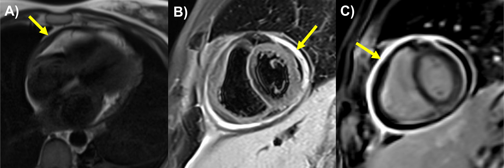
Zhou Z, Zhang N, Azhe S, et al. Myocardial perfusion impairment in children with Kawasaki disease: assessment with cardiac magnetic resonance first-pass perfusion. Quant Imaging Med Surg. 2024;14(7):4923–35. https://doi.org/10.21037/qims-23-1802. [published Online First: 2024/07/18].
Article
PubMed
PubMed Central
Google Scholar
Muthusami P, Luining W, McCrindle B, van der Geest R, Riesenkampff E, Yoo SJ, Seed M, Manlhiot C, Grosse-Wortmann L. Myocardial perfusion, fibrosis, and contractility in children with Kawasaki disease. JACC Cardiovasc Imaging. 2018;11(12):1922–4. https://doi.org/10.1016/j.jcmg.2018.06.009. Epub 2018 Aug 15.
Article
PubMed
Google Scholar
Bratis K, Chiribiri A, Hussain T, Krasemann T, Henningsson M, Phinikaridou A, Mavrogeni S, Botnar R, Nagel E, Razavi R, Greil G. Abnormal myocardial perfusion in Kawasaki disease convalescence. JACC Cardiovasc Imaging. 2015;8(1):106–8. https://doi.org/10.1016/j.jcmg.2014.05.017. Epub 2014 Nov 12.
Article
PubMed
Google Scholar
Friesen RM, Schäfer M, Jone PN, Appiawiah N, Vargas D, Fonseca B, DiMaria MV, Truong U, Malone L, Browne LP. Myocardial perfusion reserve index in children with Kawasaki disease. J Magn Reson Imaging. 2018;48(1):132–9. https://doi.org/10.1002/jmri.25922. Epub 2017 Dec 12.
Article
PubMed
Google Scholar
Furuyama H, Odagawa Y, Katoh C, et al. Assessment of coronary function in children with a history of Kawasaki disease using (15)O-water positron emission tomography. Circulation. 2002;105(24):2878–84. https://doi.org/10.1161/01.cir.0000018652.59840.57. [published Online First: 2002/06/19].
Article
PubMed
Google Scholar
Ohmochi Y, Onouchi Z, Oda Y, et al. Assessment of effects of intravenous dipyridamole on regional myocardial perfusion in children with Kawasaki disease without angiographic evidence of coronary stenosis using positron emission tomography and H2(15)O. Coron Artery Dis. 1995;6(7):555–9. [published Online First: 1995/07/01].
CAS
PubMed
Google Scholar
Muzik O, Paridon SM, Singh TP, et al. Quantification of myocardial blood flow and flow reserve in children with a history of Kawasaki disease and normal coronary arteries using positron emission tomography. J Am Coll Cardiol. 1996;28(3):757–62. https://doi.org/10.1016/0735-1097(96)00199-4. [published Online First: 1996/09/01].
Article
CAS
PubMed
Google Scholar


Comments (0)