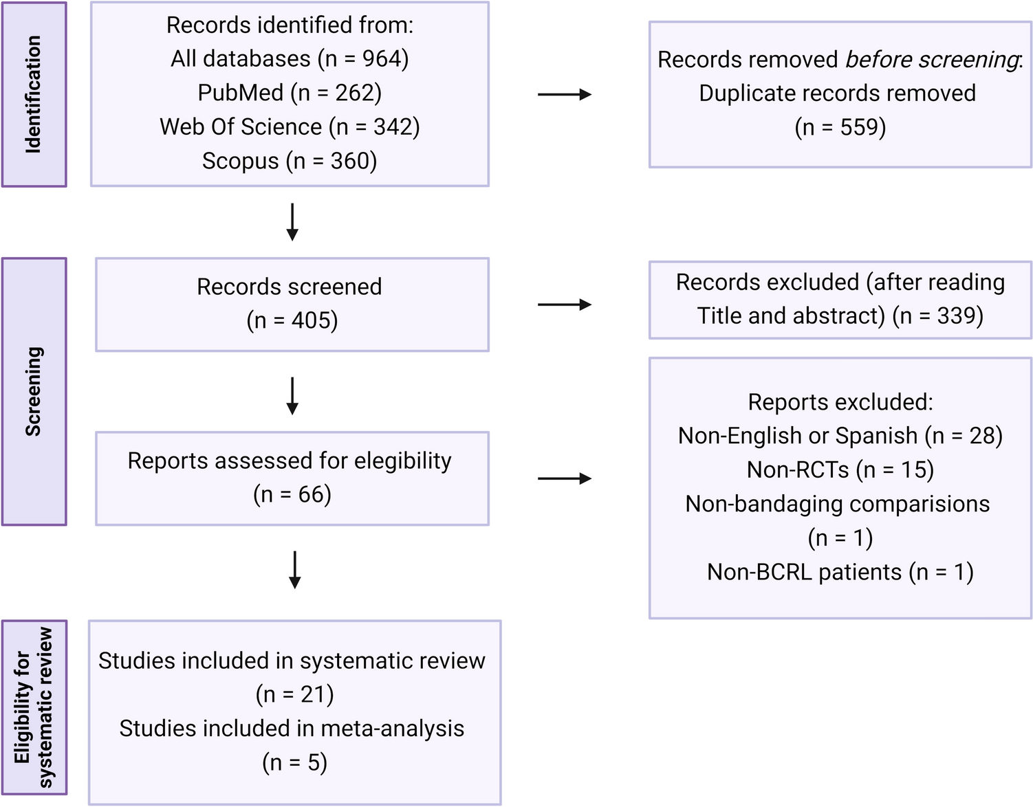
In this study, the median TMB scores were 4.00 and 3.90 (mut/Mb) for the ILC and IDC groups, respectively, and these values did not differ between ILCs and IDCs when evaluated by the subtype, test panel, age, or sample collection site. Furthermore, the proportion of TMB-H cases was statistically significantly higher in the ILC than in the IDC group (18.2% vs. 10.1%, respectively). Moreover, the proportion of TMB-H cases in the ILC group was particularly high among those with the ER+/HER2− subtype and in whom metastatic lesion was the sample collection site. In addition, the proportion of TMB-H cases was higher among those with the ER +/HER2− and ER−/HER2 + subtypes than in the TNBC and to be higher in cases sampled from metastatic sites and in those aged 50 years or older. On the other hand, when comparing ILC and IDC, there was no difference in the distribution of TMB scores between ILC and IDC, expect for BRCA1/2 pathogenic variant -negative cases or those tested by NOP. In other words, except for BRCA 1/2 pathogenic variant-negative cases, there is no difference in the distribution of TMB between ILC and IDC, but there is a special population of TMB-H cases that is found more frequently in ILC than in IDC. This cannot be predicted by clinical factors alone, and it is necessary to predict based on factors such as gene alterations.
A high TMB is associated with a high neoantigen load, making the tumor in high immunogenic conditions. Compared with immunogenic tumors, such as skin squamous cell carcinoma (45.2 mut/Mb), melanoma (14.4 mut/Mb), and non-small cell lung carcinoma (8.1 mut/Mb), the TMB scores in breast cancer were reportedly lower (3.6–3.8 mut/Mb) [5, 7]. TNBC has been reported to have a higher TMB score than ER+ or HER2+ cancers because of its high response to immunotherapy [8, 9]. Reportedly, TMB score is also high in ER+ HER2− breast cancer [10]. Herein, the proportion of TMB-H cases was higher in those with the ER+/HER2− subtype than in the TNBC cohort. This is thought to be due to genomic diversity in HR+HER2− breast cancer. Similarly, it was higher in the ILC than in the IDC group, which is consistent with the result of a previous study that included breast cancer cohorts [9]. The proportion of TMB-H was not affected by the treatment, but the proportion of TMB-H cases was found to be high in the TNBC cohort after hormone therapy. The reason why the TNBC cohort was administered hormone therapy is unknown, but this may indicate that the result of hormone therapy for HR+/HER2− breast cancer patients changing to TNBC and may be related to the intratumonal heterogeneity of breast cancer for TMB status.
More PIK3 CA mutations were observed in ILC than in IDC, and it has been reported that specific PIK3 CA mutations in ILC and metastatic lesions [1] induced mutations in APOBEC genes and that the presence of APOBEC gene mutations is related to TMB-H [11]. In this study, we examined the relationship between information obtained from clinical practice and TMB, and since we did not obtain information on gene alterations, the association between gene alterations and TMB scores was not evaluated; however, the differences in the molecular characteristics between IDC and ILC led to the difference in the proportion of TMB-H.
Meanwhile, when comparing by the sample collection site, the proportion of TMB-H was higher in the brain metastasis of lung cancer than in other metastatic sites [12, 13]. Although differences in the proportion of TMB-H may vary depending on the site from which the specimen was taken and on the type of cancer, the TMB is often higher at metastatic sites than at the primary site, even within breast cancer [9, 13, 14]. In this study, although the number of cases of brain metastasis was not enough to make comparisons, the proportion of TMB-H in metastatic lesions was higher than in primary tumors, indicating that there may be differences depending on the sample collection site. In patients without the pathogenic variant of gBRCA1/2, the TMB was higher in ILC than in IDC, whereas in those with the pathogenic variant, no difference was observed. However, in the multivariate analysis, the status of gBRCA1/2 did not affect the proportion of TMB-H. Breast cancer with the BRCA1/2 gene mutations is thought to have relatively high TMB [15]; however, the number of ILC cases with gBRCA1/2 pathogenic variants is not enough to allow for sufficient consideration. ILC has more germline CDH1 variants than IDC, still only around 0.54% [1]. Patients without pathogenic variants of gBRCA1/2 should also include those with other germline gene variants associated with hereditary breast cancer; however, further investigation on the association between such germline variants and TMB-H is warranted.
The KEYNOTE-158 study confirmed the efficacy of pembrolizumab in the treatment of solid tumors with TMB-H, with an overall response rate of 29% [4]; however, it did not include patients with breast cancer. The Checkmate 848 trial was a phase II study that randomly assigned patients with tumor TMB-H and/or blood TMB-H solid tumors to the nivolumab (NIVO) + ipilimumab (IPI) therapy or NIVO monotherapy. The objective response rates for t-TMB-H were 38.6% (28.4–49.6) in the NIVO + IPI group and 29.8% (17.3–44.9) in the NIVO group. Of the 211 randomized patients in this study, 15 (7.1%) had breast cancer [16, 17]. The results of the TAPUR study confirmed the efficacy of pembrolizumab in the treatment of breast cancer with TMB-H (TNBC, 46%; HR+/HER2−, 43%), with disease control and response rates of 37% and 21%, respectively [18]. These suggest that patients with breast cancer with TMB-H may also benefit from ICI, even in other than TNBC, especially in ILC patients. Although some ILCs were highly immunogenic, this high immunogenicity does not necessarily correspond to TMB-H [19]. The efficacy of ICI in patients with TMB-H may be limited in ILCs that are not immunologically “hot,” and further investigation is needed.
This study has several limitations. First, compared with IDC, the number of ILC cases was extremely small, particularly ILC cases tested using the NOP or with gBRCA1/2 pathogenic variants. Furthermore, the background of the patients who were tested may have greatly differed depending on the subtype as the tests were conducted under Japanese insurance reimbursement. For example, the proportion of TNBC patients was higher, and the proportion of HER2 + type patients was lower than the general population. Second, patients who had completed or were expected to complete the standard treatment were considered eligible for the tests. The small proportion of HER2 + types compared with the real world could be attributed to the fact that few clinicians were expected to benefit from the panel testing due to the already existing oncogene. Third, in this study, we excluded blood TMB to first elucidate tumor TMB. However, to the best of our knowledge, this is the first study to investigate in detail TMB in breast cancer patients using a public database in Japan.
In conclusion, this study demonstrated that the patients with ILC were more likely to have TMB-H than those with IDC. From the perspective of ICI therapy based on the TMB status, the findings would be invaluable in selecting treatment strategies for patients with ILC.


Comments (0)