Ethics
This study was conducted in accordance with the principles of the Declaration of Helsinki and was approved by the Institutional Review Board and Ethics Committee of Nagoya University Hospital (approval no.: 2019-0028). All patients provided written informed consent for the use of clinical specimens and data.
Sample collection
A total of 13 BC cell lines (BT-20, BT-474, BT-549, HCC1419, HCC1954, Hs578T, MCF7, MDA-MB-231, MDA-MB-361, MDA-MB-415, MDA-MB-468, SK-BR-3, and ZR-75-1) and two non-cancerous breast epithelial cell lines (MCF-10A and MCF-12A) were obtained. BT-549, HCC1419, HCC1954, and Hs578T cell lines were purchased from the Japanese Collection of Research Bioresources Cell Bank (Osaka, Japan). BT-474, MCF7, and MCF-12A cells were kindly provided by Prof. David Sidransky of Johns Hopkins University (Baltimore, MD, USA). Other cell lines were purchased from American Type Culture Collection (Manassas, VA, USA). All cells were stored using a cell preservation solution (Cell Banker; Mitsubishi Chemical Medicine Corporation, Tokyo, Japan) at − 80 °C, cultured in RPMI-1640 (Sigma-Aldrich, St. Louis, MO, USA) supplemented with 10% fetal bovine serum (FBS), and incubated in an atmosphere of 5% carbon dioxide at 37 °C [10, 11].
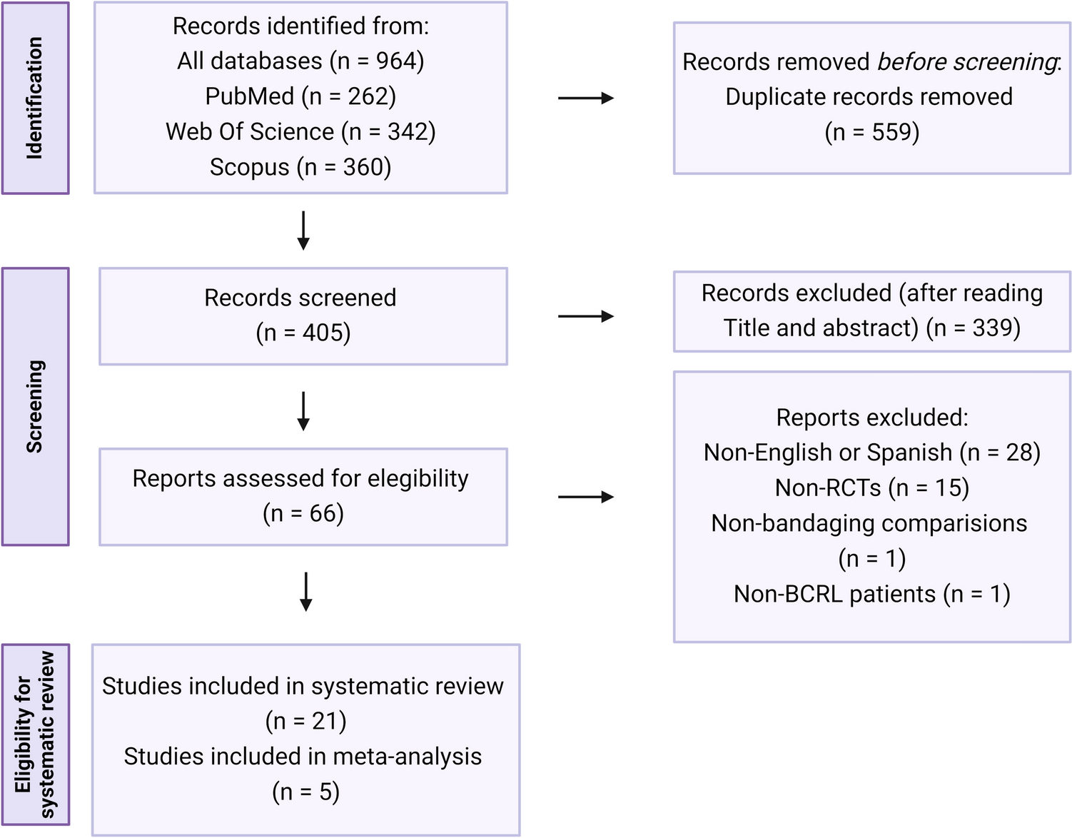
BC and non-BC tissues were collected from 156 patients who were pathologically diagnosed with BC and underwent breast surgery at Nagoya University Hospital between March 2002 and May 2007. Noncancerous tissue was collected at least 3 cm from the edge of the tumor. All harvested tissues were immediately cut into approximately 1.5 mm sections and stored at − 80 °C [12]. The BC stages were classified using the Union for International Cancer Control (UICC) staging system (8th edition). Perioperative adjuvant therapy was determined based on the patient’s general condition, pathology, subtype classification, and shared decision-making between the attending physician and patient [12].
Quantitative real-time reverse transcription polymerase chain reaction (RT-qPCR)
ATP7B mRNA expression levels were determined using RT-qPCR. RNA was extracted from the BC and non-cancerous specimens collected from 156 patients and each cell line (8.0 × 10⁶ cells per cell line). cDNA was synthesized as previously described [10, 11]. Glyceraldehyde-3-phosphate dehydrogenase (GAPDH) mRNA levels were quantified to normalize the expression levels. The specific primers for each gene were as follows: ATP7B, forward 5′-AGATCACAGCCAGAGAAGGG-3′ and reverse 5′-GCCAACATTGTCAAAAGCAA-3′, which generated a 110-bp product; and GAPDH, forward 5′-GAAGGTGAAGGTCGGAGTC-3′ and reverse 5′-GAAGATGGTGATGGGATTTC-3′, which generated a 226-bp product [12]. qRT-PCR was performed using an ABI StepOnePlus real-time PCR System (Applied Biosystems, Foster City, CA, USA), as previously described [10, 11]. The mRNA expression level of ATP7B was determined by dividing the value of each sample by the corresponding GAPDH value [10, 11].
PCR array analysis
To determine the correlation between the expression levels of ATP7B and 84 cancer-related genes in BC cell lines, PCR array analysis was performed using the RT2 Profiler PCR Array Human Oncogenes & Tumor Suppressor Genes (Qiagen, Hilden, Germany), according to the manufacturer’s protocol. The relative expression levels of these genes in each sample were determined by dividing the relevant values by their corresponding GAPDH values.
ATP7B knockdown using ATP7B-specific small interfering RNAs (siRNAs)
MDA-MB-361 and MDA-MB-415 cell lines were transfected with siRNA specific for ATP7B (designated “siATP7B”: 5′-CCAAUUGAUAUUGAGCGGUUATT-3′; Hokkaido System Science, Sapporo, Japan) to knockdown ATP7B. Fluorescein-labeled AccuTarget negative control siRNA (siControl, Cosmo Bio Co. Ltd., Tokyo, Japan) served as the nontargeting siRNA, designated “siControl.” BC cells were transfected with siRNAs via electroporation using the Neon System (Thermo Fisher Scientific, Waltham, MA, USA). The untransfected cells were electropulsed without siRNA. After the electric pulse, cells were cultured in antibiotic-free RPMI-1640 with 10% FBS for 72 h. The knockdown efficiency was determined using qRT-PCR and western blotting.
Western blotting
Western blotting was performed using a Wes Simple Western System (ProteinSimple, San Jose, CA, USA), according to the manufacturer’s instructions. Cultured cells were lysed in RIPA lysis buffer and the lysate was stored at − 30 °C. Protein concentrations were measured using the BCA protein assay kit (Thermo Fisher Scientific). Protein samples were aliquoted into assay plates and automatically detected in individual capillaries. Anti-ATP7B antibody (1:250 dilution; cat. no. ab124973; Abcam, Cambridge, UK) and anti-beta-actin antibody (1:250 dilution; cat. no. ab6276; Abcam, Cambridge, UK) were used as the primary antibodies. Streptavidin Western horseradish peroxidase and anti-mouse or anti-rabbit secondary antibodies (ProteinSimple, San Jose, CA, USA) were selected based on the corresponding primary antibody [13, 14].
Proliferation assay
Cell proliferation was evaluated using the Cell Counting Kit-8 (CCK-8) (Dojindo Molecular Technologies, Inc., Kumamoto, Japan). MDA-MB-361 (1.0 × 10⁴ cells per well) and MDA-MB-415 (1.0 × 10⁴ cells per well) cells transfected with siATP7B or siControl, or untransfected cells were seeded into 96-well plates with RPMI-1640 containing 2% FBS. Each sample was added to six wells and cultured for the indicated time periods. The optical density (450 nm) of each well was measured 2 h after the addition of 10 µL of CCK-8 solution from the start of seeding to day 5 post-seeding [12].
Invasiveness assay
Cellular invasiveness was determined using BioCoat Matrigel Invasion Chambers (pore size 8‑μm; Corning Inc., Corning, NY, USA), according to the manufacturer’s protocol. After transfection, MDA-MB-361 (3 × 10⁵ cells per well) and MDA-MB-415 (3 × 10⁵ cells per well) cells were suspended in serum-free RPMI-1640 and seeded into the upper chambers. RPMI-1640 medium supplemented with 20% FBS was added to the bottom row of the wells. After 72 h of incubation, cells on the membrane surfaces were fixed and stained with Diff Quik (cat. no. 16920; Sysmex, Kobe, Japan) solutions I and II for 5 s at room temperature. Cells on the membrane were counted in 10 randomly selected fields of view using an upright microscope (Olympus Corporation) at ×100 magnification [12].
Migration assay
The migration of MDA-MB-361 and MDA-MB-415 cells was determined using a wound-healing assay. After transfection, MDA-MB-361 (5.6 × 10⁴ cells per well) and MDA-MB-415 (5.6 × 10⁴ cells per well) cells were seeded in each well of Culture-Insert 2 Well (Ibidi, Martinsried, Germany), which were attached to 24-well plate using RPMI-1640 containing 10% FBS. After 24 h, the insert was removed and replaced with FBS-free RPMI-1640 medium and the 24-well plate was placed in an IncuCyte SX5 analysis system (Sartorius, Gottingen, Germany). The same sites were automatically photographed at 0, 12, 24, 48, and 72 h. Wound widths were measured 20 times per well at 100-μm intervals [12].
Immunohistochemistry
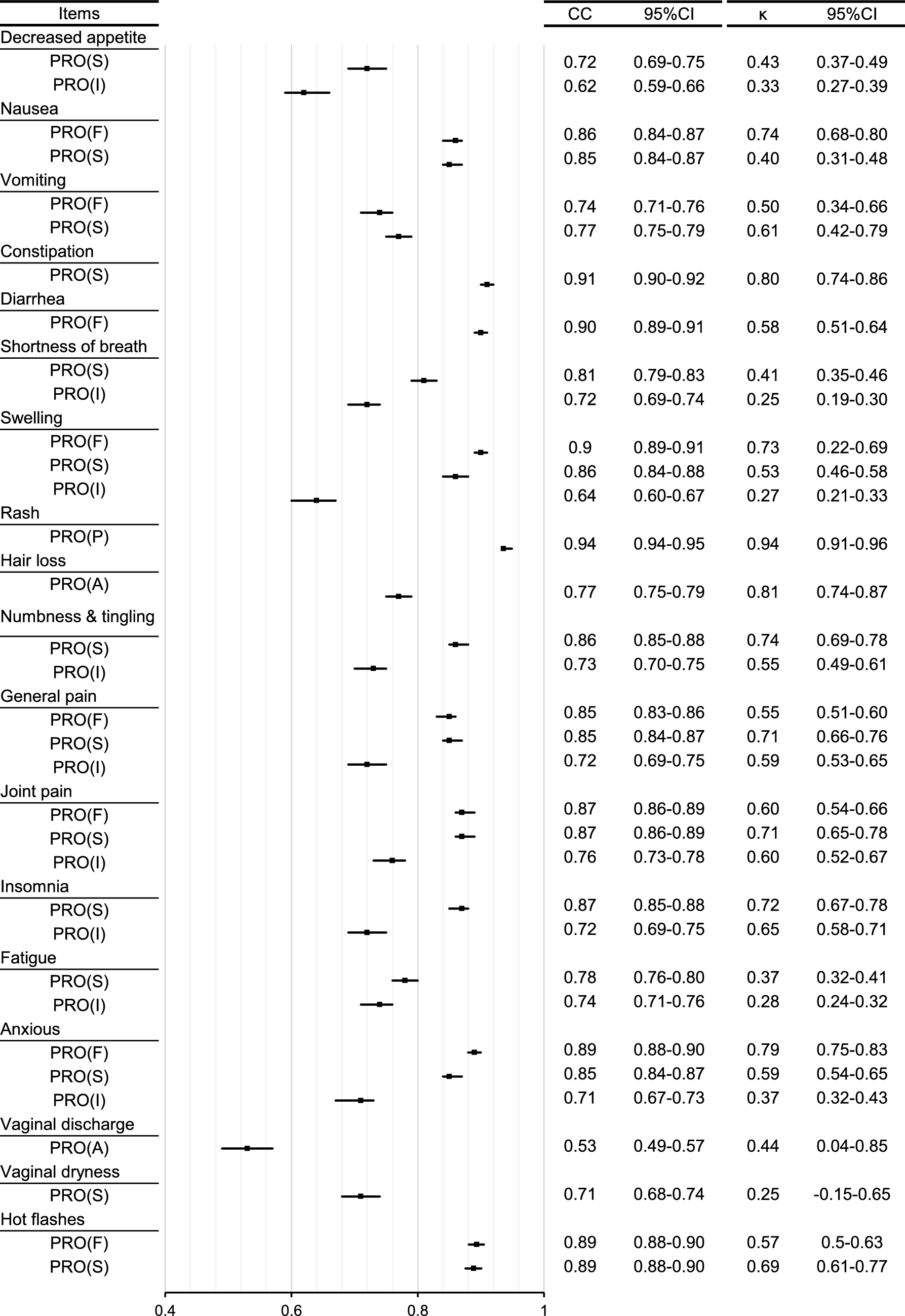
Of the 156 patients mentioned above, specimens from 152 were available for immunohistochemical analysis. Formalin‑fixed, paraffin‑embedded sections (4‑μm thick) were constructed from blocks of resected specimens. The ATP7B rabbit polyclonal antibody (1:500 dilution) (cat. no. NB100-360; Novus biologicals, LLC., Centennial, CO, USA) was used for immunohistochemistry, and sections were incubated overnight at 4 °C. The EnVision + System- HRP Labelled Polymer Anti-Rabbit (cat. no. K4003; Dako North America Inc. Carpinteria, CA, USA) was used as the secondary antibody and the sections were incubated for 30 min at room temperature. The cancerous area of each section was observed under an upright light microscope (Olympus Corporation; ×40, ×100, and ×400 magnification). The intensity of staining (IS) in the cytoplasm of cancer cells was evaluated and divided into four levels, ranging from 0 (negative) to 3 (strong). The percentage of staining (PS) was evaluated for whole cancers and divided into 11 levels, ranging from 0 to 100% in 10% increments. The IP score was assigned by multiplying the IS by the PS.
Public datasets of BC cell lines and patients
The mRNA expression levels of ATP7B in 59 BC cell lines were obtained from the Cancer Cell Line Encyclopedia (CCLE) database (https://sites.broadinstitute.org/ccle/). The data were accessed on August 28, 2022. The Kaplan–Meier plotter website (http://kmplot.com/analysis/index.php?p=background) was used to analyze relapse-free survival (RFS) and overall survival (OS) of patients with BC based on ATP7B expression levels. Patients were divided into two groups based on their median expression levels [15]. The data were accessed on May 3, 2021.
Statistical analyses
Numerical variables between the two groups were compared using the Mann–Whitney test; comparisons between multiple groups were performed using ANOVA followed by Tukey’s post hoc test. The correlation between ATP7B and cancer-related gene expression levels in PCR array analysis was assessed using Spearman’s rank correlation test. The associations between mRNA or protein expression levels of ATP7B and clinicopathological factors were analyzed using the χ2 test. Disease-free survival (DFS) and OS were calculated using the Kaplan–Meier method, and survival curves were compared using the log-rank test. Multivariate analysis was performed using the Cox hazard model. All statistical analyses were performed using JMP 16 software (SAS Institute Inc., Cary, NC, USA), and statistical significance was defined as p < 0.05.


Comments (0)