To the best of our knowledge, this study is the first to investigate the risk factors associated with rectal perforation during various surgical approaches for presacral cysts. Our findings emphasize that only cyst rupture and CRD may influence rectal wall perforation. Interestingly, the surgical approach selection does not appear to have a significant impact on the risk of perforations.
MRI is renowned for its superior visualization of pelvic organs, facilitating detailed assessments of topographic-anatomical relationships with adjacent structures and signs of local invasions. Presacral cysts appear as thin-walled structures that are hypointense on T1-weighted images. Some researchers, however, prefer CT scans for better navigation in case of potential malignant transformations [10, 16, 17]. In our study, a majority of patients (67.1%) underwent pelvic MRI, and an increase in CRD was associated with a substantially reduced risk of intestinal perforation. Therefore, patients should be well informed, and surgeons should be well prepared to perform a diverting colostomy when necessary. Notably, neither MRI nor CT demonstrated marked effectiveness in preventing damage to the rectal wall preoperatively.
Considering the rarity of presacral cysts, much of the existing literature consists of case reports [13, 14]. To determine the most suitable approach, most authors consider factors such as the cyst’s size, its presacral location relative to the sacral vertebrae, and its involvement of surrounding structures [7, 8, 10, 18].
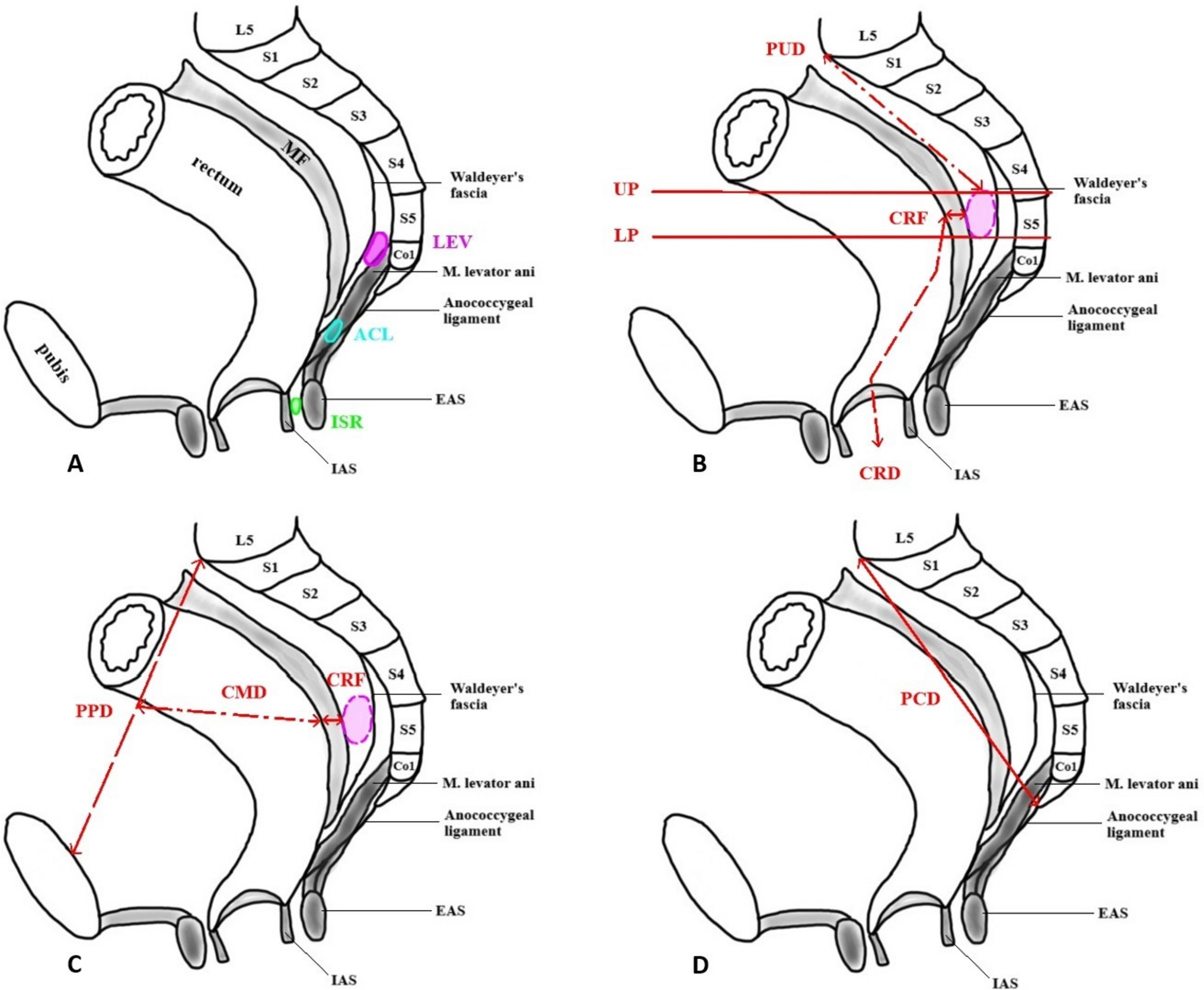
The perineal approach, as described by Kraske et al. [19], is typically favored for smaller cysts, generally those less than 10 cm, especially in the absence of invasion into surrounding structures. The upper pole location of the cyst relative to the sacral vertebrae has been reported to vary from S2 to S4 [7, 9, 10]. However, in our study, the upper pole of the cyst was most often situated at the coccyx level in 53% of patients, differing from published data. Advantages of this approach include low invasiveness, reduced bleeding risk, and enhanced visualization of the retroretcal space [4, 11, 20]. In our study, coccyx resection was performed in 23 (68%) patients to improve the surgical access. Nevertheless, the proximity of the wound to the anus raises the likelihood of postoperative infections. Sakr et al. [13] reported a 72.7% postoperative complication rate following the perineal approach. In our cohort, seroma formation was the most frequent complication, occurring in 3 (9%) patients, while the lowest incidence rates of cyst damage and unintentional rectal wall perforation were 24% and 8.8%, respectively.
The transabdominal approach is associated with fewer complications, faster bowel function recovery, shorter hospitalization, less postoperative pain, reduced blood loss, and better cosmetic outcomes [4, 21]. This approach is recommended for cyst localized above the S3–S4 level [7, 8, 22]. In our study, 12 (43%) patients had cysts located between S3 and the coccyx, again deviating from the literature. Although laparoscopy may lead to higher rates of cyst perforations compared to robotic techniques, the latter increased the risk for rectal perforation in case of cyst rupture. Overall, the transabdominal route showed a higher risk of cyst perforation, with comparable conversion rates across methods; notably, no conversions to alternative accesses were reported when executing the robotic procedure. However, the transabdominal approach had a higher rate of postoperative complications compared to the perineal approach.
A combined approach is generally used for cyst situated above the S2 vertebra, particularly those with larger volumes, involvement of pelvic organs, or spread to the anococcygeal ligament [4, 11, 23]. In our clinic, patients with cyst volumes averaging 101.7 (56.8, 226.3) cm3 were treated with this approach. While robotic methods have been previously documented, laparoscopic approaches remain common. A systematic review of minimally invasive treatments for presacral cysts indicated similar rates of surgical site infections and rectal wall injuries across both approaches [24].
The chosen surgical method did not increase the risk of rectal perforation; however, the selection was based on cyst placement and the positions of the upper and lower poles. Cyst perforation poses the risk of surgical field infection, and in case of malignant cysts, there is a potential for cancer cell spillage, raising the risk of local recurrence. Consequently, many experts advise against preoperative percutaneous or transrectal biopsies [4, 11].
One of the serious complications is the rectal wall perforation. According to the Chinese consensus on the treatment of presacral cysts, suturing the defect with omentum fixation is recommended, and if this fails, an ileostomy is suggested [11]. The incidence of rectal perforation does not exceed 10% [13, 14, 23]. In our study, rectal injury was observed in 9 (12.3%) patients, with the highest rate seen during combined and transabdominal approaches. Of those with rectal perforation, 5 (6.8%) underwent suturing of the rectal defect followed by diverting colostomy, with no postoperative complications observed. In contrast, 4 (5.5%) patients were treated only with suturing without diverting stoma, and all required reoperation due to leakage.
This study has several limitations, including its retrospective nature, which may lead to selection bias. As a single-center analysis, the findings may have limited generalizability.


Comments (0)