Animal handling
Male db/db mice and their normoglycemic heterozygous littermates (db/+), obtained from The Chinese University of Hong Kong, were kept in the Central Animal Facility of The Hong Kong Polytechnic University, housed in groups of two in plastic cages with free access to standard rodent diet and water under a 12 h/12 h light–dark cycle. The experimental protocol was approved by the Animal Subjects Ethics Sub-committee (ASESC) of The Hong Kong Polytechnic University (Approval No. 19-20/85-SO-R).
Evaluation of diabetes-related indices
The db/db mice are a model of type 2 diabetes with a spontaneous mutation of the leptin receptor gene, which leads to polyphagia, obesity, insulin resistance, and sequential diabetes and have been reported to commence development of diabetes at 4–8 weeks of age [20, 21]. At 9 weeks of age, blood glucose levels of the mice in the two groups were measured with a digital blood glucometer (Accu-Chek® Performa, Roche Diagnostics, Basel, Switzerland) after overnight fasting and db/db mice with a fasting blood glucose level ≥ 13.9 mmol/L were included in the study. Subsequently, the fasting blood glucose levels were measured every four weeks until 25 weeks of age. Hemoglobin A1c (HbA1c) levels, which reflect diabetic status over three to four months, were measured at 9 and 25 weeks of age using DCA® Vantage Systems (Siemens Healthcare GmbH, Erlangen, Germany) with blood samples collected from the tail veins. All assessments were conducted between 9 a.m. and 10 a.m. Bodyweight was monitored weekly. Food and water consumption was measured thrice weekly.
Electroretinography (ERG)
The in-vivo retinal functions were assessed with scotopic full-field ERGs (ffERG) as described previously [22] at 9, 13, 17, and 25 weeks. The mice were dark-adapted overnight (at least 16 h) in the experimental room prior to the ERG with all subsequent handling and preparations performed under dim red light. The animals were anaesthetized with a weight-based intraperitoneal injection of a solution containing Ketamine 100 mg/ml (Alfasan International BV, Woerden, Holland) and Xylazine 20 mg/ml (Alfasan International BV). The corneas of both eyes were anesthetized with Proxymetacaine 0.5% (Provain-POS®, Ursapharm, Saarbrücken, Germany), and the pupils dilated using a drop of the solution mixed with Tropicamide 0.5% and Phenylephrine 0.5% (Mydrin®-P, Santen, Osaka, Japan). The animals were then placed on a warm table maintained at 37 °C. A gold ring electrode was placed in contact with the cornea as the active electrode. Two platinum needle electrodes were inserted subcutaneously at the base of the tail and the forehead as the ground and reference electrodes, respectively. The impedances of the active and reference electrodes were less than 10 kΩ. A drop of 3% Carbomer 974P gel (Lacryvisc®, Alcon, Geneva, Switzerland) was applied to the cornea to prevent corneal dehydration. After positioning the electrodes, the animals were allowed to remain in complete darkness for 5 min before the start of the experiment.
Visual stimuli with white light-emitting diodes were delivered by a Ganzfeld bowl (Q450, Roland Consult, Brandenburg, Germany). Stimulation and data recording were performed using the RETI-Port system® (Roland Consult) according to a customized protocol, with stimulus intensities varying from − 4.32 log cd·s/m2 to + 1.3 log cd·s/m2. The signals were amplified and band-pass filtered from 1 to 30 Hz and 1 to 1000 Hz for scotopic threshold response (STR), which is suggested to originate from the proximal retina [23], and scotopic a- and b-waves, respectively. The b-wave of the scotopic ERG reflects the function of photoreceptors and the post-receptor pathway [24]. At the lowest intensity, 40 sweeps of response with a stimulus frequency of 0.5 Hz were averaged. The number of sweeps and stimulus frequency was reduced at higher flash intensity levels. The stimulus intensities were converted to the unit photoisomerizations/rod (R*/rod), where 1 scot cd/m2 = 516 R*/rod/s, after being calibrated by a photometer (ILT1700, International Light Technologies, Inc, Peabody, MA, USA), to determine the light zone to which the stimuli belonged, according to Abd-El-Barr et al. [25]. The responses triggered by the stimuli that were within the operation range of the rod cells (less than − 0.3 log cd·s/m2) were fitted with a sigmoidal curve, using the Naka-Ruston equation to determine the maximum b-wave response (Bmax).
Oscillatory potentials (OPs) were isolated by digital filtering of the raw signal recorded at + 0.3 log cd·s/m2 using a fast Fourier transform (FFT) and subsequent inverse FFT with an algorithm computed in the free software environment R (R Development Core Team, v.4.0.4). The raw data was first converted from the time domain to frequency domain. Spectral components beyond the cut-off frequency (65 to 300 Hz) [26] were then eliminated. The inverse FFT was performed to reconstruct the OP waveform in the time domain. Implicit times of OP were measured from the stimulus onset to the peak of the OP. Individual OP amplitude was measured from the peak to the adjacent trough. The first four major OP wavelets were included for analysis.
Optical coherence tomography (OCT)
Spectral domain-optical coherence tomography (SD-OCT), which is emerging as a reliable tool for non-invasively evaluating structural changes in the retina in both clinical and research fields [27,28,29], was conducted as an in-vivo assessment of retinal morphology. Change of retinal thickness, measured by OCT, was used to reflect the extent of cell degeneration. The experimental time points and preparation procedures, including anesthetization and pupil dilation, were the same as those described for the ERG measurement. To maximize the image quality, the cornea was maintained moist with lubricant containing 0.3% propylene glycol and 0.1% polyethylene glycol 400 (Systane® ultra, Alcon) throughout the measurement.
OCT was performed using Bioptigen Envisu™ SD-OCT (R2210, Leica Microsystems, Morrisville, NC, USA). OCT rectangular scans (a-scans/b-scans: 1000 lines; b-scans: 100 scans; frame/b-scans: 10 frames) that covered an area of 0.8 × 0.8 mm were performed. The optic disc was located at the center of the scan and served as a reference point to ensure the scanning location was the same across animals and between different time points. The retinal thickness was analyzed by the built-in software InVivoVue Diver (v.3.0.8, Bioptigen Inc., Morrisville, NC, USA).
Evaluation of retinal histology
The retinas of the mice in the two groups were processed for immunohistochemistry (IHC) and 4′,6-diamidino-2-phenylindole (DAPI) staining according to the procedures described previously [30]. At 25 weeks, the animals were euthanized by cervical dislocation. One eye from each animal (randomly chosen) was enucleated and dissected to isolate the whole retina, which was then incubated in 4% paraformaldehyde in phosphate buffer at room temperature for 1 h for fixation. The whole retinas were processed into vertical retinal sections (35 μm) using a microtome (Vibratome VT1200S, Leica Microsystems, Deer Park, IL, USA). The samples were then blocked with 10% donkey serum in TBS (0.5% Triton X-100 and 0.1% Sodium Azide in Dulbecco's phosphate-buffered saline, pH7.2) at 4 °C overnight to reduce non-specific labelling.
The samples were incubated with primary antibodies (anti-GNAT2: LifeSpan BioScience, Seattle, WA, USA; LS-C321680, dilution 1:75; anti-PKCα: Santa Cruz Biotechnology, Dallas, TX, USA; sc-8393, dilution 1:50; anti-bassoon: Cell Signaling Technology, Danvers, MA, USA; D63B6, dilution: 1:200) at 4 °C in TBS with 3% donkey serum for four days. Following incubation, the samples were washed several times and transferred to a 3% normal donkey serum-TBS solution containing donkey-host secondary antibodies conjugated with Cy3 (Millipore Sigma, Burlington, MA, USA, AP192C, dilution 1:200) or Alexa Fluor 488 (Invitrogen, A21206, dilution 1:1000) at 4 °C overnight. DAPI (Invitrogen, D1306) was used to stain the nuclei in the retina to evaluate the extent of neural cell survival by quantifying the number of viable cells.
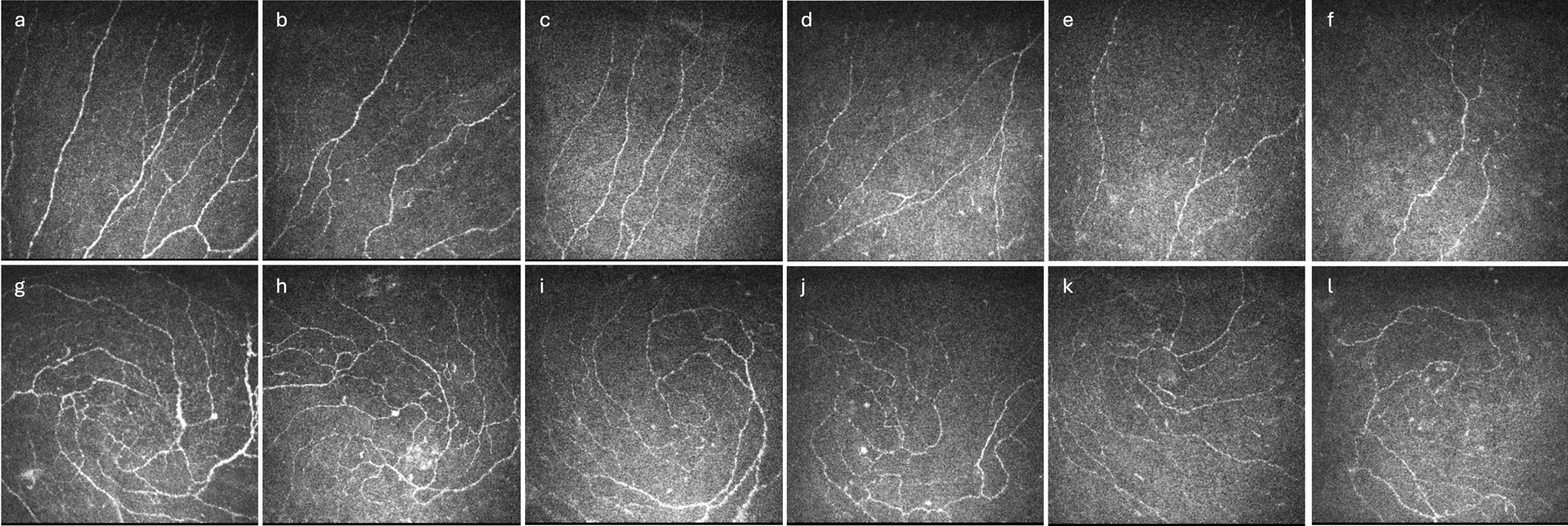
A confocal laser scanning microscope (LSM800, Zeiss, Oberkochen, Germany) was used to capture the confocal micrographs of the specimens using a 20×, 40×, or 63× objective. To measure the number of cone photoreceptors and rod bipolar cells (and their synaptic terminals), GNAT2 and PKCα positive cells were counted in the whole imaged area (i.e., a 160 μm-segment), respectively, with a cell counter in ImageJ (v.1.53k, National Institutes of Health, Bethesda, MD, USA). To determine the number of photoreceptor cells (including both rods and cones), the DAPI stained nuclei in the outer nuclear layer were counted in half of the image area (an 80 μm-segment). For the number of rod-bipolar dendritic boutons, the number of PKCα positive puncta in the out plexiform layer was counted over a 20 μm-segment. The dimensions of the outer nuclear layer and the inner nuclear layer were analyzed by ImageJ. The data of each individual animal was averaged from three retinal sections, with each group consisting of at least five animals.
Measurement of retinal mitochondrial bioenergetics
The mitochondrial bioenergetics of the retina of the db/db mice and db/+ mice were assessed by measuring the oxygen consumption rate (OCR) using the Seahorse XFe24 Extracellular Flux Analyzer (Agilent Technologies, Santa Clara, CA, USA) with reference to the protocol described by Millman et al. [31]. The Seahorse XF Analyzer evaluates the mitochondrial function of cells or tissues by serially injecting several chemicals that target the electron transport chain complexes throughout the assay to derive different key parameters of mitochondrial function.
One eye from each animal was enucleated and dissected to harvest the whole-mount retina in ice-cold PBS. Three 1.5 mm diameter punches were obtained from the neural retina with a biopsy puncher (Miltes Instrument, Integra LifeSciences, Mansfield, MA, USA). Each retinal punch, obtained adjacent to the optic nerve head to minimize variation in cell density, was carefully placed in the well of an XF24 Islet capture microplate (Agilent Technologies) with the ganglion cell layer facing up and covered with Islet Fluxpak mesh inserts (Agilent Technologies). Prior to the measurement of OCR, Seahorse XF DMEM medium (103335-100, Agilent Technologies) containing 5.5 mM glucose (G6152, Sigma-Aldrich, St Louis, MS, USA) and 1 mM sodium pyruvate (11360070, Thermo-Fisher, Waltham, MA, USA) was added to each well, and the retinal punches were incubated at 37 °C in a non-CO2 incubator for 60 min to allow the temperature and pH to reach equilibrium. The OCR was then measured under basal conditions and after serial injection of 1 µM carbonylcyanide-4-(trifluoro-methoxy) phenylhydrazone (FCCP; 15218, Cayman Chemical Company, Ann Arbor, Michigan, USA)), and a mixture of 10 µM rotenone (13995, Cayman) and 20 µM antimycin A (A8674, Sigma-Aldrich) to determine the values for basal respiration, maximal respiration, spare respiratory capacity, and non-mitochondrial oxygen consumption. After the assays, the retinal punches in each well were lysed with EB2 lysis buffer, containing 7 M urea, 2 M thiourea, 30 mM Tris, 2% (w/v) CHAPS, and 1% (w/v) ASB14 with protease inhibitor cocktail (Roche Applied Science, Basel, Switzerland), and placed on ice. The protein concentrations were determined using the Bradford Protein Assay (Bio-Rad Laboratories Inc., Hercules, CA, USA) according to the manufacturer’s guidelines for normalization of the OCR values.
Statistical analysis
Data are presented as mean ± SEM. Shapiro–Wilk’s test was used to check the normality of data distribution prior to the use of a parametric test. For longitudinal comparison, mixed-model ANOVA was used to analyze the within-group, between-group, and interactive effects. Independent sample t-test (with Welch correction when appropriate) was used for comparison between the two groups. JASP (JASP Team, 2022, v.0.16.3) Amsterdam, Netherlands) was used for the statistical analysis. Differences with a P value less than 0.05 were considered significant.


Comments (0)