The purpose of this study was to analyze the skeletal and dental expansion patterns following SARME and to investigate the role of expander type in the occurrence of asymmetric expansion. The investigators hypothesized that there were no differences between hyrax and TPD in terms of asymmetric transverse dental and skeletal expansion. The specific aims of the study were: (1) to quantify the anterior and posterior expansion after SARME; (2) to calculate the incidence and magnitude of dental and skeletal asymmetry; (3) to compare the achieved expansion and asymmetry between the two types of expanders; (4) to identify predictor variables correlated with asymmetric expansion.
The study results have confirmed the main hypothesis: no differences in the amount of expansion and in the incidence and pattern of asymmetric expansion were found between the tooth-borne (hyrax) and bone-borne (TPD) expanders. The type of expander appeared not to be a strong predictor of asymmetric expansion. Regarding the specific aims of the study, the following can be stated: (1) It was noticed that after a Sarme, more expansion was achieved at the dental level than at the skeletal level, both with the tooth-borne devices and the bone-borne TPD. (2) dental asymmetry occurred more frequently than skeletal asymmetry, and the amount of dental asymmetry was larger than the amount of skeletal asymmetry; (3) the amount of expansion and incidence of asymmetry was comparable between the tooth-borne (hyrax) and bone-borne (TPD) expanders; (4) no reliable predictors were found for the occurrence of asymmetric expansion, though posterior dental asymmetry was associated with posterior skeletal asymmetry.
The magnitude of transversal maxillary expansion found in this study was not affected by the type of expander. This finding was in line with previous studies [2, 3, 9, 18]. Koudstaal et al. reported a 2.9 mm net change of the palatal width at premolar level and 2.6 mm at molar level with a bone-borne expander and a 2.6 mm and 2.5 mm net change with a tooth-borne expander, which were comparable to the present findings. A recent systematic review by Khosravi et al. also confirmed this finding and found no differences between the two devices in amount of maxillary expansion, dental tipping and stability [9]. This was confirmed by Gogna et al. regarding the stability after significant expansion on dental and skeletal level following SARME [19]. It should be noted that stability and possible relapse is influenced by postoperative orthodontic treatment resulting in a stable dental occlusion [18]. However, in order to maximize skeletal transversal expansion comparing to dental expansion a bone-borne device should be favored [20].
Two recent studies reported and quantified asymmetric expansion following SARME [8, 14]. Compared to the present study, Huizinga et al. analyzed patients with bone-borne distracters only, whereas Nada et al. only reported the transverse skeletal asymmetry. Nada et al. found an asymmetry in posterior skeletal expansion of more than 1.5 mm in 17.6% of their patients, compared to 35% reported by Huizinga et al. Our findings showed that 9 out of 30 patients (30%) had a skeletal asymmetry in the posterior segments more than one millimeter using a hyrax and 12 out of 30 (40%) using a TPD. Huizinga reported that most transverse asymmetries occurred in the anterior maxilla in 50% of the patients, which was higher than the incidence of 37% (11 out of 30) in the TPD group of the present study. Concerning the incidence of asymmetric maxillary expansion, the results of the present study coincided with that of Nada et al., both stating that no difference existed between the hyrax and TPD group.
Comparing skeletal asymmetry with dental asymmetry, it can be said, that the asymmetry was more frequent and more severe on dental level. Also, the amount of achieved dental expansion (situated more caudally) was significantly larger than skeletal expansion (located more cranially). These findings pointed out that despite osteotomizing the midpalatal suture and bone at Le Fort I level, the expansion achieved by SARME is not a bodily parallel movement of the maxillary segments, but is rather a tipping of the segments. Even the application of a bone-borne expander that exerted expansion forces directly onto the palatal bone was not able to reduce the tendency of having more dental expansion than skeletal expansion. This indicates that the center of resistance is located cranially to the apices of the maxillary teeth and the attachment of the bone-borne expander. Previous studies [6, 8] have also implicated, that more dental expansion was induced following SARME compared to skeletal expansion. This tipping of the maxillary segments seemed to be more present posteriorly as a posterior dental asymmetric expansion was correlated to a posterior skeletal asymmetric expansion, while such a correlation was not found between anterior dental and skeletal asymmetry.
The amount of expansion might play a role in asymmetric expansions as the resistance was expected to increase as the expansion enlarged. The amount of soft tissue resistance has a substantial influence on the differences in expansion of maxillary parts [6]. Thus, a larger expansion was associated with more asymmetry. Another reason might be the orthodontic movement. Since the movements of the skeletal parts are more restricted by softs tissues, the chances might be higher to have uncontrolled orthodontic movements rather than skeletal movements. Carvalho et al. found that asymmetry and incorrect expansion occurred in 4.47% after SARME and often needed additional surgery for correction. Furthermore, they suggested that a slow activation rate and the absence of a pterygo-maxillary disjunction could be associated with asymmetric expansion [21]. The observation that anterior expansion was significantly larger in the asymmetric group compared to the symmetric group may also be explained by the increasing soft tissue resistance as the amount of expansion increases, as result of soft tissue traction from the alar base or contact between the osteotomized anterior nasal spine and the maxilla. Minor differences in osteotomy length or height between the two maxillary sides may alter resistance during expansion, contributing further to the occurrence of asymmetry.
Regarding transverse asymmetry, it is known from previous studies, that the orientation and the placement of the expander might play a major role. According to the theory postulated by Mommaerts the location of the expander and the pterygoid disjunction were believed to play a significant role determining whether a parallel or bilateral asymmetric expansion will occur [22]. The anterior asymmetry can be due to the traction from alar base and interference between the osteotomized anterior nasal spine and the maxilla. Possible explanations for posterior transverse asymmetry might be the difference in the resistance between the maxilla and pterygoid plate [6]. Carvalho et al. also noted that incomplete pterygomaxillary disjunction and rapid activation protocols may increase the risk of asymmetric expansion [20]. By decreasing the bony resistance in posterior maxilla, larger posterior palatal expansion and more parallel bony expansion could be achieved. These findings favour the use of pterygomaxillary disjunction in SARME despite the increased risk of bleeding. Furthermore, the midpalatal osteotomy might also influence the degree of posterior asymmetry, since the osteotomy is not performed all the way back and it is usually positioned paramedianly rather than exact in the midpalatal suture.
From the esthetic and occlusal point of view, a dental asymmetry following SARME may have more clinical implications compared to a skeletal asymmetry as it occurs more frequently and that the amount of dental asymmetry is usually higher as stated earlier. Fortunately, mild to moderate dental asymmetry can be managed by orthodontics successfully. Clinicians do need to be aware of the fact that dental compensation in case of a skeletal asymmetry can lead to displacement of roots outside the alveolar process, which may lead to periodontal problems such as gingival recession and tooth loss.
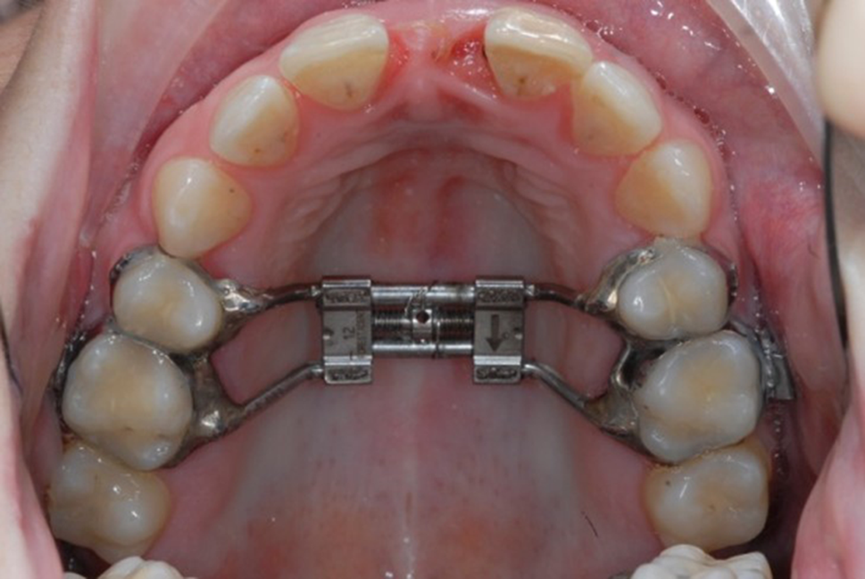
The present study has classified the maxillary anterior-posterior expansion following SARME into three categories: (1) a parallel expansion; (2) an unilateral asymmetric expansion, and (3) a bilateral asymmetric expansion. From the clinical point of view, a parallel expansion is the most desired as it maintains the dental midline and optimizes the transverse interdigitation by correction of a crossbite. An unintentional bilateral asymmetric expansion can be managed by restricting the arch width by orthodontics. An unilateral asymmetric expansion is esthetically less appreciated because it creates a dental midline shift in conjunction with a unilateral over- or under correction of the transverse maxillary dental arch dimension and a yaw of the maxillary segments. If a dental midline shift is less than 2 mm, it can usually be corrected orthodontically. If a more severe dental midline shift takes place, one may need to consider a Le Fort I osteotomy and lateral translation of the maxilla to correct the midline shift. In cases of severe yaw of the maxillary segments, the width of smile line (the distance from the central incisor to canine/bicuspid) on one side can appear optically larger than the contralateral side. Such a deformity may need a Le Fort I osteotomy of the maxilla to de-rotate the maxilla and correct for the yaw deformity.
In the present study, a 1-mm threshold was used to define asymmetry. Previous studies [8, 14] used a one and a half or three millimeter threshold. The question arises whether the 1-mm threshold is too restrictive as it tended to report a higher incidence of asymmetry compared to a higher threshold. A recent study by Borba et al. that investigated the role of different threshold values in the assessment of surgical outcomes by using 2D and 3D images underlined that 3D accuracy studies should no longer rely on a 2-mm level of discrepancy but rather on a 1-mm level [12]. Previously used threshold values in 2D imaging studies should therefore be applied with caution to 3D studies. Also the clinical relevance of possible soft tissue changes after (asymmetric) skeletal movement should be further investigated as Karabiber at al. stated that a significant asymmetric expansion of the apertura nasalis anterior did not lead to a asymmetry in the nasal regio [23].
The limitation of this study was the lack of CBCT scans at the beginning and at the end of expansion. With these scans, it would be possible to determine the influence of many factors that would have played a role in inducing an asymmetrical maxillary expansion, such as the position of the TPD/hyrax, the degree of pterygoid disjunction and the length of midpalatal osteotomy. The absence of such CBCT scans makes it difficult to investigate in-depth the causative factors of asymmetric expansion.
Another shortcoming of the study is the use of conventional landmark-based measurements as in most of the previous studies [6, 14, 24]. The measurements were under the influence of landmark identification errors, which would vary from 0.88 mm to 1.26 mm [17]. To minimize the error, only previously validated landmarks were being used in this study. Baan et al. published a new method (OrthoGnathicAnalyser) to quantify jaw movements based on regional voxel-based registrations, which eliminated the use of anatomical landmarks and subsequent landmark identification errors [25]. However, this method was focused on describing 3D movements of maxilla and mandible rather than describing movements of different teeth (or from different landmarks), making it less suitable for the present study. Point-based measurements should be incorporated into OrthoGnathicAnalyser to allow quantification of 3D movements from different cephalometric landmarks. In this way, landmark-associated measurement errors can be eliminated from future studies, while maintain the ability to generate conventional landmark-based measurements from 3D data so that study results can be compared with previous landmark-based studies.
Furthermore, changes due to the post-SARME orthodontic treatment could not have accounted for. Ideally, the post-SARME CBCT scan should be acquired directly after the completion of the active maxillary expansion. However, this approach would expose patients to unnecessary radiation. As the post-SARME orthodontics is aimed to decompensate the dental arches and to close the central diastema, the main orthodontic effect would be a general improvement of an asymmetric expanded maxilla and not a worsening of the asymmetry. The assessment of dental midline discrepancy, buccal corridor, posterior dental asymmetry and soft tissue changes in general after completion of orthodontic treatment was also not investigated and could possibly play in important role in patient satisfaction. The displacement in other dimensions was studied by Lin et al. [26] They found a significant forward and clockwise downward movement of the maxilla after SARME. This should also be investigated further as there is little consensus regarding the sagittal and vertical displacement and the effect on soft tissue.
The findings reported in the presented study, based on CBCT scans with a mean of 647.0 (Sd = 236.4) days after SARME, would rather be an underestimation of asymmetry that was present directly after SARME. Furthermore, the effect of relapse could not be considered, as the postoperative CBCT scans were taken after the completion of the maxillary expansion.
Lastly, only one surgical technique (bipartite median osteotomy) that is commonly used in our institution, was performed in this patient group. Other surgical methods, such as the three segment (bilateral paramedian) technique could be an alternative. This technique is surgically more demanding, but would lead to a larger transverse and more symmetrical expansion compared to the traditional two segment (median) technique [24]. It is important to note that since the introduction of mini-screw assisted rapid maxillary expansion (MARPE) in our department in 2018, more and more SARMEs are being replaced by the less invasive MARPE procedure, which has been reported to achieve effective and stable maxillary skeletal expansion in post-adolescent patients [27, 28]. Future studies should compare different techniques (SARME with hyrax, SARME with TPD and MARPE) using a similar patient group to find the ideal clinical indications, taking into account the advantages and disadvantages for each technique.


Comments (0)