Ethics and study design
Ethical approval was granted by the Cantonal Ethics Commission in Zurich, Switzerland (2022-D0090). All voluntary participants provided written informed consent to participate in this study in accordance with the Declaration of Helsinki and its subsequent revisions and allowed that their images would be used anonymized in publications.
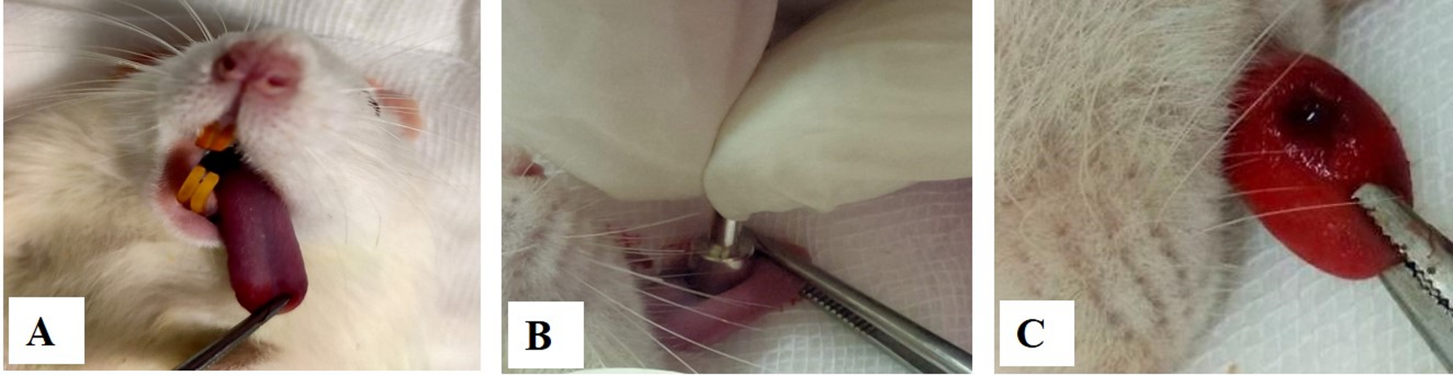
This prospective study recruited healthy volunteers from the Center of Dental Medicine and the University Hospital Zurich, Department of Cranio-Maxillofacial and Oral Surgery, during annual routine examinations between May and December 2022. Inclusion criteria were: (1) age over 18 years and (2) absence of clinical symptoms related to head and neck pathologies. Exclusion criteria included: (1) history of surgical procedures in the past three months (2), acute infections in the maxillofacial region (3), trigeminal nerve damage (4), pregnancy, and (5) standard contraindications to MR imaging. Participants in this study underwent MRI examinations conducted by trained clinical staff and research personnel.
Scan acquisition and reconstruction
All recruited individuals underwent 3 T MRI scans using a MAGNETOM Skyra system (release VE11E, Siemens Healthineers, Erlangen, Germany). The imaging protocols included gradient specifications of 45 mT/m and 200 T/m/s, employing a dedicated 15-channel mandibular coil (NORAS MRI Products, Hoechberg, Germany). This coil is specifically designed for high-resolution dentomaxillofacial imaging, offering a field of view of 32 × 16 × 16 cm. It features a 14 + 1 receiver coil array integrated into a positioning system optimized for detailed anatomical visualization. The curved phased array coil measures 12 × 38 cm2 and incorporates 14 elements arranged between two support bars. Built-in fixation components ensure precise head positioning, while designated openings are provided for the nose and mouth. The central section between these openings is aligned above the upper lip to maintain optimal coil placement. Additionally, the outer wings are flexible, allowing for customized adaptation to the patient’s anatomy. To enhance patient comfort and minimize motion artifacts, optional head fixation, and a mirror system can be employed, which is particularly beneficial for individuals prone to claustrophobia [23].
In-house specifically for dentomaxillofacial imaging optimized Black Bone and CT-like MRI protocols were acquired with sub-millimeter isotropic resolution. The following five protocols were acquired: 3D double-echo steady-state (DESS), 3D fast spin echo short-tau inversion recovery (SPACE STIR), 3D fast spin echo spectral attenuated inversion recovery (SPACE SPAIR), 3D volumetric interpolated breath-hold examination (T1-VIBE-Dixon), and a 3D ultrashort echo time (UTE) prototype protocol. To reduce potential bias related to imaging order and patient fatigue, the acquisition order of MRI protocols was systematically randomized across participants, ensuring that no single protocol was consistently performed at the end of the examination. The sequence parameters were as follows: DESS: repetition time, 11.16 ms; echo time, 4.21 ms; flip angle, 30 degrees; bandwidth, 355 Hz/Px; fat suppression, water excitation normal; Phase encoding direction, R » L; Matrix read/phase 320 × 320; total acceleration factor, off; voxel size (acquisition), 0.38 × 0.38 × 0.75 mm3; acquisition time, 12:24 min, SPACE STIR: repetition time, 3300 ms; echo time, 113 ms; flip angle, T2 var; bandwidth, 425 Hz/Px; fat suppression, none; Phase encoding direction, A » P; Matrix read/phase 256 × 256; total acceleration factor, 4; voxel size (acquisition), 0.37 × 0.37 × 0.75 mm3; acquisition time, 12:36 min, SPACE-SPAIR: repetition time, 3300 ms; echo time, 115 ms; flip angle, T2 var; bandwidth, 425 Hz/Px; fat suppression, SPAIR, strong; Phase encoding direction, A » P; Matrix read/phase 256 × 256; total acceleration factor, 4; voxel size (acquisition), 0.37 × 0.37 × 0.75 mm3; acquisition time, 12:36 min, VIBE-DIXON: repetition time, 5.81 ms; echo time, 2.46/3.69 ms; flip angle, 11 degrees; bandwidth, 660/700 Hz/Px; fat suppression, DIXON, optimal in phase; Phase encoding direction, F » H; Matrix read/phase 380 × 380; total acceleration factor, off; voxel size (acquisition), 0.8 × 0.8 × 1.0 mm3; acquisition time, 5:28 min, and UTE: repetition time, 4.62 ms; echo time, 0.04 ms; flip angle, 5 degrees; bandwidth, 1184 Hz/Px; fat suppression, none; Phase encoding direction, A » P; Matrix read/phase 384 × 384; total acceleration factor, 0; voxel size (acquisition), 0.6 × 0.6 × 0.6 mm3; acquisition time, 3:07 min. Data acquisition was conducted in an axial or coronal orientation and subsequently reformatted using multiplanar reconstruction for additional plane orientations. The same MRI protocols were also investigated in an earlier study [24].
Image analysis
MRI data were stored in DICOM format and assessed through the local Picture Archiving and Communication System (PACS) in DeepUnity Diagnost (release v.1.1.1.2, Dedalus HealthCare, Bonn, Germany). The three observers had varying experiences in dentomaxillofacial radiology and different medical specializations: Observer A (S.D.) is a resident in the Department of Cranio-Maxillofacial and Oral Surgery with 1 year of experience; Observer B (A.A.H.) is a resident in the same department with 5 years of experience; and Observer C (E.B.) is a senior physician, board-certified radiologist, and dentist at the Institute of Diagnostic and Interventional Radiology with 10 years of radiological experience. A calibration session between the observers took place with the principal investigator (A.A.H.) to standardize the rating method and reduce potential inconsistencies. To maintain consistency in image evaluation, standardized viewing conditions were implemented. Furthermore, each observer had access to windowing and zoom functions, enabling them to adjust these settings according to their individual preference. Additionally, to ensure objective and unbiased scoring, all observers were blinded to each other’s readouts and the MR protocol and scored all scans in a randomized order.
The qualitative evaluation focused on MRI-based delineation of fracture-prone regions of prevalent types in oral and maxillofacial trauma [25], including nasoseptal injuries, orbital fractures, naso-orbito-ethmoidal (NOE) injuries, zygomaticomaxillary fractures, Le Fort injuries, and mandibular fractures with a particular emphasis on condylar trauma involving the temporomandibular joint (TMJ), and dental trauma. All anatomically relevant structures were assessed using established 5-point Likert scales (described below) to evaluate protocol-specific technical image quality, artifact presence, anatomical delineation, and bone-to-soft tissue contrast.
Technical image quality and artifacts, assessing diagnostic performance, background noise, and resolution, were evaluated using the 5-point Likert scale according to Burian et al. [20] established for MRI-based oral and maxillofacial trauma assessment: 5, excellent, no restrictions for clinical use; 4, very good, containing no substantial adverse effect for clinical use; 3, average, borderline clinical use due to the image quality; 2, poor, substantial adverse effect for clinical use; 1, very poor, images not suitable for clinical use.
Anatomical delineation was evaluated using a modified 5-point Likert scale based on Sabarudin et al. [26]: 5, excellent, appropriate coverage for clinical application with fine details fully visible, providing excellent diagnostic interpretability; 4, good, relevant coverage for clinical needs, with small details visible and good diagnostic interpretability; 3, limited, coverage is present but insufficient; only broad details are visible, affecting diagnostic interpretability; 2, inadequate, coverage is inappropriate and clinically irrelevant, with significant structures not visible, which hinders diagnostic interpretation; 1, non-diagnostic, no structures are visible, and diagnostic interpretation is not possible.
Bone-to-soft-tissue contrast was assessed using the 5-point Likert scale established by Feurriegel et al. [21] for oral and maxillofacial trauma settings: 5, excellent; 4, good; 3, fair; 2, below average; 1, poor.
Statistical analysis
Descriptive statistics were employed to analyze the qualitative data on technical image quality and artifact susceptibility, anatomical delineation, and bone-to-soft tissue contrast. This included calculation of the median, interquartile range (IQR), minimum, maximum, and frequency distribution of Likert scale scores.
The agreement among observers was assessed to ensure consistency in evaluating the qualitative variables. Inter-rater reliability for the parameters was determined and reported by a two-way random effects model, as indicated in the intraclass correlation coefficient (ICC) type 2:1 analysis, along with the 95% confidence interval. The ICC values were interpreted as follows: excellent (> 0.9), good (0.75–0.9), moderate (0.5–0.75), and poor (< 0.5) [27].
All statistical analyses were conducted with a two-sided significance level of 0.05 using IBM SPSS Statistics software (version 29.0, IBM Chicago, IL, USA).


Comments (0)