Background
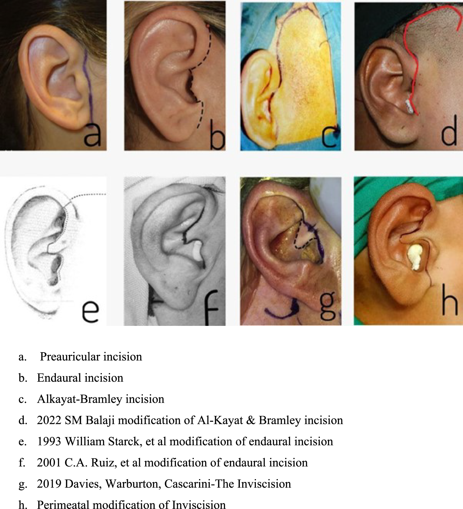
The paradigm of TMJ surgical incisions has evolved to optimize surgical access while minimizing facial disfigurement. Traditional preauricular and endaural incisions provide superior exposure but result in visible scarring. Conversely, the Inviscision technique—particularly its perimeatal modification—ensures scar concealment, offering a more esthetically favorable outcome.
Aim
To evaluate the efficacy of the perimeatal modification of the Inviscision technique in TMJ surgeries, with a focus on surgical exposure, neural injury, and esthetic outcomes.
Methods
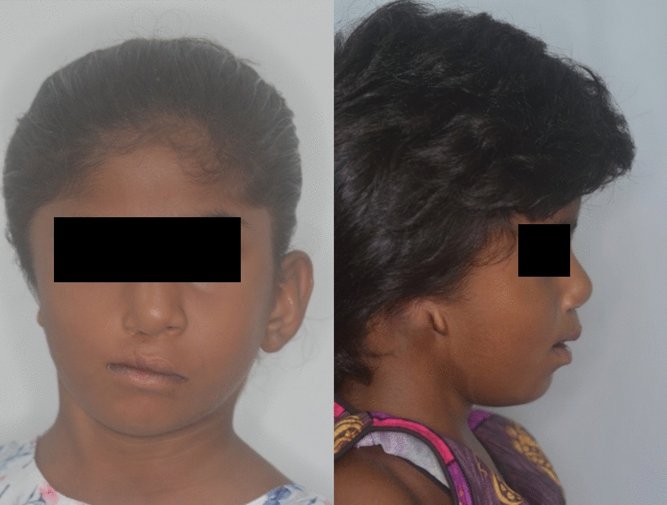
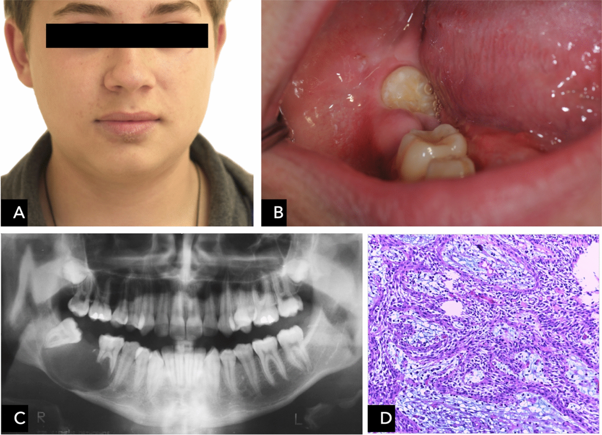
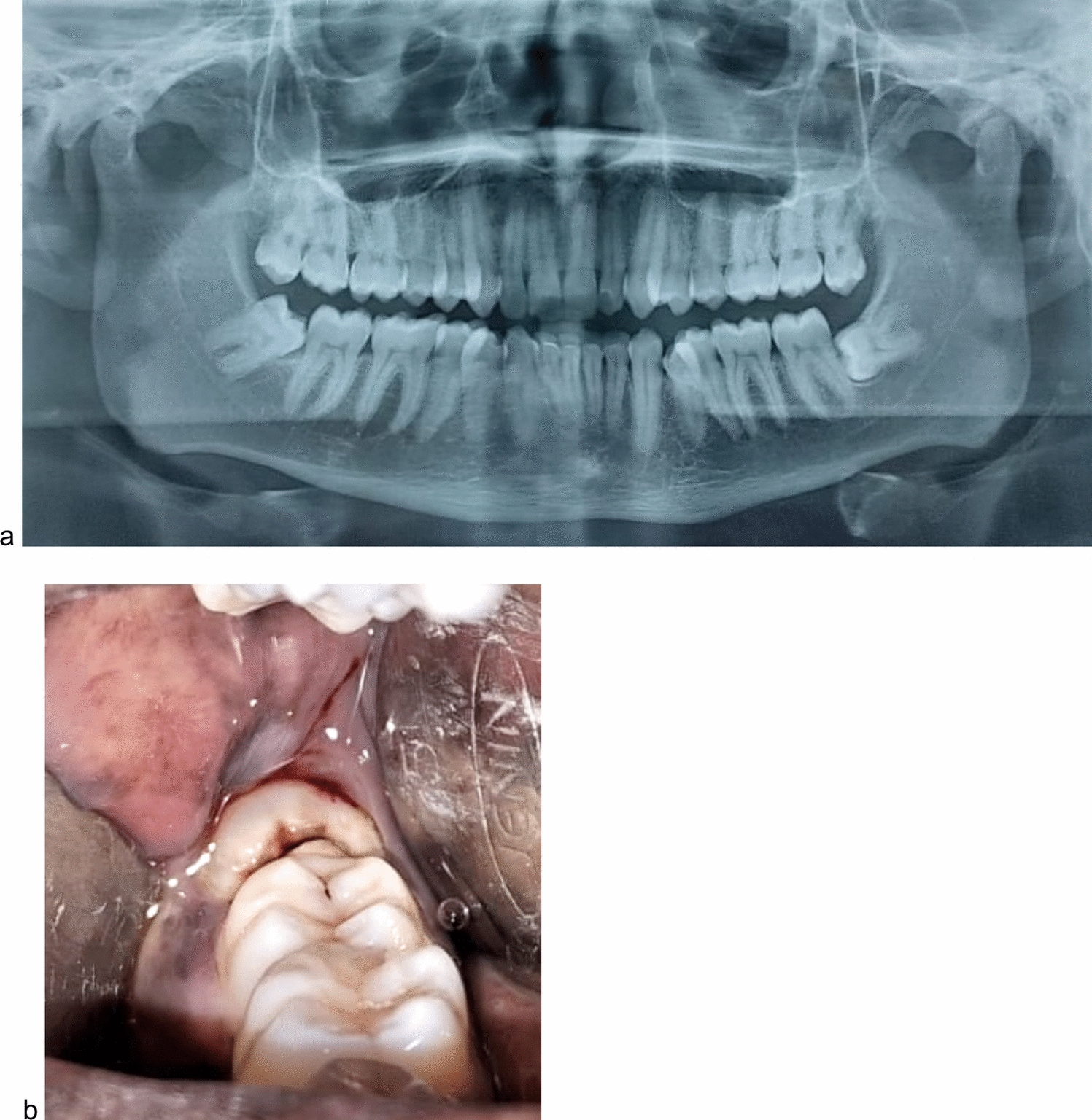
Eighty-one patients undergoing TMJ surgeries, including procedures for ankylosis, joint replacement, and condylar trauma, were retrospectively analyzed. Key variables assessed included the duration from incision to surgical exposure, adequacy of access, neural compromise, scar formation, cartilage injury or necrosis, and ear deformities. All patients were followed for a minimum of 6 months postoperatively.
Results
The mean time to achieve adequate surgical exposure was 12 min. Scarring became imperceptible on average within 40 days. No instances of hypertrophic scarring, keloid formation, cartilage necrosis, or auricular deformity were noted. Transient weakness of the temporal nerve occurred in 34% of cases, with an average recovery period of 3.4 months. The average patient satisfaction score, based on the Likert scale, was 4.
Conclusion
The perimeatal modification of the Inviscision technique provides optimal exposure in the posterior, anterior, lateral, and inferior planes while preserving critical anatomical structures. Aside from causing transient neuropraxia of the temporal nerve, this approach offers superior cosmetic results by concealing the scar within the natural anatomical folds of the ear. With skilled surgical execution, this technique yields excellent outcomes, with the added benefit of an invisible scar, making it a suitable choice for all TMJ surgical interventions.


Comments (0)