
Remember me

This retrospective case series analyzes a novel technique using autologous mandibular angle grafts as a complementary procedure to bimaxillary orthognathic surgery. The consecutive number of patients with dentofacial anomalies from the Clinica Universitaria Colombia, Bogotá, DC, Colombia, was analyzed. A search of the operated cases was conducted, and class II patients with short faces with preoperative and postoperative studies were selected. Exclusion criteria were class II patients with short faces where techniques other than those described for vertical correction were used. After analyzing the registered cases, the following ten cases were selected (Table 1).
Table 1 Summary of demographic and cephalometric dataCase 1A 20-year-old female patient was referred from the orthodontic service to the oral and maxillofacial surgery service to evaluate dentofacial anomalies. She had a history of unspecified respiratory allergies without chronic treatment. The patient’s consultation was to improve her bite and her facial appearance. Clinical examination shows a class II patient with reduced lower facial height, a horizontal mandibular plane, and marked mandibular angles. Intraorally, a class II malocclusion is observed with an increased vertical overbite where the upper incisors cover the lower incisors by approximately 90%.
Clinical findings are confirmed during virtual planning with computed tomography, and the treatment plan is defined. This is bimaxillary orthognathic surgery with a maxillary first sequence, where advancement and descent of 3 mm and a clockwise yaw correction of 1 degree are planned. The mandible is brought into occlusion with advancement and alignment. Concerning the chin, advancement, and descent of 5 mm are made. Currently, the patient has been under follow-up for 22 months without complications (Figs. 1, 2).
Fig. 1
Pre- and postoperative profile images in case 1 showing the difference in facial height and mandibular projection, and intraoral front view
Fig. 2
Preoperative virtual planning in case 1 showing ostectomies design and graft planning
Case 2A 33-year-old male patient arrives at the oral and maxillofacial surgery service for evaluation due to the sensation of a small face. He had a history of high blood pressure under study but without necessary medication. On physical examination, the patient presents a reduced lower facial height, a horizontal mandibular plane, and marked mandibular angles. Intraorally, a class II malocclusion is observed with an increased vertical overbite where the upper incisors cover the lower incisors by approximately 80%, the upper and lower dental midline coinciding.
Clinical findings are confirmed during virtual planning with computed tomography, and the treatment plan is defined. The mandibular sequence starts with a bilateral advancement of 9 mm, followed by a LeFort I osteotomy of 4 mm advancement, and finally, mentoplasty of 6 mm advancement and 3 mm descent. Approximately 5 mm of bilateral gonial angles were resected for the mentoplasty space. Currently, the patient has been under follow-up for 34 months without complications (Figs. 3, 4).
Fig. 3
Pre- and postoperative profile image in case 2 showing the difference in facial height and mandibular projection, and intraoral front view
Fig. 4
Preoperative virtual planning in case 2 showing ostectomies design and graft planning
Surgical TechniqueBimaxillary orthognathic surgery was performed with a conventional high LeFort I technique and bilateral sagittal osteotomy of the Hunsuck type of mandible. In the case where a maxilla-first sequence is performed, the approach is not sutured once the fixation of the osteosynthesis material is finished. A bilateral sagittal osteotomy is then performed using the technique described previously to mobilize the proximal segment and easily take the graft from the mandibular angles. A dislocation maneuver of the proximal segment is performed for better visualization, and the cut is made with piezosurgery-type ultrasonic cutting devices using an angled tip. These grafts are preserved in a container with Ringer’s lactate, while the mandibular surgery is completed and the chin osteotomy is performed. In mandible-first sequence cases, the procedure was similar. Sagittal osteotomy is performed to more easily obtain the mandibular angle segments by mobilizing the proximal segment. The grafts are left in Ringer’s lactate to be then placed in the maxilla and chin.
Considering the bone spaces in osteotomies to recover the lower anterior facial height, a mandibular angle ostectomy is virtually planned to profile and improve the appearance of the posterior facial height. The resected bone segment on each side is divided into two symmetrical segments of approximately 9 mm for case #1 and 5 mm for case #2. The four bone segments obtained are used as autologous grafts for interposition. They are used as follows: One on each side in the space created during the descent of the jaw. The other two bone segments are used in the space created by the descent of the chin. Autologous bone grafts promote bone healing more quickly due to contact and favor the stabilization of the mobile jaws, reducing the possibility of recurrence and bone nonunion. Both cases have a satisfactory aesthetic, functional, and occlusal result. All patients are asymptomatic, and none have presented complications (Fig. 5).
Fig. 5
Virtual planning of bilateral angular cut and interposition bone graft
The indication for mandibular angle grafts in patients with a short face depends on the exaggerated size of the gonial angle and the necessary recontouring. Depending on the resected area, these grafts are placed in the bone gap with or without additional grafts (Fig. 6). With the help of virtual planning, it is possible to determine the necessary amount of resection (bone volume of the graft) and the planned gap.
Fig. 6
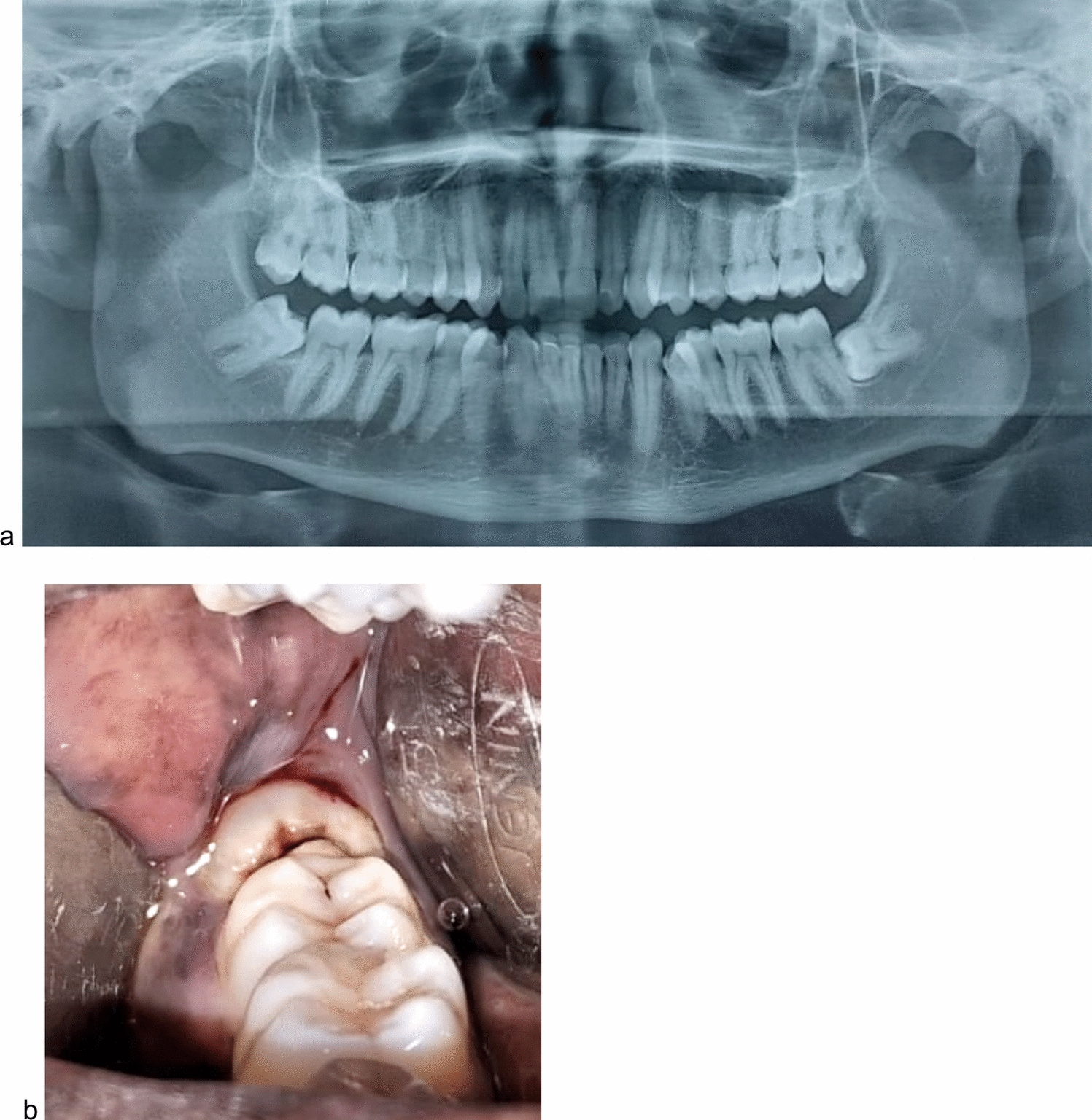
Left Diagram of how to cut the goniac angle with a piezoelectric device. Right Intraoperative view of LeFort I and menton osteotomy with bone grafts between the bone gaps. Abbreviations G, gap; M, menton; P, pyriform; BG, bone graft
Comments (0)