
Remember me


Wild-type Lactococcus lactis subsp. lactis IL1403 (Simon and Chopin 1988, strain collected from Seoul National University) was used as the host bacterium for target protein expression. Wild-type and recombinant strains were grown in M17 medium (MBcell, Korea) supplemented with 5 g/L of glucose (M17G) without antibiotics or with erythromycin (5 μg/mL) and chloramphenicol (5 μg/mL) at 30 °C, respectively.
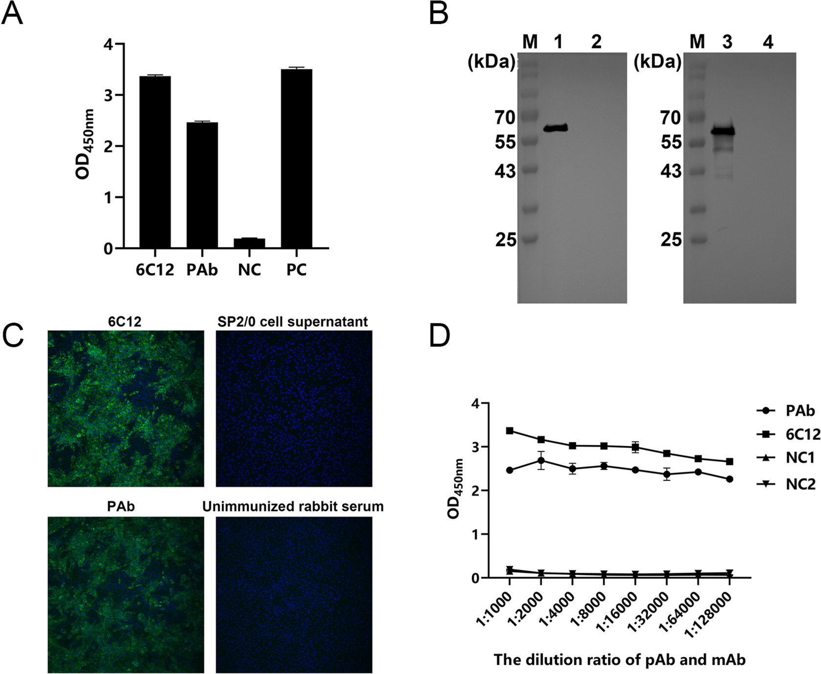
Gene synthesis and plasmid constructionTwo cRANKL amino acid (aa) sequences were obtained from the National Center for Biotechnology Information (NCBI): 400 aa (NCBI accession number: NP_001076830.1) and 318 aa (NCBI accession number: CDZ92724.1). These two proteins had the same extracellular domain sequences. Based on the results of alignment with the soluble form of mouse RANKL (mRANKL) (Knoop et al. 2009; Kim et al. 2015), the one of candidate gene fragments was selected, and the extracellular form of cRANKL was obtained from Sutton et al. (2015). To secrete the target protein from recombinant L. lactis, the signal peptide of USP45 (van Asseldonk et al. 1993) was added to the N-terminus of the target gene. The His-tag (His6x) was added to the C-terminus of the target gene to detect the target proteins. The restriction sites of NdeI and XhoI were located at the two ends for insertion and vector ligation. The vector construction is shown in Fig. 1A. The designed amino acid sequences were codon-optimised using DNAWorks v3.2.4 (Hoover and Lubkowski 2002) based on the L. lactis IL1403 codon usage table, and primers were used to synthesise the insert sequences using the overlap PCR method. The plasmid DNA pILPtuf.Mb (Kim et al. 2009) vector was used as the backbone. The insert and vector were ligated at NdeI and XhoI restriction sites and transformed into wild-type L. lactis IL1403 competent cells. All insert sequences are shown in Fig. S1.
Fig. 1
Production and secretion of recombinant cRANKL from recombinant L. lactis. A Schematic diagram for construction of pILPtuf.gExR.h vector. B Western blot for detecting recombinant cRANKL from cell extracts and cell-free culture supernatant. Lane 1, cell extracts of L. lactis IL1403; Lane 2, cell extracts of L. lactis IL1403 (pILPtuf.gExR.h); Lane 3, cell-free culture supernatant of L. lactis IL1403 (pILPtuf.gExR.h); Lanes 4–6: commercial His-tagged calmodulin (18 kDa) 1.5, 1, and 0.5 μg, respectively
SDS-PAGE and western blot assayWild-type and recombinant L. lactis were cultured in 10 mL of M17G medium without antibiotics or with antibiotics at 30 °C for 10 h, respectively. For the preparation of cell extracts, 10 mL of cultured cells was harvested by centrifugation 3,134 × g for 10 min at 4 °C, and cell pellets were washed by sterilised distilled water twice and resuspended in 200 µL of sterilised 1 × PBS. Subsequently, the solution containing the cell pellet was supplemented with 0.5 g of sterilised glass beads (0.5 mm), followed by disruption using a taco Prep Bead Beater (GeneReach, Taiwan) for 39 s. Cell debris was removed from the cell extracts by centrifugation at 12,300 × g for 5 min at 4 °C. For the preparation of secreted proteins, after centrifugation at 3134 × g for 10 min at 4 °C, the culture supernatant was filtered using the 0.2-µm filter (Minisart® Syringe Filter, Sartorius) and then precipitated with trichloroacetic acid (TCA, 4:1) at 4 °C for 1 h. The precipitates were washed twice with 1 mL of pre-chilled acetone, and centrifugation was performed at 12,300 × g for 5 min at 4 °C. The precipitated pellets were dried at 65 °C for 10 min and dissolved in 200 µL of sterilised 1 × PBS. To quantify the amount of target protein produced, commercial recombinant His-tagged human calmodulin (MERCK, Darmstadt, Germany) protein (18 kDa) was used for the construction of the standard curve with concentrations of 0.5, 1, and 1.5 µg.
The proteins from the total cell extracts or cell-free culture supernatant were separated by SDS-PAGE and transferred onto a nitrocellulose membrane (2 μm; Bio-Rad, Germany). The membrane was blocked with 2.5% (w/v) skim milk in 1 × tris-buffered saline (TBST, 0.1% Tween® 20 detergent) at 25 °C for 1 h. After blocking, the membrane was washed with 1 × TBST thrice for 10 min per wash and incubated with anti-His6x monoclonal antibody (1:500, R&D Systems, USA) at 4 °C for 12 h with shaking. After washing thrice with 1 × TBST, the membrane was visualised with ECL reagents (Bio-Rad, USA).
Validation of the bioactivity of recombinant cRANKL in vivoOne-day-old male ROSS 308 chicks were randomly assigned to three groups, namely PBS, WT_CE, and cRANKL_CE, with ten chicks in each group. The chicks in the PBS group were administered sterilised 1 × PBS. WT_CE and cRANKL_CE groups were administered cell extracts from wild-type L. lactis IL1403 and the recombinant strain L. lactis IL1403 (pILPtuf.gExR.h), respectively. For the preparation of cell extracts, 12-h-cultured wild-type or recombinant strain was harvested by centrifugation 3134 × g for 10 min at 4 °C, and cell pellets were washed by sterilised distilled water twice and resuspended in sterilised 1 × PBS. Subsequently, the solution containing the cell pellet was supplemented with 0.5 g of sterilised glass beads (0.5 mm), followed by disruption using a taco Prep Bead Beater (GeneReach, Taiwan) for 39 s. Glass beads were removed from the cell extracts by centrifugation at 12,300 × g for 5 min at 4 °C. Then, the extracted cell extracts were inoculated in M17G medium and found to contain no viable bacteria. The prepared cell extracts were administered orally to each chick using a 1 mL syringe without a needle. The dosage of cell extracts is shown in Table 1. All groups were fed for 12 consecutive days and sampled on the 13th day (Table 1, Fig. 2).
Table 1 Daily oral dosage of cell extracts in chicken experimentFig. 2
Schematic view of postbiotic-based recombinant RANKL administration, and oral immunization with infectious bursal disease (IBD) vaccine
Peyer’s patches of ileum samples were extracted to determine the expression level of the M cell marker by measuring ANXA5 mRNA expression. Total RNA was extracted using TRIzol® (Thermo Fisher, Korea) according to the manufacturer’s instructions, and cDNA was synthesised using the PrimeScriptTM RT reagent Kit (Takara Bio, Japan). Quantitative real-time PCR (qRT-PCR) was conducted using TB Green® Premix Ex Taq™ (Tli RNaseH Plus, TAKARA, Japan) with specific primers ANXA5-F:5′-AGTATACAAGAGGCACCGTG-3′; ANXA5-R:5′-GTCTCATCAAAGATACCATC-3′, and primers for the housekeeping gene GAPDH-F:5′-GTGGTGCTAAGCGTGTTATCATC-3′; GAPDH-R:5′-GGCAGCACCTCTGCCATC-3′ (Olias et al. 2014). The mRNA level was presented as 2−ΔCt, where Ct, threshold cycle for target amplification, and ΔCt, Cttaget gene (specific genes for each sample) − Ctinternal reference (housekeeping gene for each sample).
16S rRNA amplicon sequencingOn the 13th day, genomic DNA was extracted from faecal samples using a NucleoSpin Soil kit (Macherey–Nagel, Düren, Germany), according to the manufacturer’s instructions. DNA samples (5 ng) were used to amplify the 16S ribosomal RNA V4 region using Takara Ex-Taq DNA polymerase (Takara Bio) with universal primer sets (forward:5′-GGACTACHVGGGTWTCTAAT-3′ and reverse:5′-GTGCCAGCMGCCGCGGTAA-3′) (Han et al. 2018). After amplification, all samples were normalised to 50 ng per sample. A DNA library was constructed and sequenced using the Illumina MiSeq platform (Illumina, San Diego, CA, USA), generating 2 × 300 bp paired-end reads.
Bioinformatic analysis of the gut microbiomeTo analyse the microbial community, de-multiplexed and pre-processed sequence reads were imported into Quantitative Insights Into Microbial Ecology (QIIME2, version 2021.2) (Bolyen et al. 2019). Barcode and primer removal, quality control, amplicon sequence data correction, and de-replication were performed using the DADA2 software package (Callahan et al. 2016). Sequence reads were truncated to 200 bp using an in-house Perl script. Feature tables and representative sequence files were merged for downstream analysis using QIIME2. Taxonomic classification was assigned using the SILVA 132 database, with 99% identity based on the V4 16S region. All classifications were performed within QIIME2 and were assigned using the naïve Bayesian algorithm available in the Sklearn Python library. For phylogenetic diversity analysis, alpha and beta diversities were calculated using the q2-diversity plugin and included Faith’s phylogenetic diversity and weighted and unweighted UniFrac distances. Differential abundance analysis of microbiota was performed using an in-house Perl script. The relationship between relative abundance and ANXA5 mRNA expression was assessed by Pearson’s correlation efficient (r) and p values from simple linear regression. Statistical significance was set at p < 0.05.
Serum calcium assayThe serum calcium concentration was measured using a Calcium Colorimetric Assay Kit (BioVision Inc., USA). All serum samples were diluted to ten-folds (10 μL of serum and 90 μL of Chromogenic Reagent), and 60 μL of Calcium Assay Buffer was added to each well and mixed gently. The samples were incubated for 10 min at room temperature in the dark. After incubation, the optical density (OD) was analysed by measuring the absorbance of the samples at 575 nm.
Evaluation of oral vaccine efficiencyAfter 12 consecutive days of oral administration of postbiotic-based recombinant cRANKL, chickens were orally immunised with the infectious bursal disease (IBD) vaccine on the 13th day. The experimental schedule is shown in Fig. 2. The IBD vaccine (PoulShot® Gumboro) was purchased from the Central Vaccine Research Institute, Korea. The dosage of oral vaccination was based on vaccine programme guidelines. The relationship between faecal IgA and serum IgG was assessed by Pearson’s correlation efficient (r) and p values from simple linear regression. Statistical significance was set at p < 0.05.
Statistical analysisStatistical analysis was performed using an in-house Perl script and R (v4.1.4). For significance analysis, the Kruskal–Wallis test was performed followed by Dunn’s posthoc test, and the data are expressed as follows: *p < 0.05, **p < 0.01, ***p < 0.001.
Comments (0)