Design
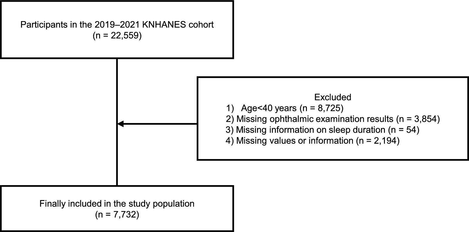
This multicenter, multi-surgeon, single-protocol, retrospective, case series study consecutively enrolled eyes that had undergone lensectomy with implantation of a trifocal IOL after a previous LCRS to treat hyperopia at our institution (all preoperative data known). To provide surgeons with significant information about outcomes, we separated and compared the results between the two groups, depending on the type of IOL implanted: group 1 included patients who received an aspheric trifocal IOL with a negative SA, and group 2 included patients who received an aspheric trifocal IOL with a neutral SA.
Subjects
Data were recorded using the central computerized clinical records system in Clinica Baviera, Spain. The study period was from 1999-11-23 (first visit before LCRS; YYYY-MM-DD) to 2021-09-30 (last available postoperative visit). Laser treatments were performed between 1999-11-23 and 2017-05-15. Lens surgeries were performed between 2016-09-19 and 2021-08-11. We have included patients who have undergone cataract surgery (no greater than NO1/NC1, C1, in LOCS III scale) or refractive lens exchange (RLE). The term “lensectomy” in the text is used to refer to both the cataract and RLE groups and therefore includes both types of phacoemulsification.
The study consists of a research on existing data. These data were recorded anonymously using identifiers linked to the subjects. Given the retrospective nature of the study, it was approved by our institutional legal and ethical committee with an exempt review (Ethical Committee of Clinica Baviera, Spain). All patients received detailed information before surgery and provided written informed consent for multifocal lensectomy after LCRS. They also provided informed consent for the use of anonymous and aggregated medical data for the study.
The study inclusion criteria were as follows: (1) lens surgery [RLE or cataract (NO1/NC1, C1) with implantation of a trifocal IOL in eyes previously treated with laser-assisted in situ keratomileusis (LASIK)] for correction of hyperopia, (2) potential visual acuity [baseline pre-LCRS logMAR corrected distance visual acuity (CDVA) < 0.5], (3) at least three months of follow-up after implantation, and (4) no corneal laser enhancement. The exclusion criteria were: (1) eyes with subnormal optics, such as corneal topographic abnormalities (small optical zones, decentered ablations, suspected ectasia), and (2) any baseline anatomical disorder (vitreoretinal or surface/anterior segment disorder) or any perioperative anatomical complications (corneal and/or lens surgeries) to rule out organic disease that could mask the functional outcomes of both refractive procedures.
Intraocular lenses
The diffractive trifocal IOLs implanted during the study period were FineVision Pod-F (PhysIOL, Liège, Belgium) in group 1 and RayOne Trifocal (Rayner, Worthing, United Kingdom) in group 2. Both IOLs were manufactured using foldable hydrophilic acrylic materials. The FineVision Pod-F (single-piece, double-C loop haptics) combines two diffractive structures adjusted to offer a + 3.50 D addition for near vision and a + 1.75 D addition for intermediate vision; it has an aspheric profile with − 0.11 µm of SA. The RayOne Trifocal IOL provides a neutral aspheric optical profile, that is, 0 µm of SA, with 16 diffractive steps, a + 3.50 D near addition, and a + 1.75 D intermediate addition. The pupil size for SA values of both lenses (RayOne and FineVision) is 6 mm.
Surgical procedures
The corneal and lens procedures were performed by experienced surgeons using homogeneous perioperative protocols. The LCRS procedure was LASIK in all cases and was performed using two microkeratomes with nasal hinges (Moria LSK-ONE and Moria ONE-USE-PLUS-SBK, Microtech Inc., Moria Ophthalmic Instruments, Anthony, France) and three excimer laser models [Technolas 217C, 217-Z-100 (Bausch & Lomb, Claremont, California, USA), Mel-80 (Carl Zeiss Meditec, Jena, Germany), and WaveLight-Allegretto Wave-Eye-Q (Alcon Laboratories, Fort Worth, Texas, USA)]. All LCRS data are available.
Patients who had previously undergone LASIK ablation to treat hyperopia returned to the clinic for lens surgery because of reduced distance and/or near visual acuity caused by presbyopia and/or cataracts. After selection of an appropriate lens, standard, uneventful phacoemulsification was performed with implantation of a trifocal IOL in the capsular bag.
The online American Society of Cataract and Refractive Surgery (ASCRS) calculator (https://iolcalc.ascrs.org) and/or the Barret True-K formula (https://www.apacrs.org/apacrsbiometry/True-K.aspx) were used for IOL calculation by entering refractive, keratometric, topographic, and biometric data based on a multiformula approach. We aimed for emmetropia, so we selected the average lens power in all cases.
Clinical evaluation
All surgical procedures were performed at our institution, using homogeneous preoperative assessment protocols. Patients underwent a complete ophthalmologic examination that included the measurement of visual acuity data, namely, Snellen distance visual acuity (Snellen auto chart projectors, Topcon Corp, Tokyo, Japan), Jaeger near and intermediate visual acuity (Runge Near Vision Card, Good-Lite, Elgin, Illinois, USA), and refraction (uncorrected and corrected, manifest, and cycloplegic). Refractive status, registered by an optometrist, included uncorrected distance visual acuity (UDVA), CDVA, uncorrected intermediate visual acuity (UIVA) at 80 cm, and uncorrected near visual acuity (UNVA) at 40 cm (visual acuities were tested under photopic conditions, at approximately 85 cd/m2). The patients also underwent topography, slit lamp biomicroscopy, ocular surface/tear film evaluation, and fundoscopy.
However, owing to the diversity in practice locations, study time points, and development of devices over time, preoperative topographic evaluation was not standardized. The three corneal topographers used during the study period were the Orbscan II (Bausch&Lomb, Claremont, California, USA), Pentacam (Oculus Optikgerate GmbH, Wetzlar, Germany), and Wavelight-Oculyzer (Alcon Laboratories, Foxworth, Texas, USA). Nevertheless, to assess the impact of previous corneal hyperopic ablation and to avoid bias when evaluating our results, the preoperative corneal Z4(0) and high-order aberration (HOA) values obtained on Pentacam were also studied and compared.
Preoperative examination for lens surgery also included endothelial cell count (SP 3000P; Topcon, Capelle, The Netherlands) and macular optical coherence tomography (SOCT Copernicus-REVO, Optopol-Tech, Zawircie, Poland). Biometric parameters were assessed using an optical biometer (IOLMaster 500; Carl-Zeiss-Meditec AG).
Refractive and visual measures
The main measurements were visual and refractive outcomes and patient satisfaction, which were obtained from the last available visit, with at least three months of follow-up after implantation. Visual outcomes included average logMAR UDVA, CDVA, UIVA and UNVA. Refractive data included postoperative sphere, cylinder, manifest refraction spherical equivalent (MRSE), and accuracy (percentage of eyes within ± 0.50 D and ± 1.00 D). Safety outcomes were defined as the percentage of eyes with a loss of ≥ 1 or ≥ 2 lines of CDVA between the time after LCRS and lens surgery; the lines presented in the graphs correspond to a change of 0.1 on logMAR scale. For instance, a patient with preoperative CDVA 0 logMAR (decimal 1.0) and postoperative UDVA 0.1 logMAR (decimal 0.8) would fall in “1 worse” on the efficacy Bar-Chart.
Efficacy outcomes were measured as the percentage of eyes with a difference between post-LCRS CDVA and post-lensectomy UDVA ≥ 0 lines. The safety index is defined as the ratio of mean preoperative CDVA to mean postoperative CDVA; and the efficacy index as mean postoperative UDVA to mean preoperative CDVA. Although the degree of cataract was not advanced enough to induce significant changes in CDVA, we have calculated Safety and Efficacy indices for the RLE group only.
A patient satisfaction questionnaire was used by our group in previous studies [4, 7].
Statistical analysis
When comparing independent groups, the distributions were assessed for outliers, normality, and homogeneity of variance. The outliers were assessed using the box plot method. Normality was assessed using the Shapiro–Wilk test and quantile-to-quantile (Q–Q) plots. Homogeneity of variances was verified using the Levene’s test. In most cases, these assumptions were met, and an independent t-test was performed. In case where the assumptions of the parametric test were not satisfied, non-parametric Mann–Whitney test was performed, and medians and quartiles were reported. Otherwise, we report usual means and standard deviations with differences tested with t-test.


Comments (0)