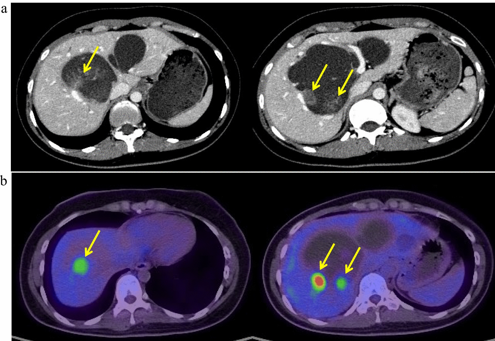
Primary angiosarcomas of the breast account for less than 0.04% of all breast malignancies, their incidence being approximately 0.0005% [3]. Chen et al. reviewed 87 reported cases. Both breasts were involved in 18 of these patients (21%) [2]. We herein report a patient with multiple angiosarcomas in both breasts.
It is difficult to diagnose angiosarcoma of the breast early. They generally present as rapidly growing, ill-defined masses, breast swelling, or asymmetry of the breasts [4]. Two thirds of these neoplasms measure > 5 cm on presentation [5]. Bilateral tumors may be derived from multifocal origins or by metastasis from a contralateral breast. Mammography often shows non-calcified masses or FADs. Ultrasonography reveals either hyperechoic or mixed hyper- and hypo-echoic masses with architectural distortion. Dynamic contrast-enhanced MRI reveals significant heterogeneous enhancement in the early phase and varying degrees of concentric enhancement in the delayed phase [6]. In the present case, the MRI findings were helpful to distinguish angiosarcomas from typical breast cancers. Well-differentiated angiosarcoma is rich in capillary networks, and the lumens are complete. The contrast agent takes a longer time to pass throughout the tumor and be washed out. Therefore, the dynamic curve exhibits a persistently enhancing or plateau pattern. CNBs are preferred for diagnosing angiosarcomas, because the false negative rate for fine needle aspiration cytology is as high as 40%. It is sometimes challenging for pathologists to diagnose such well-differentiated angiosarcomas, even by CNB, and close collaboration between clinicians and pathologists may help to make an accurate diagnosis. In the current case, the pathologist was well-informed of the patient’s clinical findings, including the MRI results indicative of malignancy, and conclusively diagnosed the bilateral tumors in the CNB specimens as well-differentiated angiosarcomas. That was more than 5 months after the patient first reported detecting a lump in her right breast.
Total mastectomy, with or without radiation therapy, has been the mainstay of treatment. However, locoregional recurrences are reported in half of all patients [7]. There have been no randomized clinical trials assessing the outcomes of breast-conserving surgery versus mastectomy, or determining the optimal margin width after resection [8]. Adjuvant radiation therapy significantly improved recurrence-free survival in a meta-analysis including 380 patients with primary angiosarcoma and 595 with secondary angiosarcoma [9]. In the current case, we performed bilateral total mastectomy, because the tumors were widespread throughout both breasts. Postoperative irradiation was administered to the bilateral chest wall soon after surgery to prevent local recurrences, because most local recurrences occur early after surgery [7]. In addition, McKay et al. reported that low-dose radiotherapy resulted in a rapid and complete response within the treated areas in a patient with recurrent angiosarcoma [10]. These findings suggest that radiation should be more widely considered for the treatment of angiosarcoma. Axillary LN metastases are uncommon at the time of primary therapy; therefore, routine axillary dissection is not indicated [7]. Herb et al. reported that 5% of patients with non-metastatic angiosarcoma of the breast had involvement of regional LNs, and their median overall survival was significantly shorter than that of LN-negative patients (15 months vs. 77 months, log-rank p < 0.001) [11]. In the current case, we performed only total mastectomy without axillary clearance, because there were no clinical findings of LN metastasis. No regional LN recurrences were detected 1 year after surgery. A prospective study to determine the indications for sentinel LN biopsy is warranted.
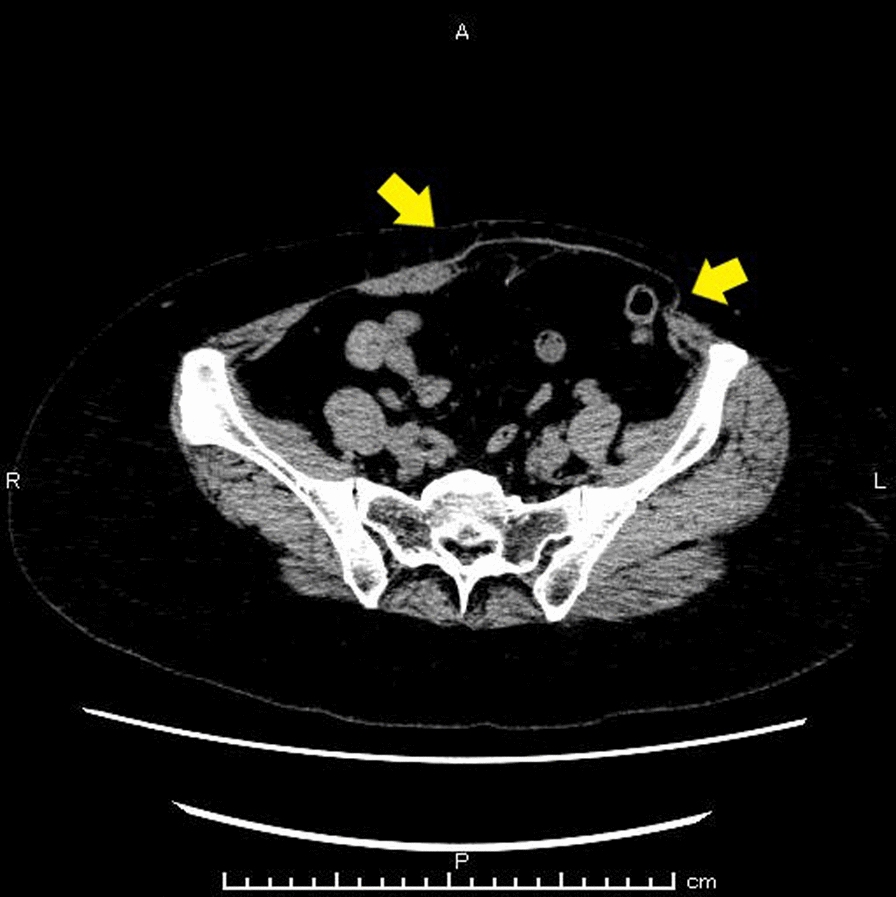
Adjuvant or neoadjuvant chemotherapy may be offered to patients with angiosarcoma of the breast. However, their effectiveness remains controversial and no chemotherapeutic regimens have yet been clearly established. Constantinidou et al. reported the use of a variety of chemotherapy regimens in adjuvant/neoadjuvant settings. The most commonly used regimens in European Organization for Research and Treatment of Cancer sarcoma centers are paclitaxel (27.9%/35.6%, respectively) and gemcitabine and docetaxel-containing regimens (25.6%/11.9%, respectively) [12]. We administered paclitaxel to the present patient, because we suspected that the multiple, small, subcutaneous nodules detected by CT in her chest wall were angiosarcoma metastases. Patients with metastatic angiosarcoma of the breast have had good responses to paclitaxel in a number of studies [7, 13]. According to the National Comprehensive Cancer Network guidelines, there is level 2A evidence that paclitaxel and anthracycline- or gemcitabine-based regimens are the most effective regimens against angiosarcoma [14]. Sher et al. [7] reported an overall response rate of 48% for cytotoxic chemotherapy for metastatic angiosarcoma. Our patient’s small metastases shrank after 2 months of paclitaxel treatment. This relatively high response rate may be partly attributable to the finding that a high proportion of cells in small, fast-growing tumors are killed by doses of drugs that have very little effect on larger, slower-growing masses of otherwise identical cancer cells [15].
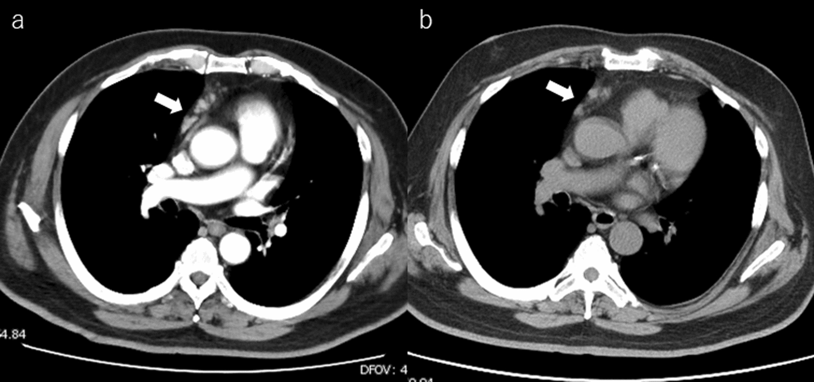
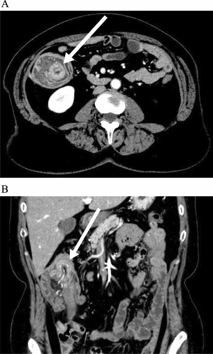
Tumor dissemination occurs early via the bloodstream. The most common sites of metastases are bone, lung, liver, contralateral breast, skin, brain, and ovary [2]. Several molecular pathways are reportedly involved in development of angiosarcomas. Loss of function of p53 and overexpression of mouse double minute 2 and vascular endothelial growth factor (VEGF) are alterations in molecular pathways that have been found to be involved in the development of angiosarcomas [16]. The PIK3CA/AKT/mTOR pathway is either directly or indirectly involved in the development of breast angiosarcomas [17]. It has been postulated that restoration of wild-type p53 expression may be an effective therapeutic strategy. PI3K inhibitor and mTOR inhibitor are possible targets for angiosarcoma therapy. A small subset of these tumors shows mutations in PLCG1 and KDR, which are involved in the VEGFR2 signaling pathway [18]. Inhibition of the VEGF pathway is a potentially effective approach to treating angiosarcomas. Comprehensive genome profiling of our patient’s tumor cells did not yield any leads to targeted therapy.

Comments (0)