
Remember me


In the middle of the last century the pursuit of arterial angiography for diagnosis and treatment of vascular and other anatomic pathology seemed more and more practical; the x-ray had been in clinical use since 1896, iodinated contrast was conceived in the 1920s (1), Werner Forssmann performed the first right atrial catheterization in 1929, and in 1950 a Mayo Clinic anesthesiology resident named David Massa described the Rochester plastic needle (2), the first percutaneous disposable IV catheter for IV therapy which was remarkably similar to contemporary IVs and an improvement from surgical cutdowns and metal IV needles.
Seemingly, all the pieces were right there for developing the technique. However, the challenge of introducing contrast upstream from regions of interest remained. Before this, angiography was described in various invasive methods, including direct abdominal aortic (1931), and carotid (1949) puncture, surgical cutdowns for radial (1948) or femoral (1950) catheter placement, transesophageal aortic arch injection (1949), and direct biventricular intracardiac injection (1951). These efforts were stymied by issues of guiding the injection site with catheters that were at times nonradiopaque or stiff needles that could injure the arterial endothelium.
A young radiology resident at the Karolinska Institutet in Solna, Sweden decided to take on this technical challenge in 1952 (3). His family ran the Mechanical Workshop in his hometown of Mora and had a long tradition as technical geniuses. He had the idea that central arterial catheterization was possible by puncturing and entering a catheter close to its distal end such that the needle emerged out of the tip. He could obtain arterial access with the assembly, then replace the needle with a guidewire and feed the catheter to its destination. Before this, catheters were fed through needles but the initial arteriotomy exceeded the diameter of the catheter so needle removal led to leakage. A smaller needle for initial arteriotomy could avoid this and adding a guidewire ensured sufficient stiffness (4). However, his grand attempt utterly failed and the resident, Sven-Ivar Seldinger, MD, stood in the laboratory, “…disappointed and sad with the three things in my hand: the needle, the guide wire, and the catheter… and I had a severe attack of common sense.” As the circumstances were, again all the elements were there. Seldinger (4) invented his eponymous technique on the spot and revolutionized vascular access, seeing broad international acceptance. Within a few years, he and his contemporaries had mapped the angiographic appearance of the human arterial system, and in 3 decades, his technique had helped an estimated 50 million people. Puzzlingly at the time, the technique that bore his name was not deemed significant enough for a doctoral thesis and 13 years later, he defended one on percutaneous cholangiography as a modification of his vascular access technique.
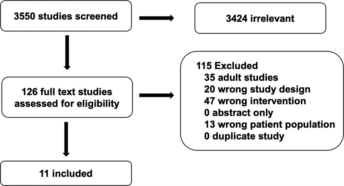
The first central venous catheter (CVC) was placed in 1967 at the Children’s Hospital of Philadelphia in an infant with small bowel atresia (5,6) and it was tunneled to reduce the chance of infection. Since then, tunneled venous catheterization became established as a mainstay for vascular access in cases where prolonged access is required. This has been where the technical challenges of tunneling are outweighed by the benefits of increased device stability and reduced infection risk (Fig. 1). Historically, the procedure has been the purview of surgeons and interventional radiologists in sterile procedural environments. Advancements in bedside vascular access, particularly using ultrasound guidance, have increased the repertoire of clinicians at the ICU bedside. In this issue of Pediatric Critical Care Medicine, Mills et al (7) report on a series of 182 tunneled femoral venous catheters placed in 161 children placed by cardiac intensivists. Although the technique is similar to procedures performed with specialized equipment and interventional radiologists at the bedside (8,9), Mills et al (7) proposes a methodology possible with common peripherally inserted central catheter (PICC) equipment by intensivists and reports contemporary outcome measures. The rate of central line associated bloodstream infection in this population was overall favorable at 0.75 per 1,000 line-days. Although this is similar to rates seen for nontunneled PICC catheters placed in the same clinical setting, it is lower than the rate of central line-associated bloodstream infection reported among 22 hospitals in the Pediatric Cardiac Critical Care Consortium (PC4) registry in 2018 (10) which also included all central catheters. In addition, the tunneled catheter observed thrombosis rate was 6% in the study by Mills et al (7) compared with 14% of all tunneled catheters also reported in 23 PC4 registry hospitals in 2020 (11). Quality assurance metrics require institutional baseline data, such as standardized infection rates for comparison, so that the effect of the intervention evaluable in the context of the local practice environment. At best pooled collaborative outcome data paints a broad picture for clinical performance though impact requires a more incisive prospective perspective. Statistical comparisons are necessary to appreciate whether these differences are significant; however, the potential for improving CVC safety with this methodology seems possible.
 Figure 1.:
 With a common protocol, team acceptance and implementation, and interspecialty collaboration, standardizing tunneled central venous catheter is a big but practical undertaking in the PICU. PICC = peripherally inserted central catheter.
Additional benefits of the technique could include performing the procedure at the ICU bedside where a patient is being actively supported and resuscitation resources are readily available. Considering the procedural complications observed; apnea, arterial thrombus, arrhythmia, and bleeding, one can imagine that proximity to the ICU and no need for therapy disconnection is advantageous in these scenarios. The methodology described also does not use fluoroscopy thereby limiting radiation exposure and patient access limitations a fluoroscope is associated with. Last, the device selected can fit the exact needs of the cardiac ICU in terms of the variety of materials, coatings, sizes, and lumen numbers available. A previous study in cardiac neonates describes creating tunnels for femoral catheters by puncturing the skin in line with the femoral vein (12), and the tunnel is a longer but straight approach to the venipuncture site. The lateral tunneling method proposed by Mills et al (7) confers the benefit of potentially lower risk of displacement as well as greater freedom in planning where the catheter exits the skin as is seen in adult data on similar methods (9,13).
Limitations of this technique are approached by the authors include single-center nature of the work in an area frequently affected by patient and institutional factors that potentially were not evaluated. The retrospective nature of the study will likely encourage necessary prospective work in the area evaluating the direct impact of the intervention while controlling for confounders that could result from study timing and population attributes. Effects of industry-standard versus adaptive practices such as drawing blood from small catheters, not generally approved by device manufacturers but a clinical necessity at times, are important factors that require prospective quality improvement assessments of clinical practice. Another consideration is that the methodology described is uniquely suited to the infant population. In older patients equipment in PICC sets is likely too short and inflexible for tunneling to a desired exit point. Limitations of PICC sets are also a potential reason why accurate upper extremity access might be challenging using the described technique. In addition to a requirement for tip detection equipment before completion of the tunnel, and lung interference with ultrasound methods for identifying the catheter, positioning the sheath dilator used in the method proposed by Mills et al (7) is potentially difficult in an infant’s neck.
Mills et al (7) explains a straightforward, patient-adaptable protocol possible for infants with equipment in the armamentarium of most ICUs. As described, the procedure requires the use of ultrasound guidance, a PICC set and no other vascular access equipment. Although questions persist as to how one might modify this technique for older patients or accessing other vascular structures or tunnel directions, these possibilities are potentially explorable with the myriad vascular access devices ICU proceduralists have at their disposal, growing literature on the topic, and the willingness to adapt this procedure to the bedsides in their own units. All the pieces are right here in front of us.
1. Quader MA, Sawmiller CJ, Sumpio BE: Radio contrast agents: History and evolution. In: Textbook of Angiology. Chang JB (Ed). New York, NY, Springer, 2000, p 775 2. Southorn PA, Narr BJ: The Massa or Rochester plastic needle. Mayo Clin Proc. 2008; 83:1165–1167 3. Greitz T: Sven-Ivar Seldinger. AJNR Am J Neuroradiol. 1999; 20:1180–1181 4. Seldinger SI: Catheter replacement of the needle in percutaneous arteriography; a new technique. Acta Radiol. 1953; 39:368–376 5. Wilmore DW, Dudrick SJ: Growth and development of an infant receiving all nutrients exclusively by vein. JAMA. 1968; 203:860–864 6. Gow KW, Tapper D, Hickman RO: Between the lines: The 50th anniversary of long-term central venous catheters. Am J Surg. 2017; 213:837–848 7. Mills M, Chanani N, Wolf M, et al.: Durable Vascular Access in Neonates in the Cardiac ICU: A Novel Technique for Tunneled Femoral Central Venous Catheters. Pediatr Crit Care Med. 2023; 24:919–926 8. Michel F, Dejode JM, Vialet R, et al.: Tunneled central venous catheter for neonates: A simple technique for prolonged indwelling central catheters in intensive care. Pediatr Crit Care Med. 2007; 8:37–39 9. Woerner A, Wenger JL, Monroe EJ: Single-access ultrasound-guided tunneled femoral lines in critically ill pediatric patients. J Vasc Access. 2020; 21:1034–1041 10. Alten JA, Rahman AKMF, Zaccagni HJ, et al.: The epidemiology of healthcare-associated infections in pediatric cardiac intensive care units. Pediatr Infect Dis J. 2018; 37:768–772 11. DiPietro LM, Gaies M, Banerjee M, et al.: Central venous catheter utilization and complications in the pediatric cardiac ICU: A report from the Pediatric Cardiac Critical Care Consortium (PC4). Pediatr Crit Care Med. 2020; 21:729–737 12. Shostak E, Tzeitlin Y, Shochat T, et al.: Bedside durable tunneled femoral central venous catheter is feasible and safe in high-risk infants in the pediatric cardiac intensive care unit. J Intensive Care Med. 2023; 38:307–312 13. Elli S, Cannizzo L, Giannini L, et al.: Femorally inserted central catheters with exit site at mid-thigh: A low risk alternative for central venous catheterization. J Vasc Access. 2022 Nov 2. [online ahead of print]
Comments (0)