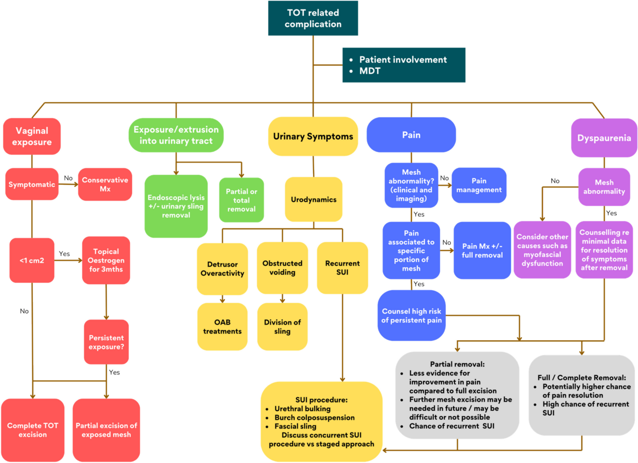
Good MM, Solomon ER. Pelvic floor disorders. Obstet Gynecol Clin North Am. 2019;46(3):527–40.
Article
PubMed
Google Scholar
Verbeek M, Hayward L. Pelvic floor dysfunction and its effect on quality of sexual life. Sex Med Rev. 2019;7(4):559–64.
Article
PubMed
Google Scholar
Haylen BT, et al. An International Urogynecological Association (IUGA)/International Continence Society (ICS) joint report on the terminology for female pelvic floor dysfunction. Neurourol Urodyn. 2010;29(1):4–20.
Article
PubMed
Google Scholar
Spence-Jones C, et al. Bowel dysfunction: a pathogenic factor in uterovaginal prolapse and urinary stress incontinence. Br J Obstet Gynaecol. 1994;101(2):147–52.
Article
CAS
PubMed
Google Scholar
Swift S, et al. Pelvic Organ Support Study (POSST): the distribution, clinical definition, and epidemiologic condition of pelvic organ support defects. Am J Obstet Gynecol. 2005;192(3):795–806.
Article
PubMed
Google Scholar
Persu C, et al. Pelvic Organ Prolapse Quantification System (POP-Q) - a new era in pelvic prolapse staging. J Med Life. 2011;4(1):75–81.
CAS
PubMed
PubMed Central
Google Scholar
Barber MD. Symptoms and outcome measures of pelvic organ prolapse. Clin Obstet Gynecol. 2005;48(3):648–61.
Article
PubMed
Google Scholar
Lau T, et al. Low back pain does not improve with surgical treatment of pelvic organ prolapse. Int Urogynecol J. 2013;24(1):147–53.
Article
PubMed
Google Scholar
Albanesi G, et al. Computed-tomography image segmentation and 3D-reconstruction of the female pelvis for the preoperative planning of sacrocolpopexy: preliminary data. Int Urogynecol J. 2019;30(5):725–31.
Article
PubMed
Google Scholar
Zilberlicht A, et al. Characterization of the median sacral artery course at the sacral promontory using contrast-enhanced computed tomography. Int Urogynecol J. 2017;28(1):101–4.
Article
PubMed
Google Scholar
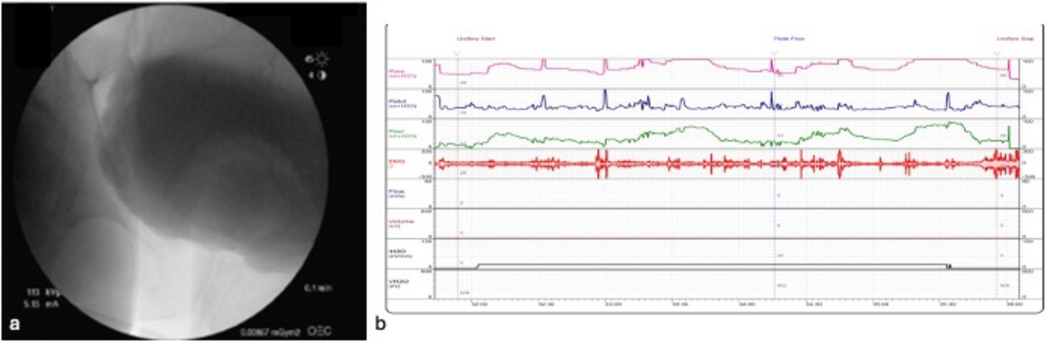
Palmer SL, et al. Dynamic fluoroscopic defecography: updates on rationale, technique, and interpretation from the Society of Abdominal Radiology Pelvic Floor Disease Focus Panel. Abdom Radiol (NY). 2021;46(4):1312–22.
Article
PubMed
Google Scholar
Flusberg M, et al. Multimodality imaging of pelvic floor anatomy. Abdom Radiol (NY). 2021;46(4):1302–11.
Article
PubMed
Google Scholar
Speed JM, et al. Trends in the diagnosis and management of combined rectal and vaginal pelvic organ prolapse. Urology. 2021;150:188–93.
Article
PubMed
Google Scholar
Vellucci F, et al. Pelvic floor evaluation with transperineal ultrasound: a new approach. Minerva Ginecol. 2018;70(1):58–68.
PubMed
Google Scholar
Dietz HP. Ultrasound in the assessment of pelvic organ prolapse. Best Pract Res Clin Obstet Gynaecol. 2019;54:12–30. References article explaining different ultrasound approaches (US) to evaluated pelvic organ prolapse (POP). Citation provided for readers for further reference, if interested in detailed descriptions of the US approaches.
Article
PubMed
Google Scholar
Pedersen L, Glavind-Kristensen M, Bor P. Clinical relevance of routine transvaginal ultrasound in women referred with pelvic organ prolapse. BMC Womens Health. 2021;21(1):26.
Article
PubMed
PubMed Central
Google Scholar
Chamie LP, et al. Translabial US and dynamic MR imaging of the pelvic floor: normal anatomy and dysfunction. Radiographics. 2018;38(1):287–308.
Article
PubMed
Google Scholar
Gao Y, et al. Diagnostic value of pelvic floor ultrasonography for diagnosis of pelvic organ prolapse: a systematic review. Int Urogynecol J. 2020;31(1):15–33. Citation provided as background information for correlation of POP-Q with US findings.
Article
PubMed
Google Scholar
Khatri G, de Leon AD, Lockhart ME. MR imaging of the pelvic floor. Magn Reson Imaging Clin N Am. 2017;25(3):457–80.
Article
PubMed
Google Scholar
Stothers L, et al. Standing open magnetic resonance imaging improves detection and staging of pelvic organ prolapse. Can Urol Assoc J. 2021;16:E20.
Article
PubMed Central
Google Scholar
Abdulaziz M, et al. Relevance of open magnetic resonance imaging position (sitting and standing) to quantify pelvic organ prolapse in women. Can Urol Assoc J. 2018;12(11):E453–60.
Article
PubMed
PubMed Central
Google Scholar
Grob ATM, et al. Underestimation of pelvic organ prolapse in the supine straining position, based on magnetic resonance imaging findings. Int Urogynecol J. 2019;30(11):1939–44.
Article
PubMed
PubMed Central
Google Scholar
El Sayed RF. Magnetic resonance imaging of the female pelvic floor: anatomy overview, indications, and imaging protocols. Radiol Clin North Am. 2020;58(2):291–303.
Article
PubMed
Google Scholar
Memon B, et al. Dynamic magnetic resonance imaging: an aid to preoperative-planning of pelvic organ prolapse. J Ayub Med Coll Abbottabad. 2021;33(3):382–5.
PubMed
Google Scholar
Rechi-Sierra K, et al. Magnetic resonance imaging to evaluate anterior pelvic prolapse: H line is the key. Neurourol Urodyn. 2021;40(4):1042–7.
Article
PubMed
Google Scholar
Kobi M, et al. Practical guide to dynamic pelvic floor MRI. J Magn Reson Imaging. 2018;47(5):1155–70.
Article
PubMed
Google Scholar
Swamy N, et al. Pelvic floor imaging with MR defecography: correlation with gynecologic pelvic organ prolapse quantification. Abdom Radiol (NY). 2021;46(4):1381–9.
Article
PubMed
Google Scholar
Mahoney C, et al. MR scan evaluation of pelvic organ prolapse mesh complications and agreement with intra-operative findings. Int Urogynecol J. 2020;31(8):1559–66.
Article
PubMed
Google Scholar
van IJsselmuiden IMN, et al. Dynamic magnetic resonance imaging to quantify pelvic organ mobility after treatment for uterine descent: differences between surgical procedures. Int Urogynecol J. 2020;31(10):2119–27.
Article
PubMed
Google Scholar
Moalli PA, et al. Methods for the defining mechanisms of anterior vaginal wall descent (DEMAND) study. Int Urogynecol J. 2021;32(4):809–18.
Article
PubMed
Google Scholar
Wyman AM, et al. Cost-effectiveness of a preoperative pelvic MRI in pelvic organ prolapse surgery. Int Urogynecol J. 2020;31(7):1443–9.
Article
PubMed
Google Scholar
Yoon I, Gupta N. Pelvic prolapse imaging, in StatPearls. Treasure Island (FL); 2022.
Google Scholar
Wang X, et al. Multi-label classification of pelvic organ prolapse using stress magnetic resonance imaging with deep learning. Int Urogynecol J. 2022;33(10):2869–77. Citation provided to reference new studies regarding new development so of machine learning for POP evaluation.
Article
PubMed
Google Scholar
Feng F, et al. Feasibility of a deep learning-based method for automated localization of pelvic floor landmarks using stress MR images. Int Urogynecol J. 2021;32(11):3069–75.
Article
PubMed
PubMed Central
Google Scholar

Comments (0)