Drugs and reagents
Compound JNJ7777120 (JNJ; 1-[(5-chloro-1H-indol-2-yl) carbonyl]-4-methylpiperazine), an H4R antagonist (Janssen Research & Development, USA), has been used at a concentration of 2 mg/kg of body weight (b.wt.) dissolved in PBS with 1% DMSO. To induce pulmonary fibrosis, bleomycin (Merck-Millipore, Burlington, MA, USA) was used at a concentration of 0.05 IU for each mouse and dissolved in 50 μl of saline. Drug doses and frequency of administration were selected based on previous publications [10].
Animals
The experimental protocol was carried out on male wildtype (WT) C57BL/6 and their littermate GILZ knock-out (KO) mice [22] weighing ~ 25–30 g and of 8–9 weeks of age. All animals received a standard diet and water ad libitum and were housed at 22 °C with a 12-h light/12-h dark cycle for at least 48 h before the experiments. The study protocol was in accordance with the European Economic Community (86/609/CEE) and the Declaration of Helsinki guidelines on animal experimentation and received approval by University of Florence (Florence, Italy) Animal Ethical and Care Committee and by the Italian Health Ministry (Authorization n° 874/2017-PR). Experiments were approved and performed following the ARRIVE guidelines at the Centre for Laboratory Animal Housing and Experimentation (CeSAL), University of Florence [23].
Surgery and treatments
A total of 15 C57BL/6 WT mice and 15 GILZ KO mice were used for the study. Ten mice of both groups were injected with a single bleomycin dose (0.05 IU diluted in 50 µl of saline), while the other 5 WT and 5 GILZ KO mice were treated similarly by intra-tracheal injection with 50 µL of saline (negative controls, Naïve) [24].
Prior to the surgical procedure, the animals were anesthetized with zolazepam/tiletamine, 50/50 mg/ml (Zoletil, Virbac Srl, Milan, Italy, at a dose of 50 µg/g b.wt. dissolved in 100 µl of saline i.p.). An incision was made along the line of the neck and the trachea was exposed. The injection of bleomycin solution or saline was done through the gap between two tracheal cartilage rings with a syringe with a needle of 30 gauge.
Starting from day 0, each mouse was treated with continuous infusion of the drug by osmotic micropumps (Alzet, Cupertino, CA, USA). Pumps were filled with 100 ml of PBS, pH 7.4, containing JNJ7777120 (JNJ, 2 mg/kg b.wt.) or vehicle (1% DMSO in PBS, fibrotic control, Vehicle). The micropumps, that release 2.64 μl of solution per day, were implanted subcutaneously into a dorsal poach at day 0 and maintained for 21 days. The mouse's body weight was measured daily during 21 days of treatment in order to exclude any toxic effects of the drug treatment.
Functional assay of fibrosis
After 21 days of treatment, all mice were subjected to the measurement of airway resistance to inflation, a functional parameter indicative of fibrosis-induced lung stiffness (pressure at airway opening, PAO). The measurement was performed using mechanical ventilation method of a constant volume and respiration rate per min [25, 26]. Briefly, a 22-gage cannula (Venflon 2; Viggo Spectramed, Windlesham, UK, 0.8 mm diameter) was introduced into the trachea of each anesthetized mouse. The mouse ventilation was controlled using a small-animal respirator (Ugo Basile, Comerio, Italy) adjusted to provide a tidal volume of 0.8 ml with a respiratory rate of 20 strokes/min. A high-sensitivity P75 type 379 pressure transducer (Harvard Apparatus Inc., Holliston, MA, USA), with settings of gain 1 and chart speed 25 mm/s, and a recording polygraph (Harvard Apparatus Inc. Edenbridge, UK) with settings of gain 1, chart speed 25 mm/s, were used to register the changes in lung resistance to inflation, i.e., the pressure at airway opening (PAO). Changes in lung resistance to inflation, registered on the polygraph for at least 3 min and expressed as mm, were determined for 40 or more consecutive tracings of respiratory strokes before averaging [24].
Tissue sampling
After functional measurements of parameters for fibrosis-induced lung stiffness, the animals were killed using anesthetic drugs at a lethal dose. The gross appearance of the lungs was examined, they were excised, and lung wet weight was determined. No macroscopic alterations of this organ were observed. The whole left lungs were excised and fixed by immersion in 4% formaldehyde in PBS for histological analysis. The right lungs were divided into two pieces, one was quickly frozen and stored at – 80 °C, and the other one was submerged in RNA later solution (Thermo Fisher Scientific, MA, USA) for the consecutive extraction of total RNA.
For biochemical analysis, the lung samples were thawed at 4 °C, homogenized on ice in RIPA buffer, and then centrifuged for 30 min at 10,000g at 4 °C, unless otherwise indicated. The homogenized supernatants were stored for the following biochemical determinations.
Myeloperoxidase (MPO) activity determination
Frozen lung samples were weighed and homogenized (10 ml/mg of tissue) in 0.2 M PBS (pH 6), supplemented with protease inhibitors (1 mM PMSF and 1 × Protease Inhibitor Cocktail, Sigma-Aldrich, MO, USA) and were centrifuged at 10,000g at 4 °C for 30 min. MPO was measured in the supernatants with a specific immunoassay kit (CardioMPO; PrognostiX, Cleveland, OH, USA), according to the manufacturer’s instructions. Total protein concentration in the lung tissue samples was determined over an albumin standard curve. The results are expressed as pmol/mg of protein. Values are means ± SEM of individual mice from different experimental groups.
Histological and morphometrical analysis
Paraffin-embedded lung samples were cut into 5 μm thick histological sections. All sections were stained in a single session to minimize artifacts during the staining process. A light microscope was used to register photomicrographs of the histological slides. The microscope was equipped with objectives at different magnifications and connected to a digital camera. Optical density (OD) and surface area were measured using the free-share ImageJ 1.53 image analysis program (https://imagej.nih.gov/ij/) to quantitatively assess the stained sections. Values are expressed as mean ± SEM of individual mice (20 images each) from the different experimental groups.
Lung tissue sections were stained with hematoxylin/eosin in order to quantify the morphometric parameters of smooth muscle layer thickness, a marker of airway remodeling. Microphotographs of small-sized bronchi were randomly recorded digitally. The bronchial smooth muscle layer thickness was measured in the digitized images using the ImageJ analysis software.
Periodic acid-Schiff (PAS) staining was used in order to quantify the bronchial goblet cell number, another marker of airway remodeling. Total bronchial epithelial cells and PAS-stained goblet cells in the bronchial sections were counted, and the goblet cell percentage was calculated [24].
The assessment of lung collagen deposition was obtained by staining the histological sections with a simplified Azan method for collagen fibers [27], omitting azocarminium and orange G to reduce parenchymal tissue background. OD measurements of the aniline blue-stained collagen fibers were performed after selection of a correct threshold to eliminate aerial air spaces and bronchial/alveolar epithelium [28].
Pro-fibrotic cytokine TGF-β evaluation
The levels of TGF-β, the major profibrotic cytokine involved in fibroblast activation, were quantified and expressed as pg/ml in plasma aliquots (100 μl) using a TGF-β1 mouse ELISA kit (ThermoFisher Scientific, Monza, Italy), following the manufacturer's protocol. Values are expressed as mean pg/μg ± SEM of total proteins determined over an albumin standard curve.
Immunofluorescence analysis
Immunofluorescence staining was performed as previously reported [3]. Briefly, lung sections were deparaffinized and boiled for 10 min in sodium citrate buffer (10 mM, pH 6.0, purchased from Bio-Optica, Milan, Italy) for antigen retrieval. Successively, the sections were immune-stained with rabbit monoclonal anti-α-SMA antibody (1:200; Abcam, Cambridge, UK) or rabbit monoclonal anti-NF-kB p65 antibody (1:400, Cell Signaling Technology, Danvers, MA, USA) and goat anti-rabbit Alexa Fluor 568-conjugated IgG (1:300; Invitrogen, San Diego, CA, USA). The negative control was the section in which non-immune rabbit serum was substituted for the primary antibodies. DAPI was used as counterstaining, and representative images were acquired by an Olympus BX63 microscope (Milan, Italy) equipped with Olympus CellSens Dimension Imaging Software version 1.6. The markers’ expression was quantified by densitometric analysis of the intensity of the fluorescence signal in digitized images with ImageJ software. Values are expressed as mean ± SEM of the OD measurements (arbitrary units) of individual mice (20 images each) from the various experimental groups.
Protein extraction and Western blot determination
Lung tissues were lysed with Cell Lysis Buffer 1X (Cell Signaling Technology, MA, USA) containing Protease Inhibitors Cocktail, Phosphatase Inhibitor Cocktail, and 1 mM PMSF (Sigma-Aldrich, MO, USA). The total proteins (100 mg) were evaluated with the use of Micro BCA Protein Assay Kit (Thermo Fisher Scientific, MA, USA), were subjected to 10% SDS-PAGE, transferred to nitrocellulose membranes, and incubated overnight (4 °C) with monoclonal antibody anti-pSMAD3 and SMAD3 (1:1000, Cell Signaling Technology, Danvers, MA, USA), or anti-H4R polyclonal antibody (1.5 mg/ml, Abcam, Cambridge, UK) or anti-p65-NF-κB monoclonal antibody (1:1000, Cell Signaling Technology, Danvers, MA, USA) or anti-Anx-A1 polyclonal antibody (1:2000, Invitrogen, IL, USA) in 5% bovine serum albumin (BSA) in TBS-T. After several rinses with TBS-T, membranes were incubated with a secondary antibody (1:2000 in 2% BSA in TBS-T) at RT for 2 h. The loading transfer of equal amounts of proteins was ascertained by reblotting the membrane with an anti-beta actin antibody (Sigma-Aldrich, MO, USA). The bands were visualized by enhanced chemiluminescence (ECL, BioRad, USA) and quantified by densitometric analysis with the ImageJ software.
Nuclear/cytoplasmic proteins fractionation
Cytosolic and nuclear extracts were prepared as follows: Fresh lung tissue (100 mg) was homogenized at 10% (wt/vol) with a Tissue Lyser (Qiagen, Hilden, Germany) using a homogenization buffer containing 20 mM HEPES (pH 7.9), 1 mM MgCl2, 0.5 mM EDTA, 1% Nonidet P-40, 1 mM EGTA, 1 mM DTT, 0.5 mM PMSF, and 1 μl/ml of PIC. Homogenates were centrifuged at 1300 g for 5 min at 4 °C. Supernatants were removed and centrifuged at 16,000 g at 4 °C for 40 min to obtain supernatant containing the cytosolic fraction. The pelleted nuclei were re-suspended in extraction buffer (1/3 volume of the homogenation buffer) containing 20 mM HEPES (pH 7.9), 1.5 mM MgCl2, 300 mM NaCl, 0.2 mM EDTA, 20% glycerol, 1 mM EGTA, 1 mM DTT, 0.5 mM PMSF, and 1 μl/ml of PIC and incubated on ice for 30 min, followed by centrifugation at 16,000 g for 20 min at 4 °C. The resulting supernatants containing nuclear proteins were carefully removed. Protein content was determined by the BCA assay, and extracts were stored at − 80 °C until use.
RNA isolation and qPCR analysis
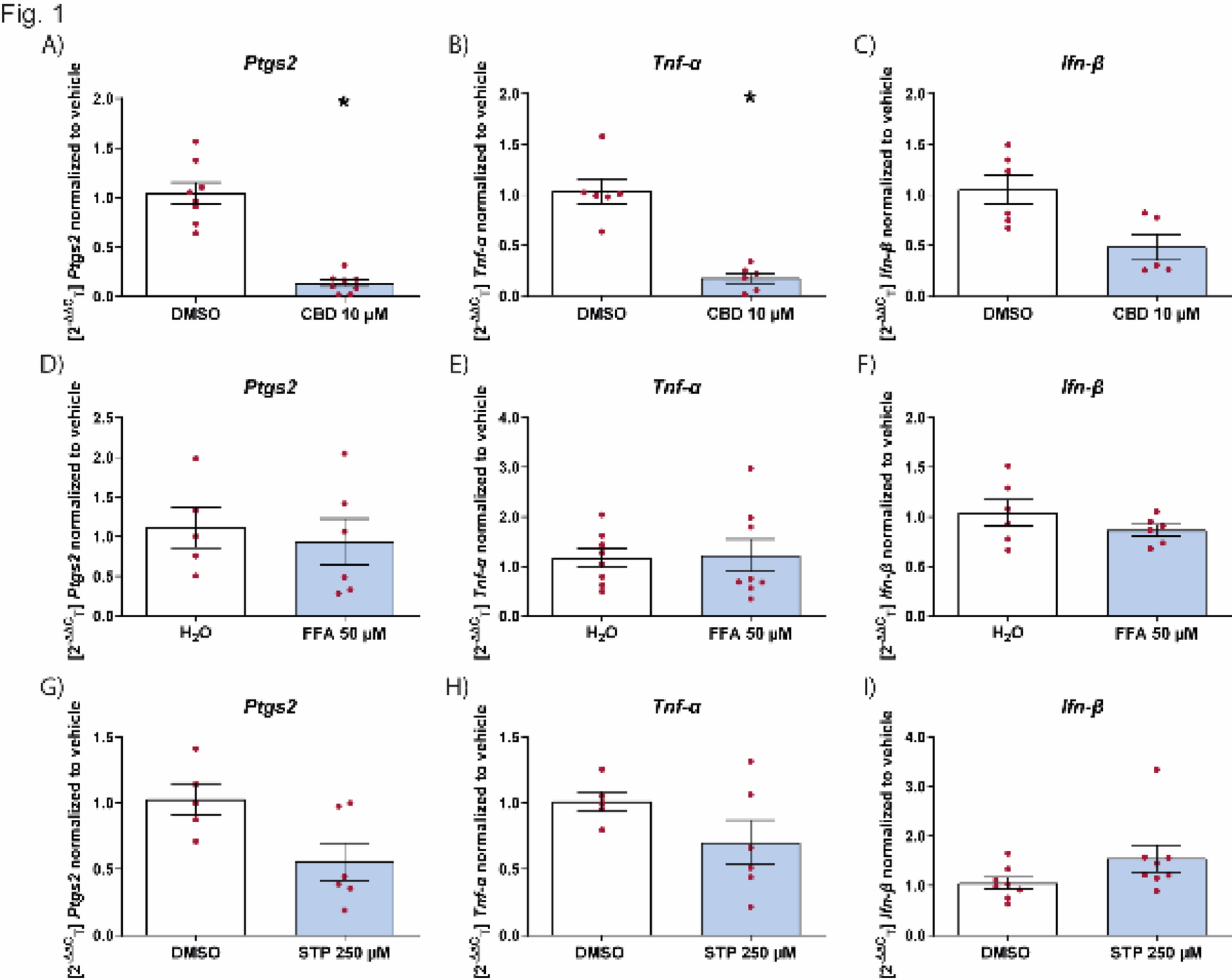
RNA was isolated from whole tissue fragment using RNA-XPress™ Reagent (MB601, HIMEDIA) and reverse-transcribed using PrimeScript RT reagent Kit, with gDNA Eraser (Perfect Real-Time-TAKARA). Quantitative real-time PCR (qPCR) was performed using the 7300 Real-Time PCR System (Applied Biosystems), SYBR™ Select Master Mix (Applied Biosystems), and TaqMan™ Gene Expression Master Mix (Applied Biosystems). The qPCR TaqMan probes (Applied Biosystems) were as follows: TGF-β Mm00441724_m1, TNF-α Mm00443258_m1, IL-6 Mm00446190_m1, IL-1β Mm00434228_m1, INF-γ Mm00801778_m1, IL-13 Mm00434204_m1. Relative expression levels are normalized using βact Mm02619580_g1 and GAPDH Mm99999915_g1.
Primers used in amplification using SYBR® Green method are listed in Table 1.
Table 1 Sequences of primers used for SYBR Green qPCR analysisStatistical analysis
The reported data are expressed as mean ± SEM of individual average measures of the different animals per group, for each assay. Significance of inter-group differences was evaluated by one-way ANOVA or two-way ANOVA followed by Bonferroni post hoc test for multiple comparisons. For qPCR analyses, significance is assessed by nonparametric Mann–Whitney U test. Calculations were done using Prism 6 statistical software (GraphPad Software, Inc., USA). The results are statistically significant when p < 0.05.


Comments (0)