Obesity is characterized by a chronic subclinical inflammation, commonly referred to as meta-inflammation believed to originate in adipose tissue and intricately connected to the progression of obesity-related comorbidities (Gomes et al. 2021). Obesity associates with macrophage infiltration in adipose tissue. Within this context, infiltrating macrophages interact with adipocytes, collectively contributing to a systemic state of chronic metabolic inflammation, in which the adipocytes and macrophages produce, secrete, and respond to their own stimuli, each cellular type interacts with each other, affecting the lipid and carbohydrate metabolism (Gomes et al. 2021; Xie et al. 2020). The proximity of adipocytes and macrophages in obese individuals’ adipose tissue, triggered the interest for the bidirectional interaction between these cells (Gomes et al. 2021), since regulation of these interactions and their affected transcriptomes and secretomes, may be considered as a therapeutic alternative in the control of metabolic disease.
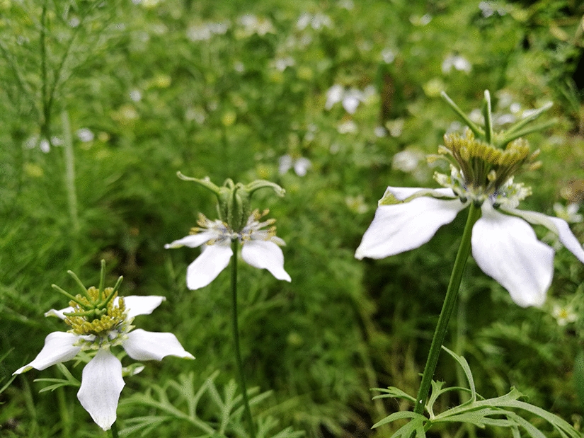
We have demonstrated in previous studies that the aqueous extract from fruit of C. ficifolia induces hypoglycemic, hypolipidemic, antioxidant, and anti-inflammatory effects in different in vivo and in vitro experimental models, including 3T3-L1 adipocytes and C2C12 myocytes (Roman-Ramos et al. 2012; Diaz et al. 2012; Fortis et al. 2013; Rosiles et al. 2022). The present study aims to investigate the effects of C. ficifolia on the transcriptomes and secretomes involved in the crosstalk between adipocytes and macrophages, using conditioned mediums exchange and indirect simultaneous co-cultures.
We hypothesized that C. ficifolia could regulate the communication between macrophages and adipocytes, thereby influencing cytokine production and release, glucose, and lipid transporters, as well as the expression of PPARs.
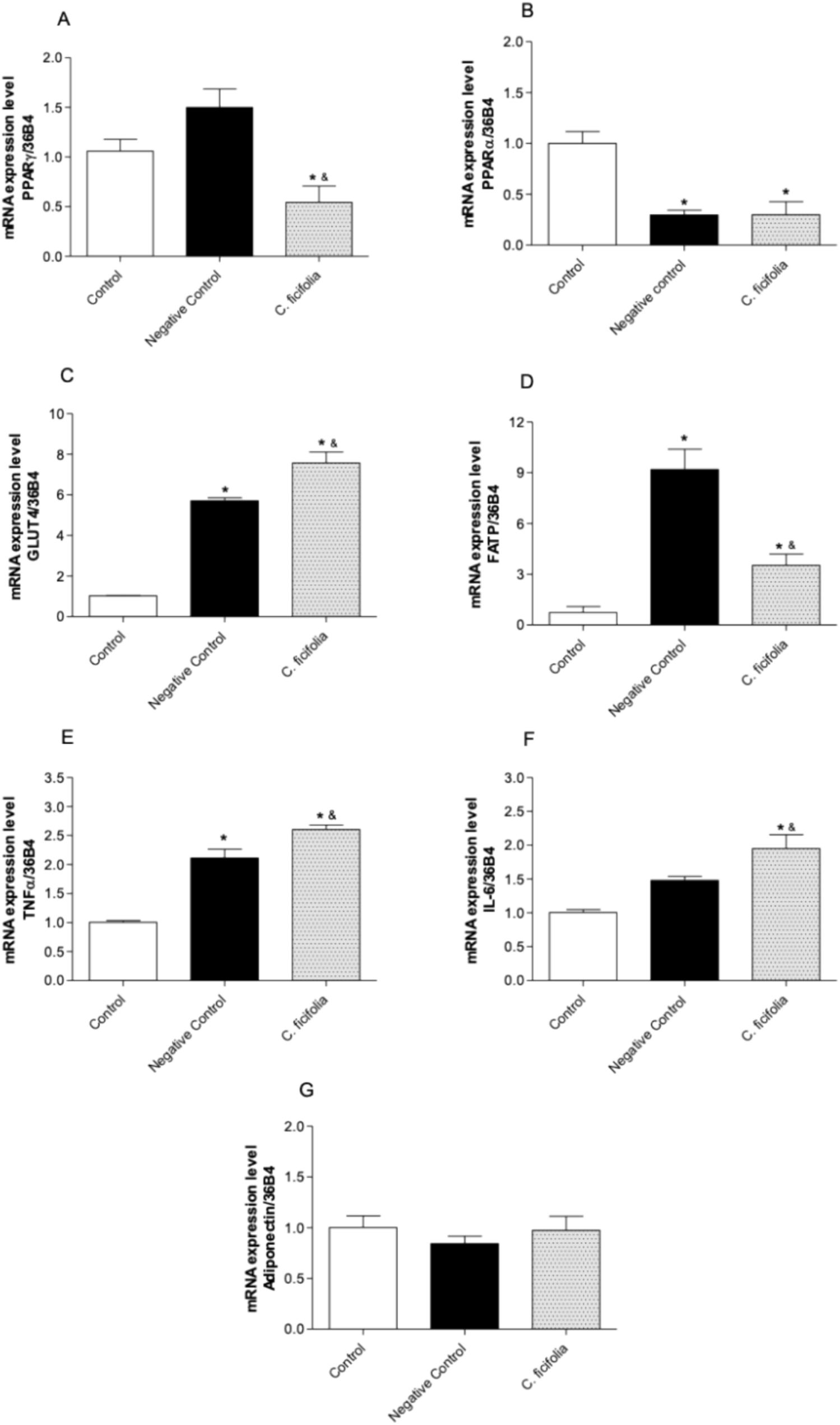
To test this hypothesis, we employed a conditioned mediums exchange model. In this model, 3T3-L1 adipocytes were exposed to the secretome from RAW 264.7 macrophages, both with and without treatment of C. ficifolia extract. The objective was to study the effects on the transcriptome and secretome associated with inflammatory factors in adipocytes, including PPARγ, PPARα, GLUT-4, FATP-1, TNF-α, IL-6 and AdipoQ. In other model, the cells were reverted, where RAW 264.7 macrophages were exposed to secretome from 3T3-L1 adipocytes with and without treatment of C. ficifolia extract, also studding the transcriptome and secretome associated with inflammatory factors of macrophages, like TNF-α, IL-6, IL-10, IL-1β, and TLR-4. Finally, we investigated an indirect simultaneous co-culture model involving both cell types after treatment with C. ficifolia extract and salicin, one of the main components in the extract. In general, extract decreased the secretomes of RAW 264.7 macrophages and 3T3-L1 adipocytes in conditions of exchange of conditioned mediums and simultaneous co-culture, causing significant modifications in TNF-α, IL-6, IL-1β, and PPARγ.
The differential impact of C. ficifolia on RAW 264.7 macrophages and 3T3-L1 adipocytes in these experimental conditions was consistent with previous observations that C. ficifolia plays a role in the cytokine secretion (Fortis et al. 2013). The conditioned medium from RAW 264.7 macrophages (negative control) did not impact the PPARγ, IL-6 nor AdipoQ transcriptome of adipocytes. However, it significantly decreased PPARα while increasing GLUT-4, FATP-1, and TNF-α (p < 0.05). In conjunction, these results suggest a pro-inflammatory profile induced by macrophages on adipocytes (Fig. 6). Interestingly, the conditioned medium from macrophages on adipocytes reduced the secretome, particularly for TNF-α, IL-6, and IL-10, while increasing IL-1β. Considering the decrease in IL-10 and the increase in IL-1β, these data suggest a certain promotion of the inflammation also. However, the relative transcript concentrations in adipocytes remained unchanged when treated with the conditioned medium from macrophages incubated with C. ficifolia extract, except for PPARγ that was significantly decreased and for IL-6 that was decreased against negative control (Fig. 6). Under this condition, the secretome was decreased, particularly for IL-1β whereas the observed reductions of TNF-α and IL-10 was lesser that in negative control, improving the inflammatory profile.
In contrast, medium of adipocytes in macrophages, significantly increased only IL-10 transcript and secretome levels, without inducing significant changes in TNF-α and IL-6 against control. The conditioned medium of adipocytes (treated with C. ficifolia extract), significantly altered the transcriptome against negative control, increasing TNF-α and IL-6, whereas reducing IL-10 and TLR-4. Interestingly, the secretome was significantly reduced by C. ficifolia treatment, decreasing concentrations of TNF-α, IL-6, and IL-10 against negative control. The differences found among the transcriptome and secretome for TNF-α and IL-6, might be explained by the time of incubation. In previous studies, the C. ficifolia anti-inflammatory activity in adipocytes was reported at 24 and 48 h of (Fortis et al. 2013). Finally, the indirect simultaneous co-culture of adipocytes and macrophages with treatment of C. ficifolia extract or salicin resulted in reductions in the concentrations of proinflammatory cytokines like TNF-α, IL-6, and IL-1β, accompanied by a significant increase in PPARγ expression (Fig. 6).
Macrophage secretome (negative control) did not significantly alter the expression of PPARγ or AdipoQ in adipocytes. This is consistent with observations from previous studies conducted on the liver of STZ-induced diabetic mice and 3T3-L1 adipocytes after exposure to C. ficifolia extract (Fortis et al. 2017; Almanza et al. 2023). Adipocytes treated with the macrophages secretome exhibited reduced expression of PPARα compared to control adipocytes. Concurrently, the expression of GLUT-4, FATP-1, TNF-α, and IL6 was significantly increased. Remarkably, treatment with conditioned medium of macrophages treated with C. ficifolia extract reduced both PPARs and FATP-1, increasing GLUT-4, TNF-α, and IL-6 in adipocytes. Therefore, the effect of the secretome of macrophages in the transcriptome of adipocytes suggests deregulation of the inflammatory process and stimulation of the inflammation by C. ficifolia extract. Interestingly, although increased inflammation was observed, GLUT-4 also increased, probably as a compensatory response to insulin resistance promoted by pro-inflammatory cytokines. However, this pro-inflammatory response also depends on incubation times, which should be investigated in further experiments.
PPARγ expression only increased significantly in conditions of simultaneous coculture, whereas GLUT-4 expression was increased in adipocytes treated with conditioned medium from C. ficifolia extract treated macrophages. Despite that the conditioned medium of macrophages treated with C. ficifolia did not increase PPARγ expression in adipocytes, GLUT-4, increased significantly, both in adipocytes incubated with both mediums of macrophages, with or without treatment of C. ficifolia extract. The existence of an intermediary produced by macrophages is a possibility, which induce an increment in the expression of this glucose transporter. Compared to control adipocytes, FATP-1 expression significantly increased in adipocytes treated with the medium from macrophages (negative control) and conditioned medium; however, in comparison to the negative control, its expression was significantly reduced with treatment of C. ficifolia extract. Therefore, C. ficifolia extract alters the expression of the genes that codify for transporters associated with the metabolism of carbohydrates and lipids. Concerning the secretome of adipocytes treated with conditioned medium of macrophages free of C. ficifolia extract, TNF-α, IL6, and IL-10 were significantly decreased, whereas IL-1β was increased compared with control adipocytes. Conditioned medium of macrophages treated with C. ficifolia reversed the effects on TNF-α, IL-10, and IL-1β compared with negative control, whereas that the reduction of the IL-6 concentration was yet more prominent.
To further explore and deepen the study of the bidirectional crosstalk between adipocytes and macrophages, we reversed our experimental model. Conditioned medium from 3T3-L1 adipocytes with and without treatment of C. ficifolia, was used to explore whether these secretomes might alter the transcriptomes and secretomes of macrophages. It is well known that LPS activated macrophages release higher levels of pro-inflammatory cytokines and lower levels of anti-inflammatory adipokines/cytokines (Oliver et al. 2009). In our study, LPS was substituted by the conditioned medium of adipocytes, treated or not, with C. ficifolia. The culture medium of adipocytes contains fatty acids that stimulate TLR-4 expression, similarly to LPS.
We investigated the impact of conditioned mediums from 3T3-L1 adipocytes on the expression of TNF-α, IL6, IL-10, and TLR-4 and on the secreted factors in the medium of TNF-α, IL6, IL-10, from macrophages. Only the expression of IL-10 in RAW 264.7 macrophages was significantly increased with conditioned medium of adipocytes free of C. ficifolia extract treatment. In contrast, transcriptome of macrophages incubated with adipocytes’ medium, the conditioned medium of adipocytes treated with C. ficifolia extract significantly increased TNF-α and IL6, whereas reduced IL-10 and TLR-4 expression. A reduction of inflammatory cytokines might be expected in the transcriptome of macrophages by the conditioned medium of adipocytes, due C. ficifolia extract has anti-inflammatory effects, without changes in macrophages. Nevertheless, in previous studies, the incubation times for the anti-inflammatory effect of C. ficifolia extract in adipocytes was of 24 h, whereas for macrophages with conditioned medium was only of 0.5 h.
Regarding the macrophages’ secretome treated with medium of adipocytes, TNF-α, IL6, and IL-10 were increased compared with control adipocytes, but significantly decreased in conditioned medium of adipocytes treated with C. ficifolia extract. Therefore, the secretome certainly exhibited the anti-inflammatory effect of the conditioned medium, by incubation with C. ficifolia, on the macrophages. This effect let us see that the extract of C. ficifolia doesn’t affect the transcription level, rather it may be involved in the inactivation or inhibition of the liberation of pro-inflammatory cytokines. Finally, under conditions of simultaneous coculture, both treatments with C. ficifolia extract and salicin resulted in a significant reduction in the concentration of proinflammatory cytokines (TNF-α, IL-6, and IL-1β), which are implicated in the stimulation of the inflammatory process. Also, C. ficifolia extract significantly increased PPARγ expression.
Previous in vivo and in vitro studies have reported the effects of C. ficifolia extract on inflammatory mediators in the secretomes of adipocytes (Fortis et al. 2013, 2017). In these studies, C. ficifolia was proposed as a cytokine’s inflammatory modulator in 3T3-L1 adipocytes, which was confirmed in monosodium glutamate-induced obesity mice, increasing the expression of IFNγ and IL-10, whereas decreasing the expression of IL-6, TNF-α, and resistin, without changes in AdipoQ’s expression (Fortis et al. 2013, 2017). Also, a recent report demonstrated the regulation of PPARα in the liver, both in vivo in liver of STZ-induced diabetic mice and in vitro in HepG2 hepatocytes (Almanza et al. 2023). Although IFNγ was not considered in the present study, the reported regulation of C. ficifolia on IFNγ might indicate an increase in immune system response to counteract the inflammatory environment. Several lines of evidence support a role for IFN-γ in mediating metabolic complications associated with obesity; IFN-γ promotes lipolysis in cultured mouse adipocytes and inhibits adipocytes differentiation (Gomes et al. 2021). While we cannot exclude the role of other pro and anti-inflammatory mediators, these results suggest that non-inflammatory signaling pathways may be involved in the regulation of meta-inflammation by C. ficifolia.
The evidence demonstrates a relationship between C. ficifolia, and cellular receptors involved in metabolism, improving metabolic and inflammatory patterns. This reinforces the role of C. ficifolia in the immune and metabolic crosstalk within adipose tissue, leading to a reduction in the expression and secretion of pro-inflammatory cytokines. However, our study has some limitations. First, the same parameters were not measured in all the experimental conditions and second, the activity of the implicated proteins was not confirmed. Both conditions must be confirmed in future studies as we only measured gene expression and concentrations in the medium. In addition, the evaluation of the different molecules found in C. ficifolia such as 4-hydroxy benzoic acid, ß-sitosterol, p-coumaric acid, gallic acid, D-chiro inositol, etc. and their mixes will be a priority study. In agreement with Gomes et al., (2021), agents in C. ficifolia, like salicin and ß-sitosterol, could represent attractive candidates for this metabolic immunomodulation in inflammatory obesity (Gomes et al. 2021).
Our data support the hypothesis that C. ficifolia regulates communication between macrophages and adipocytes, reducing the secretion of proinflammatory cytokines and possibly regulating the insulin resistance. This is a unique study, because it addresses how C. ficifolia, and possibly salicin, progressively affect both macrophage and adipocyte function. This is particularly relevant, as this situation likely occurs during the development of obesity in more than 1 billion adults worldwide (Xie et al. 2020). For example, the depression of GLUT4 transcript levels in 3T3-L1 adipocytes by macrophages and macrophage cytokines, along with the observations that macrophage IL-1β and IL-6 transcript levels were altered by coculture with adipocytes treated with C. ficifolia extract (Fig. 6), reaffirm the regulatory impact of this extract on the interaction between these two cell types. Our data suggest that TNF-α, IL-6, IL-1β may be the more potent contributor under the regulation of C. ficifolia. IL-6 inhibits Akt phosphorylation and all these proinflammatory cytokines may impair the adipocyte function and would contribute to the development of insulin resistance, in where IL-1β and TNF-α act in synergy with IL-6 (Xie et al. 2020).


Comments (0)