Kaposiform hemangioendothelioma in the mandibular gingiva: a rare case report with literature review
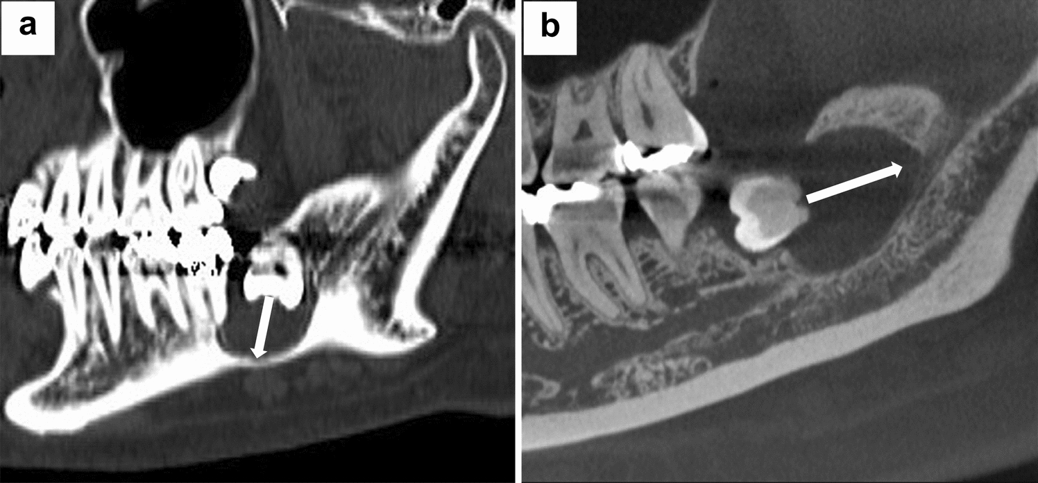
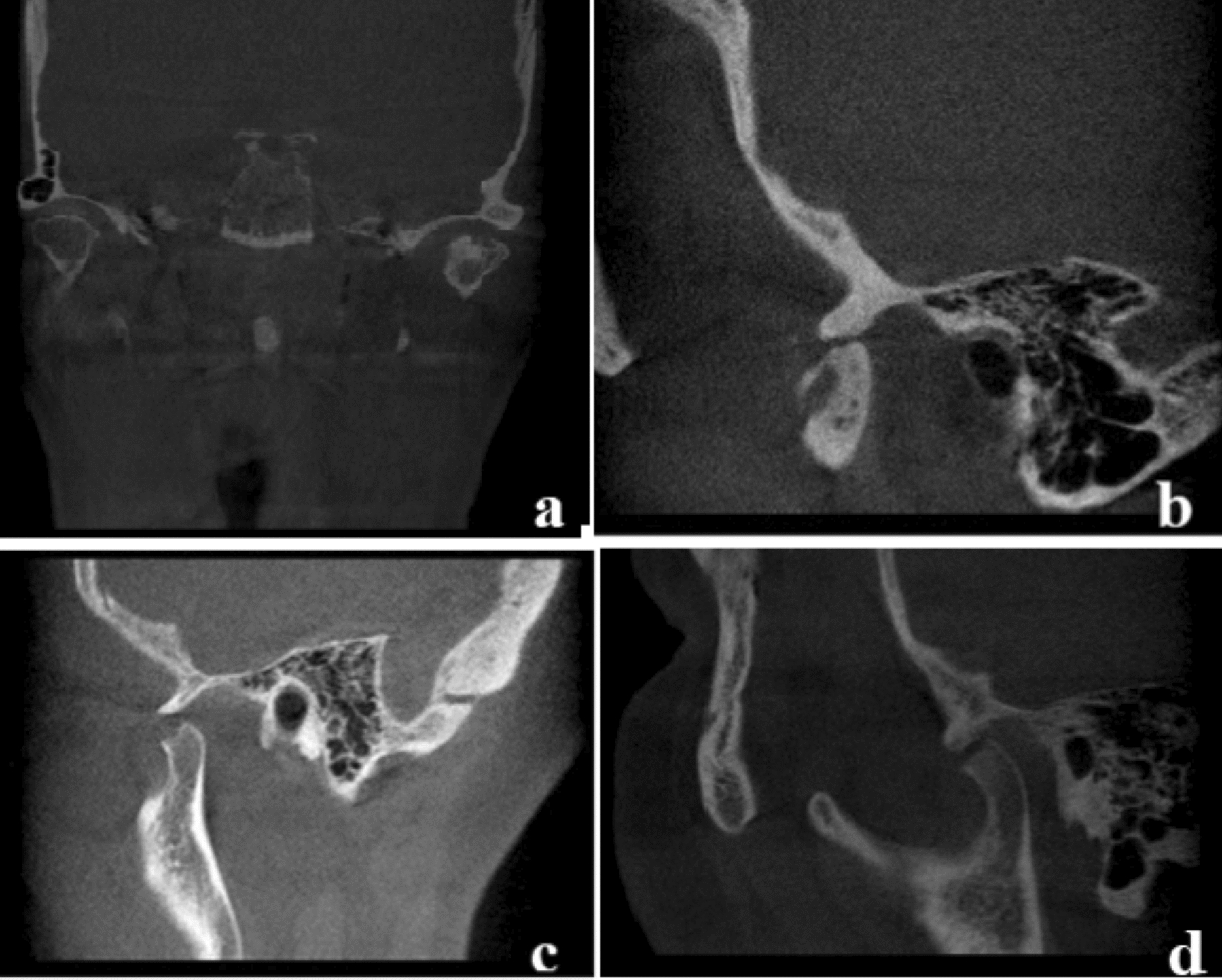
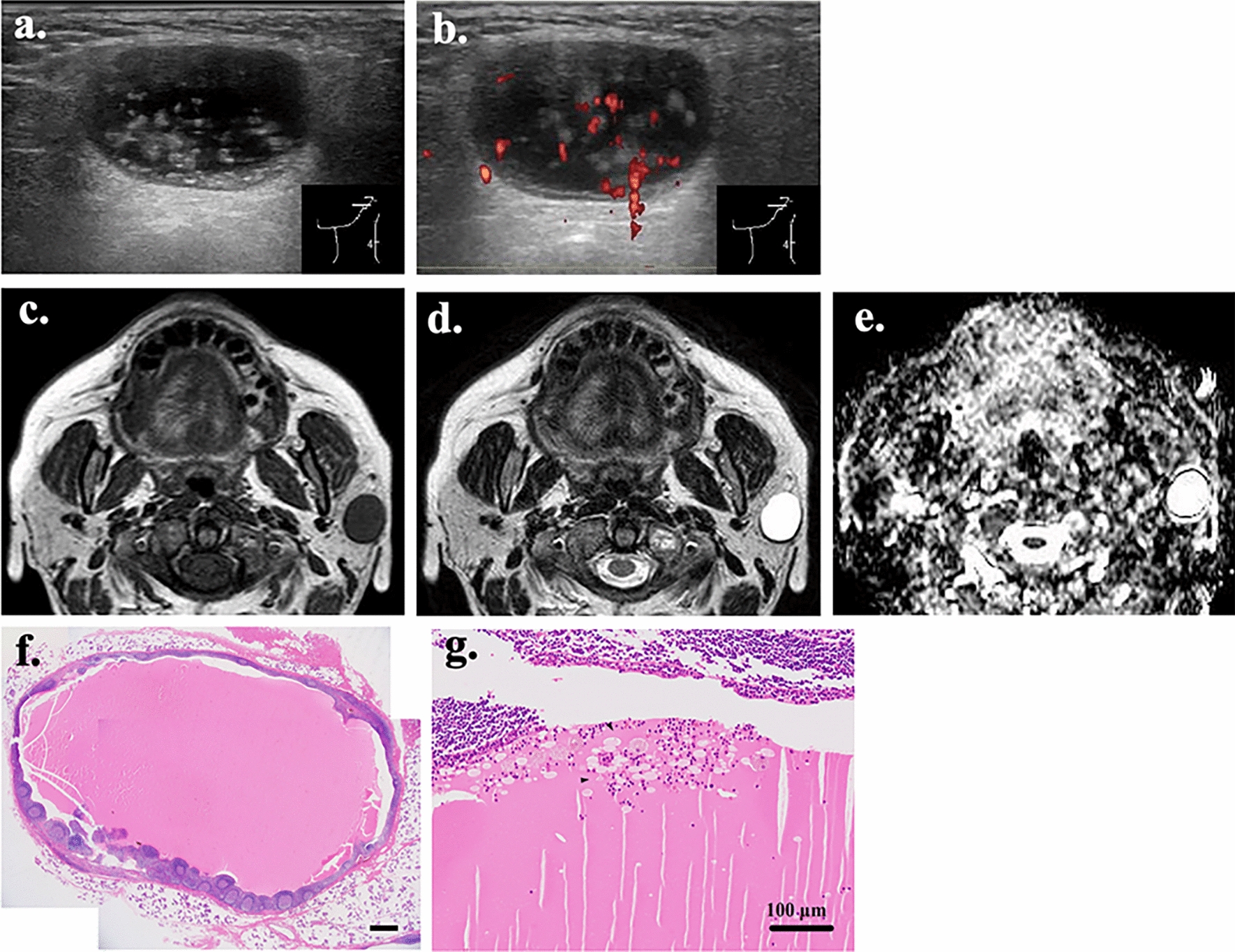
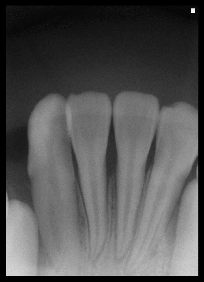
Kaposiform hemangioendothelioma (KHE) is a locally aggressive vascular tumor in neonates and children. KHE can occur anywhere in the body, but occurrence in the oral cavity is exceptionally rare. The clinical features of KHE are similar to those of oral lesions, such as infantine hemangioma, pyogenic granuloma, and chronic periodontitis. However, unlike other oral lesions, KHE invades neighboring tissue planes and recurs frequently as malignancy. Moreover, some instances of KHE are accompanied by a life-threatening condition named Kasabach–Merritt phenomenon. Correct diagnosis of KHE is very difficult despite its importance. KHE lacks not only specific clinical characteristics, but also distinct imaging findings even with advanced modalities such as computed tomography (CT) and magnetic resonance imaging (MRI). Thus, KHE is often misdiagnosed as another disease. We report a case of KHE in the mandibular gingiva. The enlarged gingival mass showed reddish granulomatous appearance with intact mucosal surface. It was misdiagnosed as another disease and underwent simple resection, but it recurred several times. In radiological examination using periapical radiography, cone beam computed tomography, and magnetic resonance imaging, no specific findings other than ill-defined alveolar bone destruction were observed. The subsequent pathological examination showed CD-31 and CD-34 positive, and D2-40 and HHV-8 negative. This report includes discussion about radiological, clinical, and histopathological features of the lesion and comprehensive literature review.

Comments (0)